Review
. 2020 Dec 20;25(24):6037.
doi: 10.3390/molecules25246037.

Revisiting the Self-Assembly of Highly Aromatic Phenylalanine Homopeptides

Affiliations
Review

Revisiting the Self-Assembly of Highly Aromatic Phenylalanine Homopeptides

Enric Mayans et al. Molecules. .

Abstract

Diphenylalanine peptide (FF), which self-assembles into rigid tubular nanostructures, is a very short core recognition motif in Alzheimer's disease β-amyloid (Aβ) polypeptide. Moreover, the ability of the phenylalanine (F or Phe)-homopeptides to self-assemble into ordered nanostructures has been proved. Within this context it was shown that the assembly preferences of this family of compounds is altered by capping both the N- and C-termini using highly aromatic fluorenyl groups (i.e., fluorenyl-9-methoxycarbonyl and 9-fluorenylmethyl ester, named Fmoc and OFm, respectively). In this article the work performed in the field of the effect of the structure and incubation conditions on the morphology and polymorphism of short (from two to four amino acid residues) Phe-homopeptides is reviewed and accompanied by introducing some new results for completing the comparison. Special attention has been paid to the influence of solvent: co-solvent mixture used to solubilize the peptide, the peptide concentration and, in some cases, the temperature. More specifically, uncapped (FF, FFF, and FFFF), N-capped with Fmoc (Fmoc-FF, Fmoc-FFF, and Fmoc-FFFF), C-capped with OFm (FF-OFm), and doubly capped (Fmoc-FF-OFm, Fmoc-FFF-OFm, and Fmoc-FFFF-OFm) Phe-homopeptides have been re-measured. Although many of the experienced assembly conditions have been only revisited as they were previously reported, other experimental conditions have been examined by the first time in this work. In any case, pooling the effect of highly aromatic blocking groups in a single study, using a wide variety of experimental conditions, allows a perspective of how the disappearance of head-to-tail electrostatic interactions and the gradual increase in the amount of π-π stacking interactions, affects the morphology of the assemblies. Future technological applications of Phe-homopeptides can be envisaged by choosing the most appropriate self-assemble structure, defining not only the length of the peptide but also the amount and the position of fluorenyl capping groups.

Keywords: molecular engineering; morphological engineering; nanostructures; peptides; staking interactions; supramolecular chemistry.


Conflict of interest statement

The authors declare no conflict of interest.

Figures

Scheme 1
Scheme 1
Chemical structures of highly aromatic N- and C-capping groups: fluorenyl-9-methoxycarbonyl (Fmoc) and 9-fluorenylmethyl ester (OFm), respectively.
Figure 1
Figure 1
Chemical structures of the peptides examined in this work.
Figure 2
Figure 2
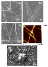
(ad) SEM and (b, right) AFM images of nano- and microtubular structures obtained from FF solutions in (a) 1:49 HFIP:water (0.1 mg/mL), (b) 1:99 HFIP:water (0.05 mg/mL), (c) 1:4 HFIP:water (1 mg/mL), and (d) 1:9 HFIP:EtOH (0.5 mg/mL) mixtures.
Figure 3
Figure 3
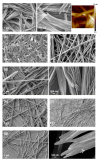
(ad) SEM and (c, right) AFM images of the structures obtained from FFFF solutions in (a) 1:99 HFIP:water (0.05 mg/mL), (b) 1:9 (0.5 mg/mL), (c) 1:4 (1 mg/mL) HFIP:water, and (d) 1:4 (1 mg/mL) HFIP:EtOH mixtures.
Figure 4
Figure 4
Sketch of the hexagonal and pseudo-hexagonal self-assembly of (a) FF and (b) FFFF, respectively. The increase of the conformational flexibility and the reduction in the density of head-to-tail electrostatic interactions led to the apparition of surface defects in FFFF.
Figure 5
Figure 5
SEM images of the structures obtained from FFF solutions in (a) 1:99 HFIP:water (0.05 mg/mL), (b) 1:9 HFIP:water (0.5 mg/mL), and (c) 4:6 HFIP:water (2 mg/mL).
Figure 6
Figure 6
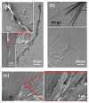
(a, left) Optical microscopy and (ac) SEM images of the structures obtained from FFF solutions in (a) 1:99 HFIP:MeOH (0.05 mg/mL), (b) 1:99 HFIP:iPrOH (0.05 mg/mL), and (c) 1:49 HFIP:iPrOH (0.1 mg/mL) mixtures.
Figure 7
Figure 7
Sketches summarizing the assemblies formed by Phe-homopeptides as a function of the co-solvent, the amount of co-solvent (i.e., the peptide concentration) and the number of Phe residues: (a) uncapped and (b) capped at both the N- and C-terminus with fluorenyl groups.
Figure 8
Figure 8
Fmoc-FF hydrogel as simultaneously reported by (a) Ulijn and co-workers [44] and (b) Gazit and co-workers [45]. (a) Glass vial containing the Fmoc-FF hydrogel, labeled as 7 in reference 44 (left), cryogenic SEM image of the hydrogel (middle) and micrographs of cells cultured on the hydrogel (right). (b) Photograph of the macroscopic hydrogel (left), TEM image of the fibrous scaffold (middle) and SEM image of the hydrogel (right). Adapted with permission from references [44,45].
Figure 9
Figure 9
(a) SEM micrographs of dried Fmoc-FFF hydrogels obtained at moderate peptide concentration (1.36 mg/mL). Taken with permission from [48]. (b) AFM height images (scale bar: 100 nm) of Fmoc-FFF made of all-L (b1), all-D (b2), LDD (b3) and DLL (b4) amino acids. Arrows indicate fibers where twisting is clearly visible. Magnified AFM height images of the twisted fibers are shown below (scale bar: 50 nm). Adapted with permission from [50].
Figure 10
Figure 10
SEM micrographs of the structures obtained from Fmoc-FFFF solutions in (a) 1:49 HFIP:water (0.1 mg/mL), (b) 1:9 HFIP:water (0.5 mg/mL), and (c) 3:47 HFIP:EtOH (0.3 mg/mL).
Figure 11
Figure 11
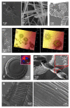
SEM micrographs of the structures obtained at 4 °C from TFA·FF-OFm solutions in (a) 4:6 HFIP:water (2 mg/mL) and (b,c) 1:9 HFIP:water (0.5 mg/mL) mixtures. The solution deposited in the coverslips was fresh in (b) while it was previously stored for 12 days at room temperature in (c). (d) AFM images (20 × 20 and 6 × 6 μm2 at the top and the bottom, respectively) and (d,e) SEM images of the structures obtained at room temperature from TFA·FF-OFm solutions in (d) 4:1 HFIP:water (50 mM KCl) (4 mg/mL) and (e) 1:9 HFIP:water (50 mM KCl) (0.5 mg/mL).
Figure 12
Figure 12
(ad) SEM micrographs and (a) AFM image (5.4 × 5.4 µm2) of the structures obtained at room temperature from TFA·FF-OFm solutions in (a) 1:4 HFIP:MeOH, (b) 1:4 DMF:MeOH, (c) 1:4 HFIP:chloroform mixtures (1 mg/mL in all cases), and (d) pure HFIP (5 mg/mL).
Figure 13
Figure 13
(ac) SEM and (b) AFM (20 × 20 µm2) images of the structures obtained from Fmoc-FF-OFm solutions in (a) 1:99 HFIP:water (0.05 mg/mL), (b) 1:9 HFIP:water (0.5 mg/mL), and (c) 4:1 HFIP:water (4 mg/mL) mixtures.
Figure 14
Figure 14
SEM and optical (inset in b) micrographs of the structures obtained from Fmoc-FF-OFm solutions in (a) 1:4 HFIP:MeOH (1 mg/mL), (b) 4:1 HFIP:iPrOH (4 mg/mL), and (c) 4:1 HFIP:acetone (4 mg/mL) mixtures.
Figure 15
Figure 15
(ag) SEM and (a) AFM (7 × 7 µm2) images of the structures obtained from Fmoc-F*F-OFm solutions in (a) 4:6 HFIP:water (2 mg/mL), (b) 1:9 HFIP:water (0.5 mg/mL), (c) 4:1 HFIP:water (4 mg/mL), (d) 4:6 HFIP:MeOH (2 mg/mL), (e) 4:6 HFIP:iPrOH (2 mg/mL), (f) 1:19 HFIP:iPrOH (0.25 mg/mL), and (g) 4:6 HFIP:acetone (2 mg/mL) mixtures.
Figure 16
Figure 16
(a,b,d,e) SEM, (c,d) optical and (c) AFM (left: 60 × 60 µm2; right: 30 × 30 µm2) images of the structures obtained from Fmoc-FFF-OFm solutions in (a) 1:99 HFIP:water (0.05 mg/mL), (b) 1:4 HFIP:water (1 mg/mL), (c) 4:1 HFIP:water (4 mg/mL), (d) HFIP (5 mg/mL), (e) 4:6 HFIP:EtOH (2 mg/mL), and (f) 1:9 HFIP:iPrOH (0.5 mg/mL) mixtures.
Figure 17
Figure 17
(ac) SEM and (df) optical micrographs of the structures obtained from Fmoc-FFFF-OFm solutions in (a) 1:9 HFIP:water (0.5 mg/mL), (b) 1:9 HFIP:EtOH (0.5 mg/mL), (c) 1:99 HFIP:EtOH (0.05 mg/mL), (d) 1:49 HFIP:EtOH (0.1 mg/mL), (e) 1:4 HFIP:EtOH (1 mg/mL), and (f) 1:9 HFIP:MeOH (0.5 mg/mL) mixtures.

References

    1. Dobson C.M. The amyloid phenomenon and its links with human disease. Cold Spring Harb. Perspect. Biol. 2017;9:a023648. doi: 10.1101/cshperspect.a023648. - DOI - PMC - PubMed
    1. Chiti F., Dobson C.M. Protein misfolding, amyloid formation, and human disease: A summary of progress over the last decade. Ann. Rev. Biochem. 2017;86:27–68. doi: 10.1146/annurev-biochem-061516-045115. - DOI - PubMed
    1. Takai E., Ohashi G., Ueki R., Yamada Y., Fujita J.I., Shiraki K. Scanning electron microscope imaging of amyloid fibrils. Am. J. Biochem. Biotechnol. 2014;10:31–39. doi: 10.3844/ajbbsp.2014.31.39. - DOI
    1. Ruggeri F.S., Sneideris T., Vendruscolo M., Knowles T.P.J. Atomic force microscopy for single molecule characterisation of protein aggregation. Arch. Biochem. Biophys. 2019;664:134–148. doi: 10.1016/j.abb.2019.02.001. - DOI - PMC - PubMed
    1. Dharmadana D., Reynolds N., Dekiwadia C., Conn E.C., Valéry C. Heparin assisted assembly of somatostatin amyloid nanofibrils results in disordered precipitates by hindrance of protofilaments interactions. Nanoscale. 2018;10:18195–18204. doi: 10.1039/C8NR02159G. - DOI - PubMed