Review
. 2021 Feb 1;71(1):3-45.
doi: 10.30802/AALAS-CM-20-000054. Epub 2021 Jan 8.

A One Health Perspective for Defining and Deciphering Escherichia coli Pathogenic Potential in Multiple Hosts

Affiliations
Review

A One Health Perspective for Defining and Deciphering Escherichia coli Pathogenic Potential in Multiple Hosts

Alexis García et al. Comp Med. .

Abstract

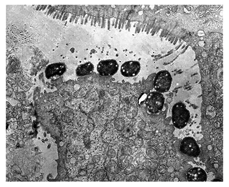
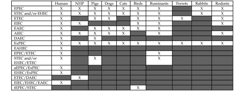
E. coli is one of the most common species of bacteria colonizing humans and animals. The singularity of E. coli 's genus and species underestimates its multifaceted nature, which is represented by different strains, each with different combinations of distinct virulence factors. In fact, several E. coli pathotypes, or hybrid strains, may be associated with both subclinical infection and a range of clinical conditions, including enteric, urinary, and systemic infections. E. coli may also express DNA-damaging toxins that could impact cancer development. This review summarizes the different E. coli pathotypes in the context of their history, hosts, clinical signs, epidemiology, and control. The pathotypic characterization of E. coli in the context of disease in different animals, including humans, provides comparative and One Health perspectives that will guide future clinical and research investigations of E. coli infections.


Figures

Figure 1.
Figure 1.
Transmission electron micrograph showing organisms consistent with E. coli and associated attaching and effacing lesions and pedestals on the cecal mucosal surface of an experimentally infected Dutch Belted rabbit.
Figure 2.
Figure 2.
Natural EPEC infections: Hosts, manifestations, and virulence determinants.*, This column includes selected virulence determinants investigated in the cited references such as E. coli attaching and effacing (eae or eaeA) gene, bundle-forming pilus (bfp or bfpA) gene, and cytolethal distending toxin (cdt) gene. Typical E. coli usually encodes the bfpA gene whereas atypical does not. EAF refers to EPEC adherence factor plasmid. †, not characterized as typical or atypical.
Figure 3.
Figure 3.
Multifocal brainstem degeneration in a Dutch Belted rabbit after experimental intravenous Shiga toxin 2 infusion (hematoxylin and eosin stain, scale bar: 200 µm). Reprinted from García A, Marini RP, Catalfamo JL, Knox KA, Schauer DB, Rogers AB, Fox JG. 2008. Intravenous Shiga toxin 2 promotes enteritis and renal injury characterized by polymorphonuclear leukocyte infiltration and thrombosis in Dutch Belted rabbits. Microbes Infect 10:650–656, with permission from Elsevier. Reference .
Figure 4.
Figure 4.
Natural STEC/EHEC infections: Hosts, manifestations, and virulence determinants. *, This column includes selected virulence determinants investigated in the cited references such as stx genes and Stx production; slt (SLT) refers to Shiga-like toxin (Shiga toxin).
Figure 5.
Figure 5.
Natural ETEC infections: Hosts, manifestations, and virulence determinants. *, This column includes selected virulence determinants investigated in the cited references.
Figure 6.
Figure 6.
Natural EIEC infections: Hosts, manifestations, and virulence determinants. *, This column includes selected virulence determinants investigated in the cited references such as genes for type III secretion system effectors including ipaH and virA.,
Figure 7.
Figure 7.
Natural EAEC infections: Hosts, manifestations, and virulence determinants. *, This column includes selected virulence determinants such as the plasmid-borne transcriptional activator gene (aggR) and also the adherence phenotype investigated in the cited references.
Figure 8.
Figure 8.
Natural AIEC infections: Hosts, manifestations, and virulence determinants. *, This column includes selected virulence determinants and phenotypic characteristics investigated in the cited references.
Figure 9.
Figure 9.
Natural DAEC infections: Hosts, manifestations, and virulence determinants. *, This column includes selected virulence determinants investigated in the cited references.
Figure 10.
Figure 10.
Natural ExPEC infections: Hosts, manifestations, and virulence determinants. *, This column includes selected virulence determinants investigated in the cited references.
Figure 11.
Figure 11.
Natural hybrid E. coli infections: Hosts, manifestations, and virulence determinants. *, This column includes selected virulence determinants investigated in the cited references.
Figure 12.
Figure 12.
(A) Ferret naturally infected with E. coli exhibiting signs of mastitis including swollen and erythematous mammary tissue. (B) Hyperemic and hemorrhagic serosa at the level of the distal cecum adjacent to the junction with the proximal colon in a Dutch Belted rabbit experimentally infected with enterohemorrhagic E. coli O157:H7; Copyright © American Society for Microbiology, [Infection and Immunity 80, pages 369-380, 2012]. (C) Global intracellular edematous swelling, increased numbers of heterophils (arrows), and decreased number of erythrocytes (“bloodless glomerulus”) in a glomerulus of a Dutch Belted rabbit experimentally infected with enterohemorrhagic E. coli O153 (scale bar: 60 µm); García and colleagues, Renal Injury Is a Consistent Finding in Dutch Belted Rabbits Experimentally Infected with Enterohemorrhagic Escherichia coli, The Journal of Infectious Diseases, 2006, volume 193, issue 8, pages 1125-1134, by permission of the Infectious Diseases Society of America. (D) E. coli-associated necrotizing suppurative metritis (pyometra) in a naturally infected “alpha V integrin+/-; alpha v fl/+; Tie 2, Cre+/-” mouse. E. coli is fluorescently labeled with a green peptic nucleic acid in situ hybridization probe that detected bacteria in the affected and luminal areas of the uterus. The nuclei of the cells are stained blue with 4’,6’-diamidino-2-phenylindole (DAPI) (no scale bar: x100); Reprinted from Microbes and Infection18(12), García A, Mannion A, Feng Y, Madden CM, Bakthavatchalu V, Shen Z, Ge Z, Fox JG, Cytotoxic Escherichia coli strains encoding colibactin colonize laboratory mice, 777-786, Copyright (2016) with permission from Elsevier. (E) Renal section of a mouse naturally infected with cytotoxic E. coli (pks+) and exhibiting multifocal subacute suppurative pyelonephritis, intraluminal bacteria, and tubular necrosis (scale bar: 1 mm). (F) Brain section of a mouse naturally infected with cytotoxic E. coli (pks+) and exhibiting focally extensive subacute necrohemorrhagic meningoencephalitis (scale bar: 200 µm). Figures 12(E) and 12(F) have been reprinted from Bakthavatchalu and colleagues (2018) Cytotoxic Escherichia coli strains encoding colibactin isolated from immunocompromised mice with urosepsis and meningitis. PLoS One 13(3): e0194443. doi: 10.1371/journal.pone.0194443, with permission through an open access Creative Commons Attribution (CC BY) license. (C, E, F are hematoxylin and eosin stained sections).
Figure 13.
Figure 13.
Reported E. coli pathotypes or hybrids and the animals in which they have been identified. EPEC, enteropathogenic E. coli; STEC, Shiga toxin-producing E. coli; EHEC, enterohemorrhagic E. coli; ETEC, enterotoxigenic E. coli; EIEC, Enteroinvasive E. coli; DAEC, Diffusely adhering E. coli; ExPEC, Extraintestinal pathogenic E. coli; EAHEC, Entero-aggregative-hemorrhagic E. coli; aEPEC, atypical EPEC; tEPEC, typical EPEC; EPEC/ETEC, STEC and/or EHEC/ETEC, aEPEC/ExPEC, EHEC/ExPEC, ETEC/DAEC, EIEC/EHEC/EAEC, tEPEC/STEC are hybrids; X, reported; Empty box, not reported.

Similar articles

Cited by

References

    1. Abaas S, Franklin A, Kuhn I, Orskov F, Orskov I. 1989. Cytotoxin activity on Vero cells among Escherichia coli strains associated with diarrhea in cats. Am J Vet Res 50:1294–1296. - PubMed
    1. Acheson DW, Reidl J, Zhang X, Keusch GT, Mekalanos JJ, Waldor MK. 1998. In vivo transduction with shiga toxin 1-encoding phage. Infect Immun 66:4496–4498. 10.1128/IAI.66.9.4496-4498.1998. - DOI - PMC - PubMed
    1. Achtman M, Mercer A, Kusecek B, Pohl A, Heuzenroeder M, Aaronson W, Sutton A, Silver RP. 1983. Six widespread bacterial clones among Escherichia coli K1 isolates. Infect Immun 39:315–335. 10.1128/IAI.39.1.315-335.1983. - DOI - PMC - PubMed
    1. Acosta GJ, Vigo NI, Durand D, Riveros M, Arango S, Zambruni M, Ochoa TJ. 2016. Diarrheagenic Escherichia coli: prevalence and pathotype distribution in children from Peruvian rural communities. Am J Trop Med Hyg 95:574–579. 10.4269/ajtmh.16-0220. - DOI - PMC - PubMed
    1. Acres SD. 1985. Enterotoxigenic Escherichia coli infections in newborn calves: a review. J Dairy Sci 68:229–256. 10.3168/jds.S0022-0302(85)80814-6. - DOI - PMC - PubMed

Substances

LinkOut - more resources