. 2021 Apr 29;57(4):2003755.
doi: 10.1183/13993003.03755-2020. Print 2021 Apr.

Proteinase release from activated neutrophils in mechanically ventilated patients with non-COVID-19 and COVID-19 pneumonia

Affiliations

Proteinase release from activated neutrophils in mechanically ventilated patients with non-COVID-19 and COVID-19 pneumonia

Seda Seren et al. Eur Respir J. .

Abstract

COVID-19 ARDS is associated with release of biologically active neutrophil elastase-related proteinases to the airways and blood at a comparable level to non-COVID ARDS https://bit.ly/3nihveh


Conflict of interest statement

Conflict of interest: S. Seren has nothing to disclose. Conflict of interest: L. Derian has nothing to disclose. Conflict of interest: I. Keleş has nothing to disclose. Conflict of interest: A. Guillon has nothing to disclose. Conflict of interest: A. Lesner has nothing to disclose. Conflict of interest: L. Gonzalez has nothing to disclose. Conflict of interest: T. Baranek has nothing to disclose. Conflict of interest: M. Si-Tahar has nothing to disclose. Conflict of interest: S. Marchand-Adam has nothing to disclose. Conflict of interest: D.E. Jenne has nothing to disclose. Conflict of interest: C. Paget has nothing to disclose. Conflict of interest: Y. Jouan has nothing to disclose. Conflict of interest: B. Korkmaz has been paid for the time spent as a committee member for advisory boards (INSMED), other forms of consulting (Neuprozyme Therapeutics Aps (Denmark), Santhera Pharmaceuticals (Switzerland)), symposium organisation (INSMED) and travel support, lectures or presentations, outside the submitted work.

Figures
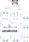
FIGURE 1
FIGURE 1
Neutrophils and proteolytically active proteinases in endotracheal aspirates (ETAs). Schematic of the workflow is shown above. ETAs collected from mechanically ventilated non-COVID-19 acute respiratory distress syndrome (ARDS) and coronavirus disease 2019 (COVID-19) ARDS patients from intensive care unit were weighed and incubated in PBS (5 mL·g−1) with 1 mM dithiothreitol (DTT) for 30 min at 4°C under gentle agitation. After centrifugation, supernatants were collected and then concentrated five times before total protein quantification by the BCA or Bradford assay. Cell pellets were filtered through a 100 µm cell strainer. Red blood cells were removed using a red blood cell lysis buffer and then cell suspensions from ETAs were passed through a 40 µm cell strainer prior to staining for flow cytometry. a) Cells were stained with antibodies against surface markers (CD45, CD14 and CD16) and viability dye. Neutrophils were defined as live CD45+ CD14 CD16+ cells. Neutrophil relative proportion in ETAs of all non-COVID-19 ARDS and COVID-19 ARDS patients is represented as percentage of live leukocytes (CD45+ cells). Individuals are depicted. The data represent mean±sem. Mann–Whitney test. b) Quantitative detection of myeloperoxidase (MPO) in ETA supernatants (dilution: 1/500) from 14 non-COVID-19 ARDS and 14 COVID-19 ARDS patients using a human MPO Elisa kit (DMYE00B, R&D Systems). Individuals are depicted. The data represent mean±sem. Mann–Whitney test. c) The concentration of active cathepsin C (CatC) was estimated by comparison of the rate of hydrolysis of FRET (fluorescence resonance energy transfer) substrate by ETA supernatants of all non-COVID-19 ARDS and COVID-19 ARDS patients and recombinant CatC. Each point represents data of two independent experiments, both in duplicate, from an individual subject. Individuals are depicted. The data represent mean±sem. Mann–Whitney test. d) The CatC activity in five times concentrated ETA supernatants (4 µg protein) was measured spectrofluorometrically at 460 nm with or without the nitrile inhibitor (L)-Thi-(L)-Phe-CN (0.5 µM final, 20 min incubation at 37°C) using Thi-Ala(Mca)-Ser-Gly-Tyr(3-NO2)-NH2 (20 µM final) as selective FRET substrate. The residual activities were inhibited by the CatC inhibitor (L)-Thi-(L)-Phe-CN which demonstrate the presence of CatC activity in samples. Inhibition experiments were performed with all ETA samples used in the study. Results with ETA samples from 13 non-COVID-19 ARDS are shown. Data represent the mean±sd of two independent experiments, both performed with duplicates of the same sample. Similar results were obtained with ETA supernatants of all COVID-19 ARDS patients. FU: fluorescence unit. e) Western blotting (WB) of concentrated ETA supernatants (30 µg of protein) from 13 non-COVID-19 ARDS (no 1–13) using an anti-CatC antibody (sc-74590, 1:1000, Santa Cruz Biotechnology) shows the presence of the CatC heavy chain. Similar results were obtained with ETA supernatants of all non-COVID-19 ARDS and COVID-19 ARDS patients in three independent experiments. f) The proteinase 3 (PR3) activity in ETA supernatants was measured at 420 nm with or without the PR3 inhibitor Ac-PYDAP(O-C6H6-4-Cl)2 (0.5 µM final, 20 min incubation at 37°C) using ABZ-VAD(nor)VADYQ-EDDnp (20 µM final) as a substrate. g) CatG activity in ETA supernatants was measured in the presence or absence of alpha-1-antichymotrypsin using ABZ-TPFSGQ-EDDnp (20 µM final) as a substrate. h) The neutrophil elastase (NE) activity in ETA supernatants was measured with or without NE inhibitor EPI-hNE-4 (0.5 µM final, 20 min incubation at 37°C) using ABZ-APEEIMRRQ-EDDnp as a substrate. The residual activities in ETA samples were inhibited by selective NSP inhibitors demonstrating the specificity of the FRET assays utilised. Inhibition experiments were performed with all ETA samples used in the study. Each point represents data of two independent experiments, both in duplicate, from an individual subject. Individuals are depicted. The data represent mean±sem. Mann–Whitney test. i) Quantitative detection of circulating NE. A human NE ELISA kit (HK319, HycultBiotech) was used for the quantitative detection of NE in sera (dilution: 1/100) from 10 non-COVID-19 ARDS and 10 COVID-19 ARDS patients. Individuals are depicted. The data represent mean±sem. Kruskal–Wallis test. ns: nonsignificant.

References

    1. Thompson BT, Chambers RC, Liu KD. Acute respiratory distress syndrome. N Engl J Med 2017; 377: 562–572. doi:10.1056/NEJMra1608077 - DOI - PubMed
    1. Sweeney RM, McAuley DF. Acute respiratory distress syndrome. Lancet 2016; 388: 2416–2430. doi:10.1016/S0140-6736(16)00578-X - DOI - PMC - PubMed
    1. Potey PM, Rossi AG, Lucas CD, et al. . Neutrophils in the initiation and resolution of acute pulmonary inflammation: understanding biological function and therapeutic potential. J Pathol 2019; 247: 672–685. doi:10.1002/path.5221 - DOI - PMC - PubMed
    1. Korkmaz B, Lesner A, Marchand-Adam S, et al. . Lung protection by cathepsin C inhibition: a new hope for COVID-19 and ARDS? J Med Chem 2020; 63: 13258–13265. doi:10.1021/acs.jmedchem.0c00776 - DOI - PubMed
    1. Korkmaz B, Caughey GH, Chapple I, et al. . Therapeutic targeting of cathepsin C: from pathophysiology to treatment. Pharmacol Ther 2018; 190: 202–236. doi:10.1016/j.pharmthera.2018.05.011 - DOI - PubMed

Substances