

Therapy-induced Deletion in 11q23 Leading to Fusion of KMT2A With ARHGEF12 and Development of B Lineage Acute Lymphoplastic Leukemia in a Child Treated for Acute Myeloid Leukemia Caused by t(9;11)(p21;q23)/ KMT2A-MLLT3
- PMID: 33419897
- PMCID: PMC7796818
- DOI: 10.21873/cgp.20242
Therapy-induced Deletion in 11q23 Leading to Fusion of KMT2A With ARHGEF12 and Development of B Lineage Acute Lymphoplastic Leukemia in a Child Treated for Acute Myeloid Leukemia Caused by t(9;11)(p21;q23)/ KMT2A-MLLT3
Abstract
Background/aim: Fusion of histone-lysine N-methyltransferase 2A gene (KMT2A) with the Rho guanine nucleotide exchange factor 12 gene (ARHGEF12), both located in 11q23, was reported in some leukemic patients. We report a KMT2A-ARHGEF12 fusion occurring during treatment of a pediatric acute myeloid leukemia (AML) with topoisomerase II inhibitors leading to a secondary acute lymphoblastic leukemia (ALL).
Materials and methods: Multiple genetic analyses were performed on bone marrow cells of a girl initially diagnosed with AML.
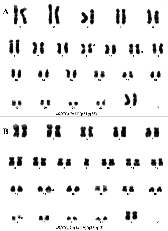
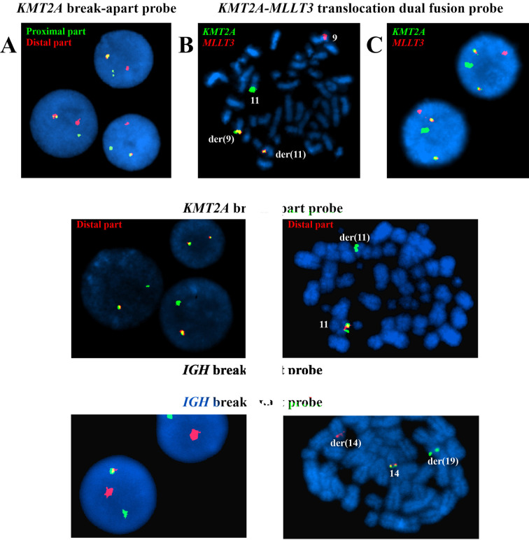
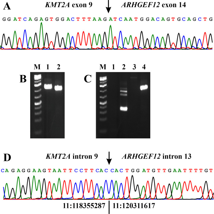
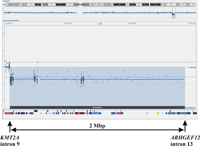
Results: At the time of diagnosis with AML, the t(9;11)(p21;q23)/KMT2A-MLLT3 genetic abnormality was found. After chemotherapy resulting in AML clinical remission, a 2 Mb deletion in 11q23 was found generating a KMT2A-ARHGEF12 fusion gene. When the patient later developed B lineage ALL, a t(14;19)(q32;q13), loss of one chromosome 9, and KMT2A-ARHGEF12 were detected.
Conclusion: The patient sequentially developed AML and ALL with three leukemia-specific genomic abnormalities in her bone marrow cells, two of which were KMT2A-rearrangements.
Keywords: ARHGEF12; KMT2A; KMT2A-ARHGEF12; KMT2A-MLLT3.; Pediatric leukemia; acute lymphoblastic leukemia; acute myeloid leukemia; chemotherapy; fusion gene.
Copyright© 2021, International Institute of Anticancer Research (Dr. George J. Delinasios), All rights reserved.
Conflict of interest statement
The Authors declare that they have no potential conflicts of interest in regard to this study.
Figures
Similar articles
-
Mutational landscape and clinical outcome of patients with de novo acute myeloid leukemia and rearrangements involving 11q23/KMT2A.Proc Natl Acad Sci U S A. 2020 Oct 20;117(42):26340-26346. doi: 10.1073/pnas.2014732117. Epub 2020 Oct 5. Proc Natl Acad Sci U S A. 2020. PMID: 33020282 Free PMC article.
-
Acute myeloid leukemia with t(10;11)(p11-12;q23.3): Results of Russian Pediatric AML registration study.Int J Lab Hematol. 2019 Apr;41(2):287-292. doi: 10.1111/ijlh.12969. Epub 2019 Jan 9. Int J Lab Hematol. 2019. PMID: 30624859
-
GAS6 Oncogene and Reverse MLLT3-KMT2A Duplications in an Infant with Acute Myeloid Leukemia and a Novel Complex Hyperdiploid Karyotype: Detailed High-Resolution Molecular Cytogenetic Studies.Cytogenet Genome Res. 2017;152(1):33-37. doi: 10.1159/000477108. Epub 2017 Jun 9. Cytogenet Genome Res. 2017. PMID: 28595195
-
Rare KMT2A-ELL and Novel ZNF56-KMT2A Fusion Genes in Pediatric T-cell Acute Lymphoblastic Leukemia.Cancer Genomics Proteomics. 2021 Mar-Apr;18(2):121-131. doi: 10.21873/cgp.20247. Cancer Genomics Proteomics. 2021. PMID: 33608309 Free PMC article. Review.
-
Recent Developments and Evolving Therapeutic Strategies in KMT2A-Rearranged Acute Leukemia.Cancer Med. 2024 Oct;13(20):e70326. doi: 10.1002/cam4.70326. Cancer Med. 2024. PMID: 39428967 Free PMC article. Review.
Cited by
-
The causal effects of 2,821 protein level ratios on non-small cell lung cancer: a two-sample Mendelian randomization study.Transl Cancer Res. 2025 Feb 28;14(2):1101-1110. doi: 10.21037/tcr-24-1523. Epub 2025 Jan 8. Transl Cancer Res. 2025. PMID: 40104726 Free PMC article.
-
Novel MYCBP::EHD2 and RUNX1::ZNF780A Fusion Genes in T-cell Acute Lymphoblastic Leukemia.Cancer Genomics Proteomics. 2023 Jan-Feb;20(1):51-63. doi: 10.21873/cgp.20364. Cancer Genomics Proteomics. 2023. PMID: 36581344 Free PMC article.
-
Autophagy-related circRNA evaluation reveals hsa_circ_0001747 as a potential favorable prognostic factor for biochemical recurrence in patients with prostate cancer.Cell Death Dis. 2021 Jul 22;12(8):726. doi: 10.1038/s41419-021-04015-w. Cell Death Dis. 2021. PMID: 34294687 Free PMC article.
-
KMT2A-ARHGEF12, a therapy related fusion with poor prognosis.Mol Biol Rep. 2021 Oct;48(10):7021-7027. doi: 10.1007/s11033-021-06621-5. Epub 2021 Aug 12. Mol Biol Rep. 2021. PMID: 34383244 Review.
-
Interstitial Deletions Generating Fusion Genes.Cancer Genomics Proteomics. 2021 May-Jun;18(3):167-196. doi: 10.21873/cgp.20251. Cancer Genomics Proteomics. 2021. PMID: 33893073 Free PMC article. Review.
References
-
- Mitelman F, Johansson B, Mertens F. Mitelman Database of Chromosome Aberrations and Gene Fusions in Cancer, 2020. Available at: https://mitelmandatabase.isb-cgc.org/
-
- Meyer C, Hofmann J, Burmeister T, Groger D, Park TS, Emerenciano M, Pombo de Oliveira M, Renneville A, Villarese P, Macintyre E, Cave H, Clappier E, Mass-Malo K, Zuna J, Trka J, De Braekeleer E, De Braekeleer M, Oh SH, Tsaur G, Fechina L, van der Velden VH, van Dongen JJ, Delabesse E, Binato R, Silva ML, Kustanovich A, Aleinikova O, Harris MH, Lund-Aho T, Juvonen V, Heidenreich O, Vormoor J, Choi WW, Jarosova M, Kolenova A, Bueno C, Menendez P, Wehner S, Eckert C, Talmant P, Tondeur S, Lippert E, Launay E, Henry C, Ballerini P, Lapillone H, Callanan MB, Cayuela JM, Herbaux C, Cazzaniga G, Kakadiya PM, Bohlander S, Ahlmann M, Choi JR, Gameiro P, Lee DS, Krauter J, Cornillet-Lefebvre P, Te Kronnie G, Schafer BW, Kubetzko S, Alonso CN, zur Stadt U, Sutton R, Venn NC, Izraeli S, Trakhtenbrot L, Madsen HO, Archer P, Hancock J, Cerveira N, Teixeira MR, Lo Nigro L, Moricke A, Stanulla M, Schrappe M, Sedek L, Szczepanski T, Zwaan CM, Coenen EA, van den Heuvel-Eibrink MM, Strehl S, Dworzak M, Panzer-Grumayer R, Dingermann T, Klingebiel T, Marschalek R. The MLL recombinome of acute leukemias in 2013. Leukemia. 2013;27(11):2165–2176. doi: 10.1038/leu.2013.135. - DOI - PMC - PubMed
-
- Meyer C, Burmeister T, Groger D, Tsaur G, Fechina L, Renneville A, Sutton R, Venn NC, Emerenciano M, Pombo-de-Oliveira MS, Barbieri Blunck C, Almeida Lopes B, Zuna J, Trka J, Ballerini P, Lapillonne H, De Braekeleer M, Cazzaniga G, Corral Abascal L, van der Velden VHJ, Delabesse E, Park TS, Oh SH, Silva MLM, Lund-Aho T, Juvonen V, Moore AS, Heidenreich O, Vormoor J, Zerkalenkova E, Olshanskaya Y, Bueno C, Menendez P, Teigler-Schlegel A, Zur Stadt U, Lentes J, Gohring G, Kustanovich A, Aleinikova O, Schafer BW, Kubetzko S, Madsen HO, Gruhn B, Duarte X, Gameiro P, Lippert E, Bidet A, Cayuela JM, Clappier E, Alonso CN, Zwaan CM, van den Heuvel-Eibrink MM, Izraeli S, Trakhtenbrot L, Archer P, Hancock J, Moricke A, Alten J, Schrappe M, Stanulla M, Strehl S, Attarbaschi A, Dworzak M, Haas OA, Panzer-Grumayer R, Sedek L, Szczepanski T, Caye A, Suarez L, Cave H, Marschalek R. The MLL recombinome of acute leukemias in 2017. Leukemia. 2018;32(2):273–284. doi: 10.1038/leu.2017.213. - DOI - PMC - PubMed
Publication types
MeSH terms
Substances
LinkOut - more resources
Full Text Sources
Other Literature Sources
Medical
Research Materials
Miscellaneous