. 2021 Jan 8;10(1):2.
doi: 10.1038/s41389-020-00294-8.

YAP/TAZ inhibition reduces metastatic potential of Ewing sarcoma cells

Affiliations

YAP/TAZ inhibition reduces metastatic potential of Ewing sarcoma cells

Lisa Bierbaumer et al. Oncogenesis. .

Abstract

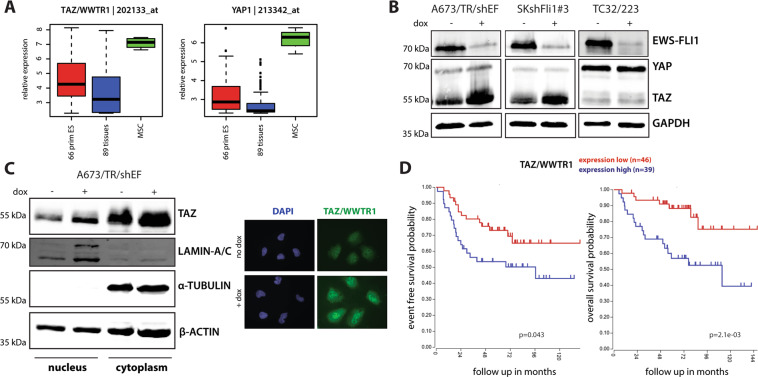
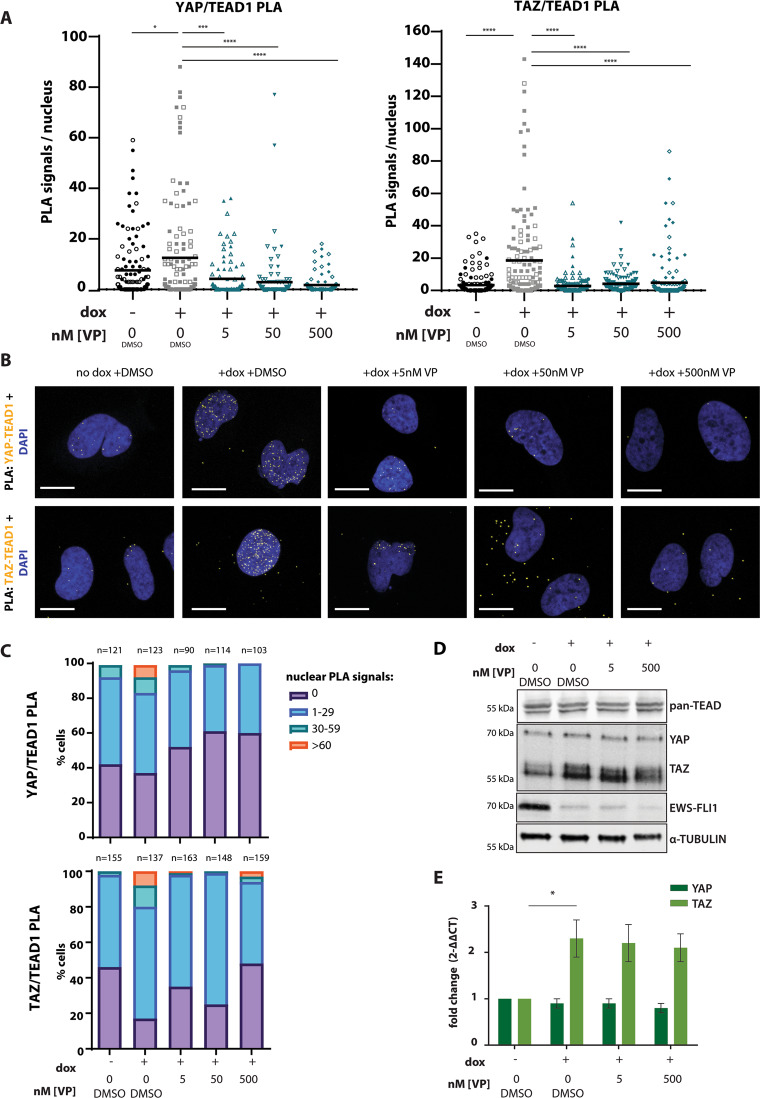
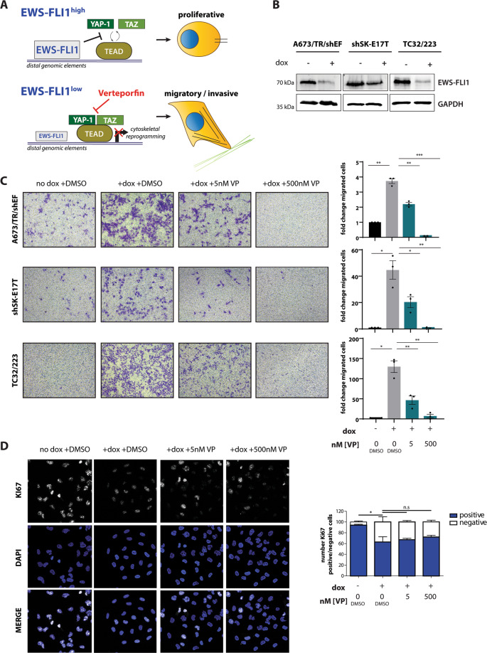
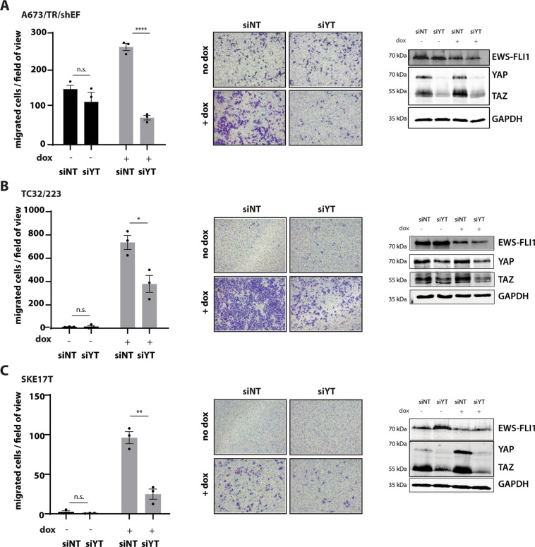
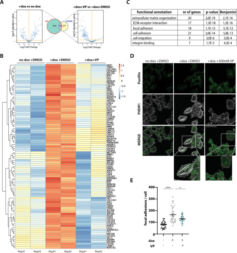
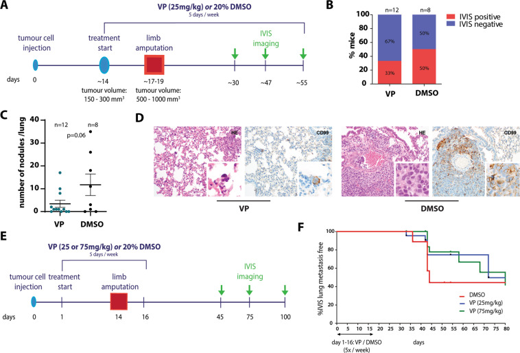
Ewing sarcoma (EwS) is a highly metastatic bone cancer characterized by the ETS fusion oncoprotein EWS-FLI1. EwS cells are phenotypically highly plastic and switch between functionally distinct cell states dependent on EWS-FLI1 fluctuations. Whereas EWS-FLI1high cells proliferate, EWS-FLI1low cells are migratory and invasive. Recently, we reported activation of MRTFB and TEAD, effectors of RhoA and Hippo signalling, upon low EWS-FLI1, orchestrating key steps of the EwS migratory gene expression program. TEAD and its co-activators YAP and TAZ are commonly overexpressed in cancer, providing attractive therapeutic targets. We find TAZ levels to increase in the migratory EWS-FLI1low state and to associate with adverse prognosis in EwS patients. We tested the effects of the potent YAP/TAZ/TEAD complex inhibitor verteporfin on EwS cell migration in vitro and on metastasis in vivo. Verteporfin suppressed expression of EWS-FLI1 regulated cytoskeletal genes involved in actin signalling to the extracellular matrix, effectively blocked F-actin and focal-adhesion assembly and inhibited EwS cell migration at submicromolar concentrations. In a mouse EwS xenograft model, verteporfin treatment reduced relapses at the surgical site and delayed lung metastasis. These data suggest that YAP/TAZ pathway inhibition may prevent EwS cell dissemination and metastasis, justifying further preclinical development of YAP/TAZ inhibitors for EwS treatment.


Conflict of interest statement

The authors declare that they have no conflict of interest.

Figures

Fig. 1
Fig. 1. TAZ expression is repressed by EWS-FLI1 in vitro and correlates with patient outcome in primary EwS.
a Box-plots showing relative expression levels of TAZ and YAP in 66 primary EwS tumours compared to 89 normal tissues and mesenchymal stem cells (MSC). Affymetrix hgu133a microarray expression data from Kauer et al.. b Representative immunoblots showing YAP and TAZ expression under dox-induced EWS-FLI1low (48 h dox) versus EWS-FLI1high conditions in different EwS cell lines. c Subcellular localization of TAZ in A673/TR/shEF cells upon EWS-FLI1high (no dox) and EWS-FLIlow (48 h dox) conditions was analysed by protein fractionation experiments and immunofluorescence microscopy. One representative immunoblot out of three biological replicates is shown. Lamin A/C and α-tubulin were used as nuclear and cytoplasmic fraction controls, respectively. Immunofluorescent pictures are shown at ×40 magnification. d Kaplan–Meier analysis of huex10t microarray expression data for event-free (chi = 4.08, p = 0.043; left graph) and overall (chi = 9.44, p = 2.1e−03; right graph) survival of 85 EwS patients in relation to TAZ expression. Data (GEO ID: gse63157) was visualized using the R2 database (http://r2.amc.nl).
Fig. 2
Fig. 2. Verteporfin prevents complex formation of YAP/TAZ and TEAD in A673/TR/shEF.
a Quantification of nuclear YAP/TEAD1 and TAZ/TEAD1 PLA signals under EWS-FLI1high (no dox) and EWS-FLI1low ( + dox) conditions and upon VP (5 nM, 50 nM, 500 nM) treatment. Pooled data from two independent biological replicates, represented by distinct symbol patterns, is shown. Mean numbers of PLA signals/cell are indicated. (b) Representative confocal images (63x objective, zoom factor 2.5) of nuclear YAP/TEAD1 and TAZ/TEAD1 PLA signals from experiments shown in (A). Scale bar: 20 µm. (c) Quantification of YAP/TEAD1 and TAZ/TEAD1 PLA signals shown in (A), represented as % of cells with corresponding PLA signal ranges per nucleus. (d) Immunoblot showing expression of pan-TEAD, YAP/TAZ and EWS-FLI1 from total protein lysates upon EWS-FLI1high (no dox) and EWS-FLI1low ( + dox) conditions and upon VP treatment. One representative experiment from three biological replicates is shown. (e) qPCR analysis of YAP and TAZ mRNA transcripts upon same experimental conditions as in (D). Expression values are shown as fold change ± s.e.m of three biological replicates relative to no dox +DMSO-control conditions. All statistics were calculated by two-sided, unpaired Student’s t-test *p ≤ 0.05, **** p ≤ 0.0001.
Fig. 3
Fig. 3. Verteporfin inhibits cell migration of EWS-FLI1low cells without affecting proliferation.
(a) Proposed model of YAP/TAZ/TEAD pathway inhibition by VP in EWS-FLI1low cells and its potential effect on EwS cell phenotype. (b) Representative immunoblots verifying dox-induced knockdown of EWS-FLI1 in A673/TR/shEF, shSK-E17T and TC32/223 cell lines at the start of the migration assay. (c) Boyden chamber migration assays using cell lines from (B) in presence and absence of different VP concentrations. Exemplary pictures of migrated cells as well as quantification (fold change of migrated cells relative to “no dox +DMSO”) of three biological replicates performed in triplicates are shown. (d) Cell proliferation of A673/TR/shEF cells was evaluated by KI67 positivity in parallel to Boyden chamber assays. KI67 positive and negative fractions from three biological replicates were determined and normalized to the total number of counted cells. Statistics were calculated by One-sample t-test for comparison to “no dox +DMSO” conditions and two-sided, unpaired Student’s t-test for all other conditions. *p ≤ 0.05, **p ≤ 0.01, ***p ≤ 0.001.
Fig. 4
Fig. 4. Combined silencing of YAP/TAZ interferes with the migratory capacity of EWS-FLI1low cells, similar to Verteprofin treatment.
Boyden chamber migration assays upon combined knockdown of YAP/TAZ (siYT), compared to cells transfected with control siRNA (siNT), under EWS-FLI1high and dox-induced EWS-FLI1low conditions in (a) A673/TR/shEF, (b) TC32/223 and (c) shSK-E17T. Efficient silencing of YAP/TAZ and EWS-FLI1 knockdown are shown by one representative immunoblot. Data are represented as mean ± s.e.m. of three biological replicates performed in triplicates. Statistics were calculated by two-sided, unpaired Student’s t-test *p ≤ 0.05, **p ≤ 0.01, ***p ≤ 0.001.
Fig. 5
Fig. 5. Verteporfin affects expression of cytoskeletal genes leading to reduced F-actin and focal-adhesion formation in A673/TR/shEF.
(a) Volcano plots and Venn diagrams showing gene expression upon EWS-FLI1low versus EWS-FLI1high ( + dox vs no dox) conditions compared to the effects of 500 nM VP treatment versus DMSO control (+dox+VP vs + dox+DMSO) in the EWS-FLI1low state. 911 genes were found to increase (Venn diagram: UP) in expression upon EWS-FLI1low versus EWS-FLI1high conditions (EWS-FLI1-anticorrelated genes). Treatment with VP downregulated gene expression (Venn diagram: DOWN) of a total of 370 genes, of these 93 genes are EWS-FLI1-anticorrelated genes. Cells were collected in parallel to performing transwell migration assays (see Fig. 3), and RNA from two biological replicates was sent for RNA-seq. Volcano plots show log2-fold changes of gene expression, Venn diagrams show genes changing with a │logFC│>0.7, p < 0.05. (b) Heatmap showing effects of 500 nM VP on EWS-FLI1-anticorrelated genes of two biological replicates (Rep#1,2). (c) DAVID functional annotation analysis of genes shown in Fig. 5b. EWS-FLI1-anticorrelated genes, which were affected by VP treatment (93 genes) are significantly enriched in cytoskeletal processes, such as communication with the extracellular matrix, adhesion and migration. (d) Confocal immunofluorescence microscopy showing effects of 500 nM VP treatment on stress fibre (TRITC-phalloidin, PHDR1) and focal-adhesion (FITC-paxillin) formation. F-actin thickness and abundance is increased in the migratory EWS-FLI1low ( + dox) state as compared to EWS-FLI1high (no dox). Number and intensity of focal adhesions is also strongly increased. Treatment with VP reduced the number of focal adhesions at the leading cell edges and led to F-actin breakdown (indicated with arrows). Representative confocal images from two biological replicates are shown (40x magnification, selection: zoom 146%, scale bar: 100 µm). (e) Quantification of paxillin-positive foci, normalized to respective cell size, in the migratory EWS-FLI1low ( + dox; ±500 nM VP) state and compared to EWS-FLI1high (no dox) cells.
Fig. 6
Fig. 6. Verteporfin treatment reduces EwS lung metastasis in a mouse xenograft model.
(a) Experimental setting 1: set-up and VP treatment scheme. Luciferase-expressing TC71 cells were injected into the tibial crest of mice. Intra-peritoneal injections of VP (25 mg/kg) or solvent control (20% DMSO) started once tumours reached a specific size. When tumours reached a volume of 150–300mm3, tumour-bearing limbs were amputated and VP and control treatments (5 days/week) were continued for a maximum of 35 days. (b) Proportions of mice with IVIS-detectable pulmonary metastases per treatment group. (c) Number of histopathologically detectable tumour nodules in lung sections of control- and VP-treated mice based on evaluation of H&E and CD99 stainings. The mean number ±s.e.m. of tumour nodules per condition is shown. P value was calculated by two-tailed Student’s t-test. (d) Exemplary H&E and CD99 stainings showing a reduced size of EwS lung metastatic nodules (200x magnification, inserts: 600x magnification). (e) Experimental setting 2: set-up and VP treatment scheme. As for setting 1 in (A), luciferase-expressing TC71 cells were injected, but VP (25 mg/kg or 75 mg/kg) and control treatments were started one day after tumour cell injection and stopped two days after limb amputation. (f) Lung metastasis free survival of control- and VP-treated mice from experimental setting 2. Although data are not statistically significant, VP-treated mice show a delay in metastatic on-set.

Similar articles

Cited by

References

    1. Lawlor ER, Sorensen PH. Twenty years on: what do we really know about Ewing sarcoma and what is the path forward? Crit. Rev. Oncogenesis. 2015;20:155–71.. doi: 10.1615/CritRevOncog.2015013553. - DOI - PMC - PubMed
    1. Toomey EC, Schiffman JD, Lessnick SL. Recent advances in the molecular pathogenesis of Ewing’s sarcoma. Oncogene. 2010;29:4504–16.. doi: 10.1038/onc.2010.205. - DOI - PMC - PubMed
    1. Herrero-Martin D, et al. Factors affecting EWS-FLI1 activity in Ewing’s sarcoma. Sarcoma. 2011;2011:352580. doi: 10.1155/2011/352580. - DOI - PMC - PubMed
    1. Grunewald TGP, et al. Ewing sarcoma. Nat. Rev. Dis. Prim. 2018;4:5. doi: 10.1038/s41572-018-0003-x. - DOI - PubMed
    1. Franzetti GA, et al. Cell-to-cell heterogeneity of EWSR1-FLI1 activity determines proliferation/migration choices in Ewing sarcoma cells. Oncogene. 2017;36:3505–14.. doi: 10.1038/onc.2016.498. - DOI - PMC - PubMed

LinkOut - more resources