. 2021 Jan 8;12(1):189.
doi: 10.1038/s41467-020-20320-y.

Mechanism of filament formation in UPA-promoted CARD8 and NLRP1 inflammasomes

Affiliations

Mechanism of filament formation in UPA-promoted CARD8 and NLRP1 inflammasomes

L Robert Hollingsworth et al. Nat Commun. .

Abstract

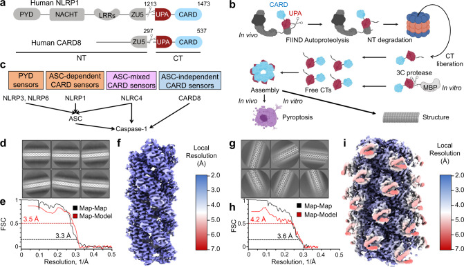
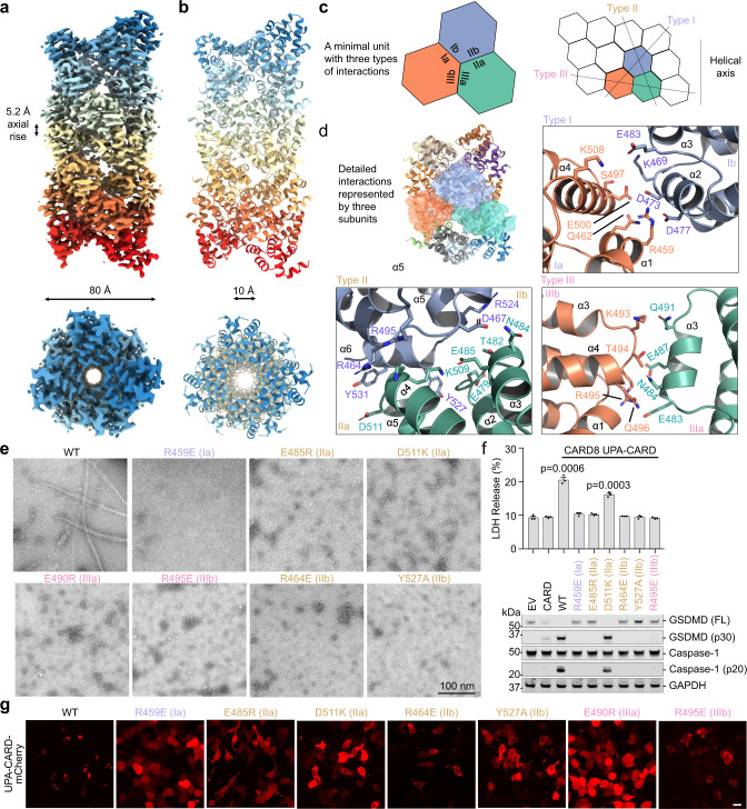
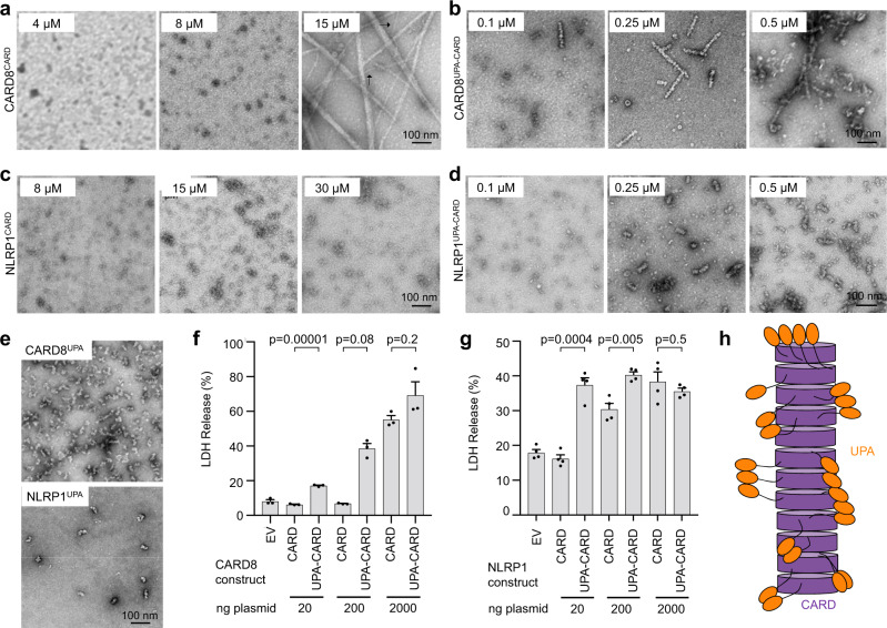
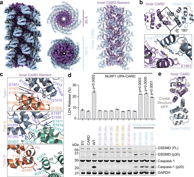
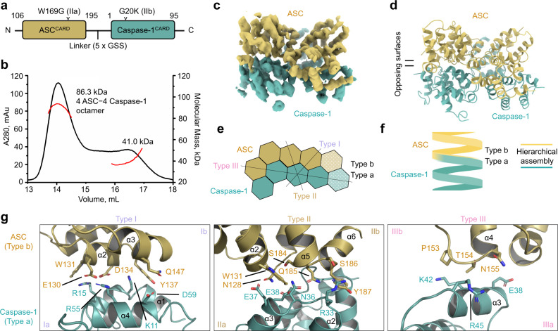
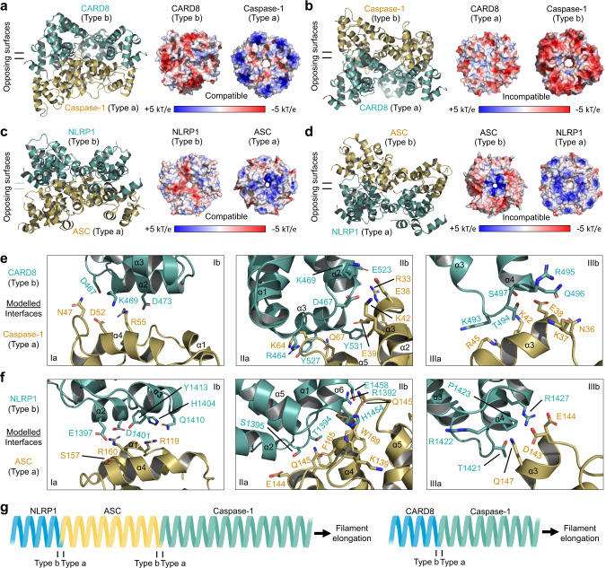
NLRP1 and CARD8 are related cytosolic sensors that upon activation form supramolecular signalling complexes known as canonical inflammasomes, resulting in caspase-1 activation, cytokine maturation and/or pyroptotic cell death. NLRP1 and CARD8 use their C-terminal (CT) fragments containing a caspase recruitment domain (CARD) and the UPA (conserved in UNC5, PIDD, and ankyrins) subdomain for self-oligomerization, which in turn form the platform to recruit the inflammasome adaptor ASC (apoptosis-associated speck-like protein containing a CARD) or caspase-1, respectively. Here, we report cryo-EM structures of NLRP1-CT and CARD8-CT assemblies, in which the respective CARDs form central helical filaments that are promoted by oligomerized, but flexibly linked, UPAs surrounding the filaments. Through biochemical and cellular approaches, we demonstrate that the UPA itself reduces the threshold needed for NLRP1-CT and CARD8-CT filament formation and signalling. Structural analyses provide insights on the mode of ASC recruitment by NLRP1-CT and the contrasting direct recruitment of caspase-1 by CARD8-CT. We also discover that subunits in the central NLRP1CARD filament dimerize with additional exterior CARDs, which roughly doubles its thickness and is unique among all known CARD filaments. Finally, we engineer and determine the structure of an ASCCARD-caspase-1CARD octamer, which suggests that ASC uses opposing surfaces for NLRP1, versus caspase-1, recruitment. Together these structures capture the architecture and specificity of the active NLRP1 and CARD8 inflammasomes in addition to key heteromeric CARD-CARD interactions governing inflammasome signalling.


Conflict of interest statement

H.W. is a co-founder of Ventus Therapeutics. The other authors declare no competing interests.

Figures

Fig. 1
Fig. 1. Structure determination of CARD8-CT and NLRP1-CT filaments.
a Domain architecture of human NLRP1 and CARD8, each with a C-terminal domain (CT) containing UPA-CARD. b CARD8 and NLRP1 activation pathway in which signals leading to NLRP1 and CARD8 activation target them to the proteasome for N-terminal degradation. This leads to the release of free CT oligomers, which assemble the inflammasome. The purification strategy bypasses N-terminal degradation by directly expressing MBP-CT fusion proteins and cleaving the tag in vitro. c Classification of different inflammasome sensors by their signaling through the adapter ASC. Human NLRP1 and CARD8 are ASC-dependent and ASC-independent, respectively. d Cryo-EM 2D classification of CARD8-CT filaments. e Gold-standard Fourier shell correlation (FSC) and map-model correlation plots of the CARD8-CT filament 3D reconstruction, which gave an overall resolution of 3.3 Å. f Local resolution of the CARD8-CT filament calculated with RELION’s local resolution estimation, and colored as indicated. g Cryo-EM 2D classification of NLRP1-CT filaments. h FSC plots of the NLRP1-CT filament 3D reconstruction, which gave an overall resolution of 3.6 Å. i Local resolution of the NLRP1-CT filament with RELION’s local resolution estimation, and colored as indicated.
Fig. 2
Fig. 2. Cryo-EM structure and mutagenesis of the CARD8-CT filament.
a, b Overall structure of the CARD8-CT inflammasome, with a resolved CARD8CARD filament. The cryo-EM density (a) and atomic model (b) are colored by subunit. c Illustration of classical CARD filament interfaces: type I, II, and III. d Detailed CARD–CARD interactions in the CARD8-CT filament structure colored as in c. e Negative stain electron microscopy snapshots of CARD8CARD wild-type (WT) and mutants that abolish filament formation in vitro. Filaments were formed at 15 µM (shown here) and 30 µM. Micrographs are representative of >3 fields of view for each sample. f Inflammasome assay of CARD8-CT-mCherry mutants transiently expressed in HEK293T cells that stably express caspase-1 and GSDMD. Filament-deficient mutants abolish inflammasome activity as measured by LDH release (top) and western blot (bottom) for activated inflammasome components. P-values are compared to empty vector (EV) by two-sided Student’s t-test. Exact P-values are provided in the Source Data File. Data are means ± SEM of n = 3 biological replicates. g Confocal imaging of HEK293T cells transiently expressing CARD8UPA-CARD-mCherry WT and mutants. Filament-deficient mutants abolish filament formation in cells. Experiments were performed with two biological replicates. Scale bar 5 µm. In e–g, mutations are labeled in the colors of the interface types as defined in c.
Fig. 3
Fig. 3. The UPA subdomain reduces the concentration required for inflammasome formation.
a, b Concentration-dependent formation of CARD8CARD (a) and CARD8UPA-CARD (b) filaments, visualized by negative staining EM. Purified monomeric MBP-fused proteins were cleaved at concentrations ranging from 0.1 to 15 µM. CARD8CARD filaments did not appear consistently until 15 µM. CARD8UPA-CARD filaments formed consistently at 0.25 µM. Arrows in a indicates single, unbundled filaments. c, d Concentration-dependent formation of NLRP1CARD (c) and NLRP1UPA-CARD (d) filaments. Purified monomeric MBP-fused proteins were cleaved at concentrations ranging from 0.1 to 30 µM. NLRP1CARD filaments did not appear even at 30 µM, whereas NLRP1UPA-CARD filaments formed consistently at 0.25 µM. For ad, filament formation was performed with two biological replicates. Micrographs are representative of >3 fields of view for each condition. e CARD8UPA and NLRP1UPA imaged by negative staining EM, representative of >5 fields of view. f Titration of CARD8CARD and CARD8UPA-CARD in HEK293T cells stably expressing caspase-1 and GSDMD. A ~100-fold higher amount of the CARD8CARD plasmid was needed to achieve a comparable level of cell death (marked by LDH release) by CARD8UPA-CARD. Data are means ± SEM of n = 3 biological replicates. g Titration of NLRP1CARD and NLRP1UPA-CARD with ASC co-expression in HEK293T cells stably expressing caspase-1 and GSDMD. A ~100-fold higher amount of the NLRP1CARD plasmid was needed to achieve a comparable level of cell death (marked by LDH release) by NLRP1UPA-CARD. P-values by two-sided Student’s t-test. Exact P-values are provided in the Source Data File. Data are means ± SEM of n = 3 biological replicates. EV empty vector. h Model of UPA-enhanced inflammasome formation. The UPA subdomain increases the multivalency of the CARD8 and NLRP1 inflammasomes, lowering the concentration required to nucleate filaments and subsequently signal.
Fig. 4
Fig. 4. Cryo-EM structure of the dimeric NLRP1 CARD filament.
a Overall structure of the NLRP1-CT inflammasome. Each subunit of the inner helical CARD filament (purple) dimerizes with an outer CARD (blue). b Dimeric interface between an inner (purple) and outer (blue) CARD pair. c Zoom-ins of type I, II, and III helical interfaces between inner CARD molecules. d Structure-guided mutations of helical interfaces and the CARD–CARD dimer interface on UPA-CARD inflammasome signaling. LDH release (top) and western blot (bottom) are shown. P-values are compared to empty vector (EV) by two-sided Student’s t-test. Exact P-values are provided in the Source Data File. Data are means ± SEM of n = 3 biological replicates. In c, d residues and mutations are labeled in the colors of the interface types as defined in Fig. 2c. e Alignment between Inner-outer CARD (purple and blue) and a dimer within the crystallographic symmetry unit of the NLRP1 CARD crystal structure (gray).
Fig. 5
Fig. 5. Purification and structure determination of an ASCCARD−caspase-1CARD octamer.
a Construct design of ASCCARD (brown) and caspase-1CARD (turquoise) linked by 5× GSS linker. b MALS data for the ASCCARD−caspase-1CARD complex, with a molecular mass of 86.3 kDa corresponding to a complex formed by four subunits of ASCCARD and four subunits of caspase-1CARD. c Cryo-EM density of the ASCCARD−caspase-1CARD octamer at 3.9 Å resolution in which a layer of ASCCARD (gold) is located on top of a caspase-1CARD layer (green). d Model of the ASCCARD–aspase-1CARD octamer. e Schematic diagram of the octamer complex with depicted interfaces. There are three type I interactions, four type II interactions, and one type III interaction between ASC and caspase-1. f Simplified illustration of ASC and caspase-1 hierarchy in inflammasome signaling. g Zoom-ins of type I, II, and III helical interfaces between ASC and caspase-1 molecules. In cg, ASCCARD and caspase-1CARD are colored as defined in c.
Fig. 6
Fig. 6. Modeled specific interfaces of the CARD8-CT and NLRP1-CT filaments for caspase-1 and ASC, respectively.
a, b Opposing arrangements of CARD8 and caspase-1 in their hypothetical modes of interaction. The electrostatic surfaces indicate that the negatively charged Type b surface of CARD8 most likely matches the positively charged Type a surface of caspase-1 in a. c, d Opposing arrangements of NLRP1 and ASC in their hypothetical modes of interaction. The electrostatic surfaces indicate that the largely negatively charged Type b surface of NLRP1 most likely matches the positively charged Type a surface of ASC in c. e Detailed modeled CARD-CARD type I–III interactions between CARD8 (green) and caspase-1 (gold). f Detailed modeled CARD-CARD type I–III interactions between NLRP1 (green) and ASC (gold). g Simplified illustration of NLRP1 and CARD8 hierarchy in inflammasome signaling. NLRP1 recruits ASC, followed by capase-1 recruitment. In contrast, CARD8 can only recruit caspase-1 directly. In af, the inflammasome sensors NLRP1 and CARD8 are colored turquoise while the downstream molecules ASC and caspase-1 are colored brown.

Similar articles

Cited by

References

    1. Martinon F, Burns K, Tschopp J. The inflammasome: a molecular platform triggering activation of inflammatory caspases and processing of proIL-β. Mol. Cell. 2002;10:417–426. doi: 10.1016/S1097-2765(02)00599-3. - DOI - PubMed
    1. Broz P, Dixit VM. Inflammasomes: mechanism of assembly, regulation and signalling. Nat. Rev. Immunol. 2016;16:407–420. doi: 10.1038/nri.2016.58. - DOI - PubMed
    1. Lu A, et al. Unified polymerization mechanism for the assembly of ASC-dependent inflammasomes. Cell. 2014;156:1193–1206. doi: 10.1016/j.cell.2014.02.008. - DOI - PMC - PubMed
    1. Shen C, Sharif H, Xia S, Wu H. Structural and mechanistic elucidation of inflammasome signaling by cryo-EM. Curr. Opin. Struct. Biol. 2019;58:18–25. doi: 10.1016/j.sbi.2019.03.033. - DOI - PMC - PubMed
    1. Hu Z, et al. Structural and biochemical basis for induced self-propagation of NLRC4. Science. 2015;350:399–404. doi: 10.1126/science.aac5489. - DOI - PubMed

Publication types

MeSH terms