. 2021 Jan 8;12(1):179.
doi: 10.1038/s41467-020-20429-0.

Predicting postoperative peritoneal metastasis in gastric cancer with serosal invasion using a collagen nomogram

Affiliations

Predicting postoperative peritoneal metastasis in gastric cancer with serosal invasion using a collagen nomogram

Dexin Chen et al. Nat Commun. .

Abstract

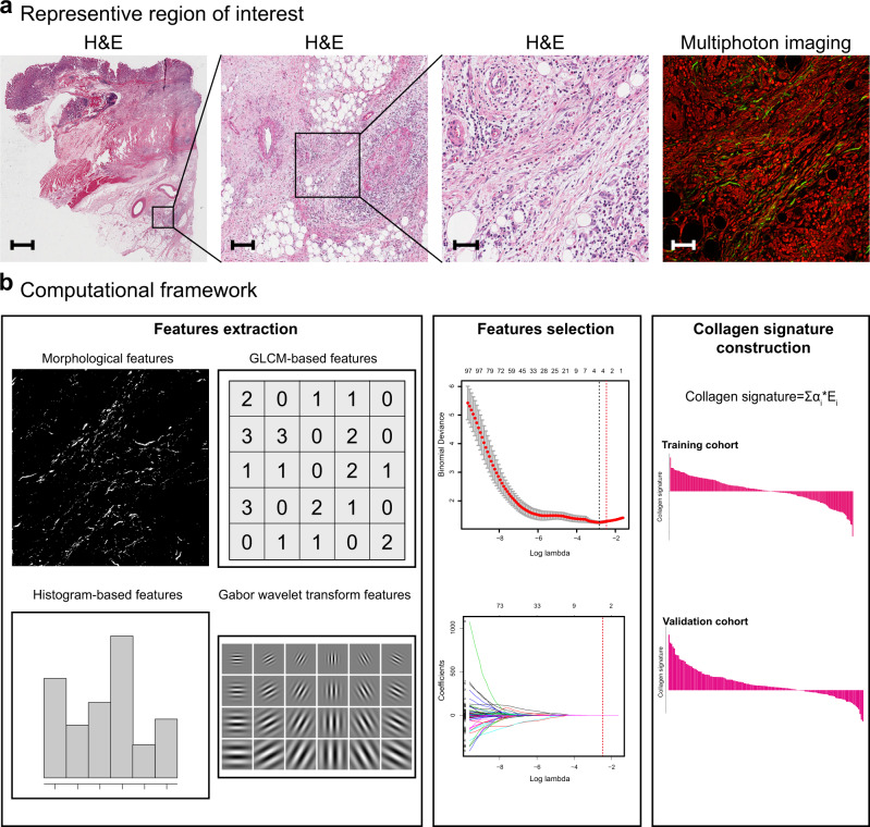
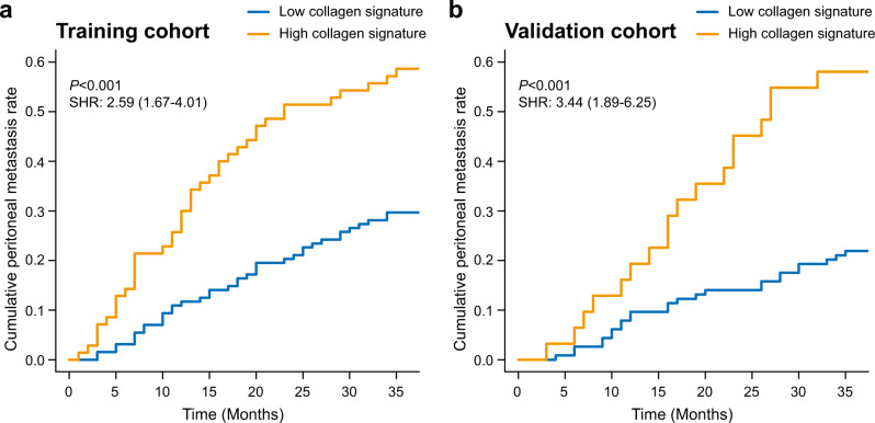
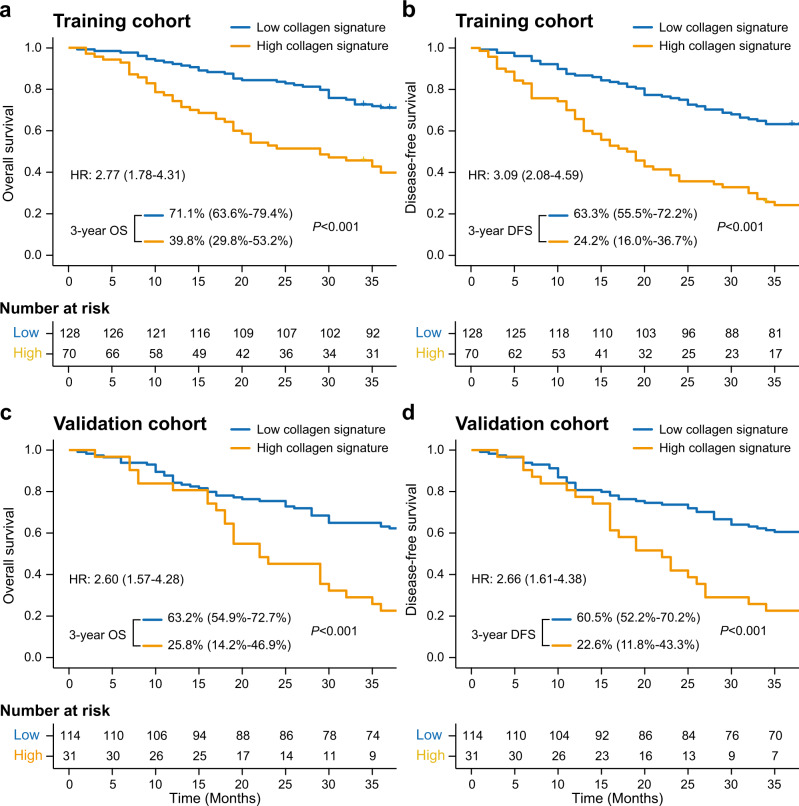
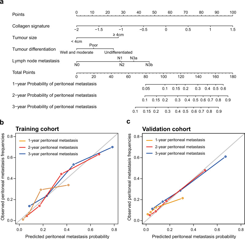
Accurate prediction of peritoneal metastasis for gastric cancer (GC) with serosal invasion is crucial in clinic. The presence of collagen in the tumour microenvironment affects the metastasis of cancer cells. Herein, we propose a collagen signature, which is composed of multiple collagen features in the tumour microenvironment of the serosa derived from multiphoton imaging, to describe the extent of collagen alterations. We find that a high collagen signature is significantly associated with a high risk of peritoneal metastasis (P < 0.001). A competing-risk nomogram including the collagen signature, tumour size, tumour differentiation status and lymph node metastasis is constructed. The nomogram demonstrates satisfactory discrimination and calibration. Thus, the collagen signature in the tumour microenvironment of the gastric serosa is associated with peritoneal metastasis in GC with serosal invasion, and the nomogram can be conveniently used to individually predict the risk of peritoneal metastasis in GC with serosal invasion after radical surgery.


Conflict of interest statement

The authors declare no competing interests.

Figures

Fig. 1
Fig. 1. Construction framework of the collagen signature.
a Selection of the region of interest by comparing H&E staining and multiphoton imaging. The samples of all enroled 343 patients are used, and five regions of interest with a field of view of 500 × 500 μm per sample within the invasive region of the gastric serosa are randomly selected for multiphoton imaging. Scale bars: 1500 μm, 150 μm, 50 μm and 50 μm, respectively. b A total of 146 collagen features for each enroled patient are extracted from multiphoton images, and predictive collagen features are selected using LASSO regression in the training cohort, and the collagen signature is constructed. The collagen signature of the validation cohort is calculated from the collagen signature calculation formula obtained in the training cohort. GLCM, grey-level co-occurrence matrix; H&E, hematoxylin and eosin; LASSO, least absolute shrinkage and selection operator.
Fig. 2
Fig. 2. Collagen signature and peritoneal metastasis in the training and validation cohorts.
Cumulative peritoneal metastasis rate stratified by the collagen signature in the (a) training and (b) validation cohorts. The comparisons of the cumulative peritoneal metastasis between two subgroups are performed using a two-sided Gray’s test. SHR, subdistribution hazard ratio.
Fig. 3
Fig. 3. Kaplan–Meier survival analysis of the training and validation cohorts grouped by the collagen signature.
a Three-year OS comparison between the high and low collagen signatures in the training cohort. b The 3-year DFS comparison between the high and low collagen signatures in the training cohort. c The 3-year OS comparison between the high and low collagen signatures in the validation cohort. d The 3-year DFS comparison between the high and low collagen signatures in the validation cohort. The comparisons of OS and DFS between two subgroups are performed using a two-sided log-rank test. OS, overall survival; DFS, disease-free survival; HR, hazard ratio.
Fig. 4
Fig. 4. Competing-risk nomogram and the corresponding calibration curve.
a Competing-risk nomogram incorporating the collagen signature, tumour size, tumour differentiation and lymph node metastasis. b Calibration curve of the competing-risk nomogram in the training cohort. c Calibration curve of the competing-risk nomogram in the validation cohort.

References

    1. Bray F, et al. Global cancer statistics 2018: GLOBOCAN estimates of incidence and mortality worldwide for 36 cancers in 185 countries. CA Cancer J. Clin. 2018;68:394–424. doi: 10.3322/caac.21492. - DOI - PubMed
    1. Yoo CH, Noh SH, Shin DW, Choi SH, Min JS. Recurrence following curative resection for gastric carcinoma. Br. J. Surg. 2000;87:236–242. doi: 10.1046/j.1365-2168.2000.01360.x. - DOI - PubMed
    1. Sasako M, et al. D2 lymphadenectomy alone or with para-aortic nodal dissection for gastric cancer. N. Engl. J. Med. 2008;359:453–462. doi: 10.1056/NEJMoa0707035. - DOI - PubMed
    1. Van Cutsem E, Sagaert X, Topal B, Haustermans K, Prenen H. Gastric cancer. Lancet. 2016;388:2654–2664. doi: 10.1016/S0140-6736(16)30354-3. - DOI - PubMed
    1. Boku T, et al. Prognostic significance of serosal invasion and free intraperitoneal cancer cells in gastric cancer. Br. J. Surg. 1990;77:436–439. doi: 10.1002/bjs.1800770425. - DOI - PubMed

Publication types