

Valproic acid-exposed astrocytes impair inhibitory synapse formation and function
- PMID: 33420078
- PMCID: PMC7794250
- DOI: 10.1038/s41598-020-79520-7
Valproic acid-exposed astrocytes impair inhibitory synapse formation and function
Abstract
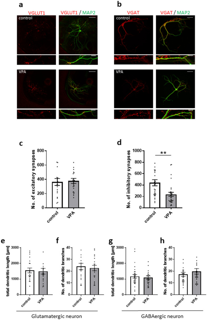
Valproic acid (VPA) is widely prescribed to treat epilepsy. Maternal VPA use is, however, clinically restricted because of the severe risk that VPA may cause neurodevelopmental disorders in offspring, such as autism spectrum disorder. Understanding the negative action of VPA may help to prevent VPA-induced neurodevelopmental disorders. Astrocytes play a vital role in neurodevelopment and synapse function; however, the impact of VPA on astrocyte involvement in neurodevelopment and synapse function has not been examined. In this study, we examined whether exposure of cultured astrocytes to VPA alters neuronal morphology and synapse function of co-cultured neurons. We show that synaptic transmission by inhibitory neurons was small because VPA-exposed astrocytes reduced the number of inhibitory synapses. However, synaptic transmission by excitatory neurons and the number of excitatory synapses were normal with VPA-exposed astrocytes. VPA-exposed astrocytes did not affect the morphology of inhibitory neurons. These data indicate that VPA-exposed astrocytes impair synaptogenesis specifically of inhibitory neurons. Our results indicate that maternal use of VPA would affect not only neurons but also astrocytes and would result in perturbed astrocyte-mediated neurodevelopment.
Conflict of interest statement
The authors declare no competing interests.
Figures


Similar articles
-
Valproic acid mediates the synaptic excitatory/inhibitory balance through astrocytes--a preliminary study.Prog Neuropsychopharmacol Biol Psychiatry. 2012 Apr 27;37(1):111-20. doi: 10.1016/j.pnpbp.2012.01.017. Epub 2012 Feb 7. Prog Neuropsychopharmacol Biol Psychiatry. 2012. PMID: 22343008
-
Valproic acid selectively suppresses the formation of inhibitory synapses in cultured cortical neurons.Neurosci Lett. 2014 May 21;569:142-7. doi: 10.1016/j.neulet.2014.03.066. Epub 2014 Apr 5. Neurosci Lett. 2014. PMID: 24708928
-
Hippocampal neurons isolated from rats subjected to the valproic acid model mimic in vivo synaptic pattern: evidence of neuronal priming during early development in autism spectrum disorders.Mol Autism. 2021 Mar 6;12(1):23. doi: 10.1186/s13229-021-00428-8. Mol Autism. 2021. PMID: 33676530 Free PMC article.
-
Cell adhesion and matricellular support by astrocytes of the tripartite synapse.Prog Neurobiol. 2018 Jun-Aug;165-167:66-86. doi: 10.1016/j.pneurobio.2018.02.002. Epub 2018 Feb 11. Prog Neurobiol. 2018. PMID: 29444459 Review.
-
Emerging mechanisms of valproic acid-induced neurotoxic events in autism and its implications for pharmacological treatment.Biomed Pharmacother. 2021 May;137:111322. doi: 10.1016/j.biopha.2021.111322. Epub 2021 Feb 16. Biomed Pharmacother. 2021. PMID: 33761592 Review.
Cited by
-
Pharmacological Investigations in Glia Culture Model of Inflammation.Front Cell Neurosci. 2021 Dec 16;15:805755. doi: 10.3389/fncel.2021.805755. eCollection 2021. Front Cell Neurosci. 2021. PMID: 34975415 Free PMC article. Review.
-
Pathology and Astrocytes in Autism.Neuropsychiatr Dis Treat. 2023 Apr 12;19:841-850. doi: 10.2147/NDT.S390053. eCollection 2023. Neuropsychiatr Dis Treat. 2023. PMID: 37077706 Free PMC article. Review.
-
The Interplay of Astrocytes and Neurons in Autism Spectrum Disorder.Adv Neurobiol. 2024;39:269-284. doi: 10.1007/978-3-031-64839-7_11. Adv Neurobiol. 2024. PMID: 39190079 Review.
-
Astrocytes in Bipolar Disorder.Adv Neurobiol. 2021;26:95-113. doi: 10.1007/978-3-030-77375-5_5. Adv Neurobiol. 2021. PMID: 34888832
-
Spontaneous Calcium Transients Recorded from Striatal Astrocytes in a Preclinical Model of Autism.Neurochem Res. 2024 Nov;49(11):3069-3077. doi: 10.1007/s11064-024-04218-5. Epub 2024 Aug 9. Neurochem Res. 2024. PMID: 39120794 Free PMC article.
References
Publication types
MeSH terms
Substances
LinkOut - more resources
Full Text Sources
Other Literature Sources
