. 2021 Jan 8;11(1):157.
doi: 10.1038/s41598-020-79592-5.

Fibrillar pharmacology of functionalized nanocellulose

Affiliations

Fibrillar pharmacology of functionalized nanocellulose

Sam Wong et al. Sci Rep. .

Abstract

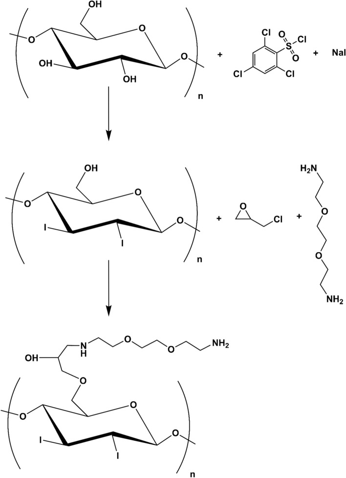
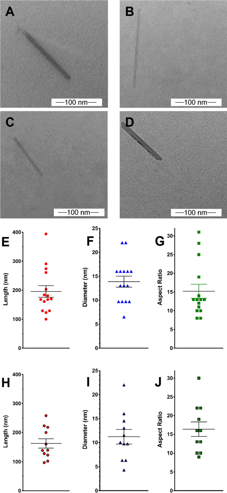
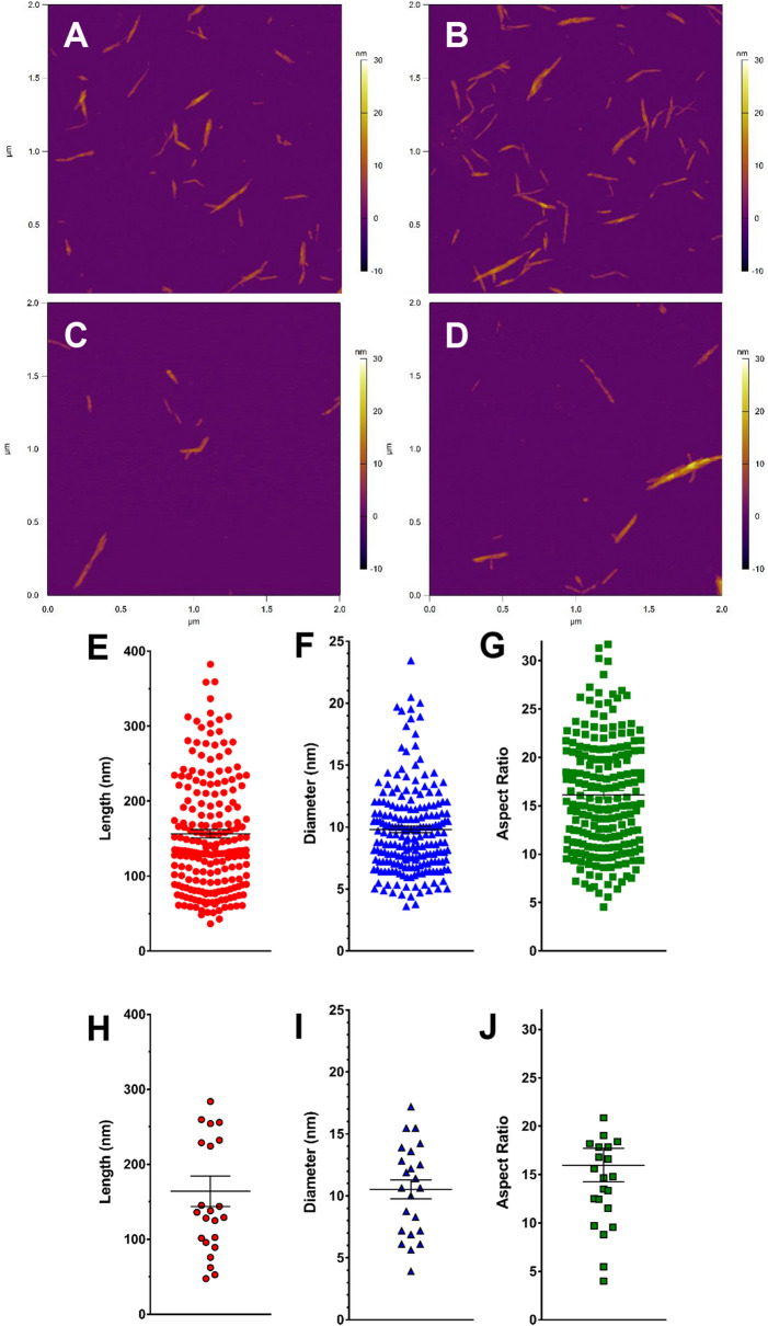
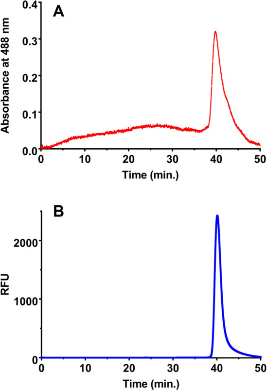
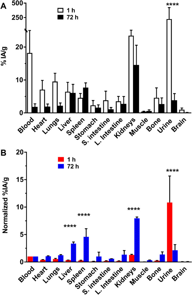
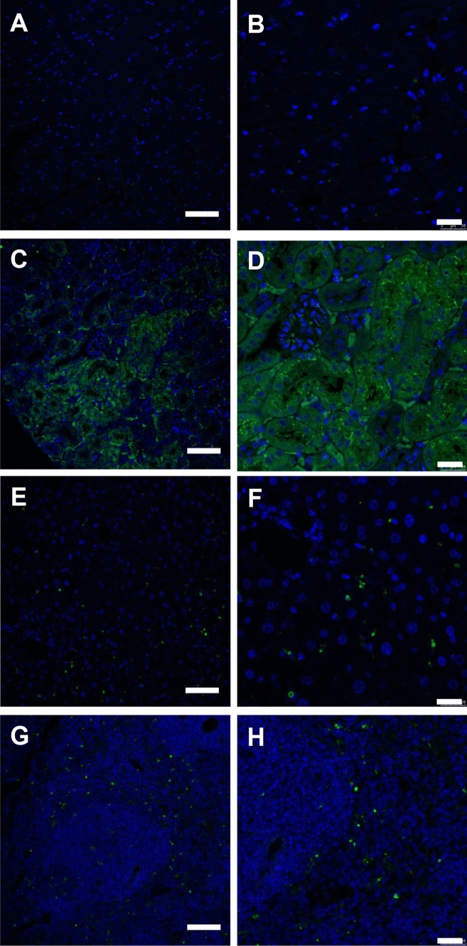
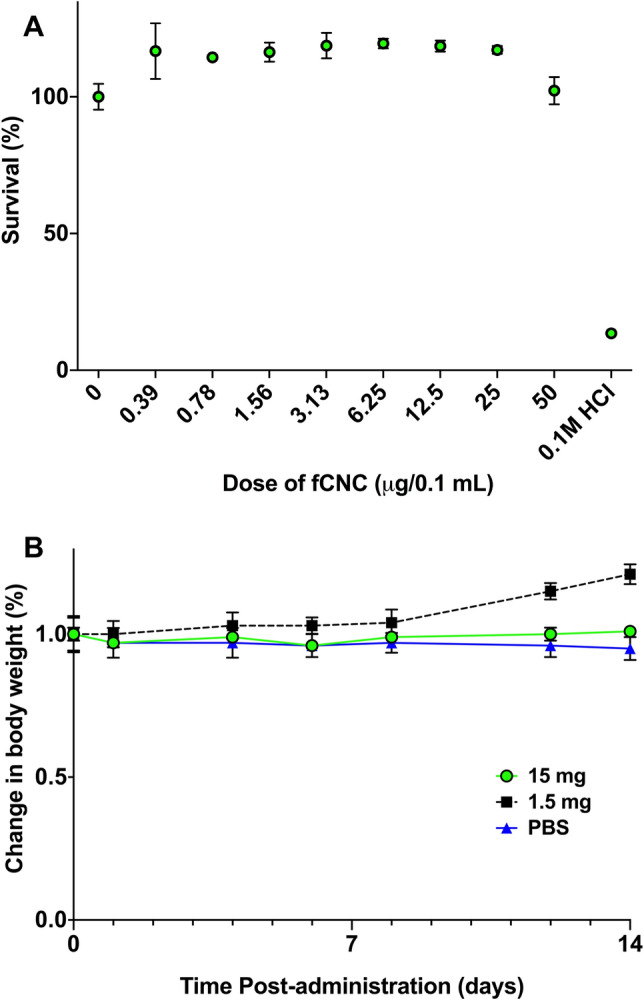
Cellulose nanocrystals (CNC) are linear organic nanomaterials derived from an abundant naturally occurring biopolymer resource. Strategic modification of the primary and secondary hydroxyl groups on the CNC introduces amine and iodine group substitution, respectively. The amine groups (0.285 mmol of amine per gram of functionalized CNC (fCNC)) are further reacted with radiometal loaded-chelates or fluorescent dyes as tracers to evaluate the pharmacokinetic profile of the fCNC in vivo. In this way, these nanoscale macromolecules can be covalently functionalized and yield water-soluble and biocompatible fibrillar nanoplatforms for gene, drug and radionuclide delivery in vivo. Transmission electron microscopy of fCNC reveals a length of 162.4 ± 16.3 nm, diameter of 11.2 ± 1.52 nm and aspect ratio of 16.4 ± 1.94 per particle (mean ± SEM) and is confirmed using atomic force microscopy. Size exclusion chromatography of macromolecular fCNC describes a fibrillar molecular behavior as evidenced by retention times typical of late eluting small molecules and functionalized carbon nanotubes. In vivo, greater than 50% of intravenously injected radiolabeled fCNC is excreted in the urine within 1 h post administration and is consistent with the pharmacological profile observed for other rigid, high aspect ratio macromolecules. Tissue distribution of fCNC shows accumulation in kidneys, liver, and spleen (14.6 ± 6.0; 6.1 ± 2.6; and 7.7 ± 1.4% of the injected activity per gram of tissue, respectively) at 72 h post-administration. Confocal fluorescence microscopy reveals cell-specific accumulation in these target tissue sinks. In summary, our findings suggest that functionalized nanocellulose can be used as a potential drug delivery platform for the kidneys.


Conflict of interest statement

The authors declare no competing interests.

Figures

Figure 1
Figure 1
Chemical modification scheme of pristine CNC to yield iodo- and ammonium-functionalized fCNC.
Figure 2
Figure 2
Representative TEM images of pristine CNC (A,B) and fCNC (C,D) show linear molecular shapes for both the starting material and the product. The characteristic high aspect ratio and rigid linear shape of the starting material is preserved following the functionalization procedures. Analysis of pristine CNC particle length (E), diameter (F), and aspect ratio (G) shows a length of 195.7 ± 20.2 nm, diameter of 13.9 ± 1.16 nm, and aspect ratio of 15.2 ± 1.87 (mean ± SEM, n = 15). Analysis of fCNC particle length (H), diameter (I), and aspect ratio (J) shows a length of 162.4 ± 16.3 nm, diameter of 11.2 ± 1.52 nm, and aspect ratio of 16.4 ± 1.94 (mean ± SEM, n = 11).
Figure 3
Figure 3
Representative AFM images of pristine CNC (A,B) and fCNC (C,D) on a mica substrate show linear shaped molecules in both the starting material and the product populations. The characteristic high aspect ratio and rigid linear shape of the starting material is preserved following the functionalization procedures and confirms the TEM results. Analysis of pristine CNC particle length (E), diameter (F), and aspect ratio (G) shows a length of 156.6 ± 5.70 nm, diameter of 9.81 ± 0.27 nm, and aspect ratio of 16.14 ± 0.43 (mean ± SEM, n = 214). Analysis of fCNC particle length (H), diameter (I), and aspect ratio (J) shows a length of 164.0 ± 20.3 nm, diameter of 10.52 ± 0.76 nm, and aspect ratio of 15.97 ± 1.73 (mean ± SEM, n = 23).
Figure 4
Figure 4
Size exclusion chromatography data of fCNC in aqueous mobile phase. (A) UV–Vis absorbance trace at 488 nm and (B) fluorescence (excitation at 495 nm and emission at 519 nm) trace of fCNC-AF488.
Figure 5
Figure 5
Biodistribution and clearance of [225Ac]fCNC in immunocompetent mice at 1 and 72 h post intravenous administration showing tissue biodistribution, blood clearance and renal elimination. (A) The percent of the injected activity per gram (%IA/g) of [225Ac]fCNC in each sample and (B) the %IA/g normalized to blood activity showing the specific accumulation of fCNC in liver, spleen, and kidney along with elimination into the urine.
Figure 6
Figure 6
Immunofluorescence images of fCNC-AF488 in mouse (A,B) heart, (C,D) kidney, (E,F) liver and (G,H) spleen tissue. Anti-AF488 staining (green) 24 h after intravenous administration of fCNC-AF488. Tissue distribution of nanomaterial was not evidenced in heart tissue but accumulation was noted in kidney, liver and spleen. Scale bars are for Panels (A,C,E,G) are 75 µm; scale bars are for Panels (B,D,F,H) are 25 µm. Nuclei (blue) are DAPI-stained.
Figure 7
Figure 7
Biocompatibility and safety of fCNC (A) in vitro and (B) in vivo. There was no evidence of human kidney epithelial cell toxicity arising from treatment with fCNC in vitro. Cell viability did not decrease across a range of fCNC doses. The fCNC was biocompatible and safe in naïve mice as indicated by absence of weight loss upon treatment. There was no signs of lethargy or absence of grooming noted in these animals.

References

    1. Chen GQ, Patel MK. Plastics derived from biological sources: present and future: a technical and environmental review. Chem. Rev. 2012;112:2082–2099. doi: 10.1021/cr200162d. - DOI - PubMed
    1. Ulery BD, Nair LS, Laurencin CT. Biomedical applications of biodegradable polymers. J. Polym. Sci. B Polym. Phys. 2011;49:832–864. doi: 10.1002/polb.22259. - DOI - PMC - PubMed
    1. Klemm D, Heublein B, Fink HP, Bohn A. Cellulose: fascinating biopolymer and sustainable raw material. Angew. Chem. Int. Ed. Engl. 2005;44:3358–3393. doi: 10.1002/anie.200460587. - DOI - PubMed
    1. Ding SY, Liu YS, Zeng Y, Himmel ME, Baker JO, Bayer EA. How does plant cell wall nanoscale architecture correlate with enzymatic digestibility? Science. 2012;338:1055–1060. doi: 10.1126/science.1227491. - DOI - PubMed
    1. Usov I, Nyström G, Adamcik J, Handschin S, Schütz C, Fall A, Bergström L, Mezzenga R. Understanding nanocellulose chirality and structure-properties relationship at the single fibril level. Nat. Commun. 2015;6:7564–7574. doi: 10.1038/ncomms8564. - DOI - PMC - PubMed

LinkOut - more resources