

Development of hematopoietic syndrome mice model for localized radiation exposure
- PMID: 33420217
- PMCID: PMC7794306
- DOI: 10.1038/s41598-020-80075-w
Development of hematopoietic syndrome mice model for localized radiation exposure
Erratum in
-
Author Correction: Development of hematopoietic syndrome mice model for localized radiation exposure.Sci Rep. 2022 Aug 2;12(1):13262. doi: 10.1038/s41598-022-17370-1. Sci Rep. 2022. PMID: 35918441 Free PMC article. No abstract available.
Abstract
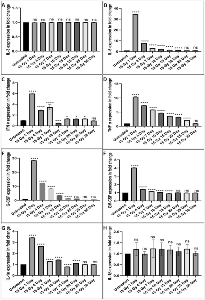
Current models to study the hematopoietic syndrome largely rely on the uniform whole-body exposures. However, in the radio-nuclear accidents or terrorist events, exposure can be non-uniform. The data available on the non-uniform exposures is limited. Thus, we have developed a mice model for studying the hematopoietic syndrome in the non-uniform or partial body exposure scenarios using the localized cobalt60 gamma radiation exposure. Femur region of Strain 'A' male mice was exposed to doses ranging from 7 to 20 Gy. The 30 day survival assay showed 19 Gy as LD100 and 17 Gy as LD50. We measured an array of cytokines and important stem cell markers such as IFN-γ, IL-3, IL-6, GM-CSF, TNF-α, G-CSF, IL-1α, IL-1β, CD 34 and Sca 1. We found significant changes in IL-6, GM-CSF, TNF-α, G-CSF, and IL-1β levels compared to untreated groups and amplified levels of CD 34 and Sca 1 positive population in the irradiated mice compared to the untreated controls. Overall, we have developed a mouse model of the hematopoietic acute radiation syndrome that might be useful for understanding of the non-uniform body exposure scenarios. This may also be helpful in the screening of drugs intended for individuals suffering from radiation induced hematopoietic syndrome.
Conflict of interest statement
The authors declare no competing interests.
Figures


Similar articles
-
Acute Radiation-induced GI-ARS and H-ARS in a Canine Model of Mixed Neutron/Gamma Relative to Reference Co-60 Gamma Radiation: A Retrospective Study.Health Phys. 2020 Sep;119(3):351-357. doi: 10.1097/HP.0000000000001215. Health Phys. 2020. PMID: 31934930
-
A Systematic Review of the Hematopoietic Acute Radiation Syndrome (H-ARS) in Canines and Non-human Primates: Acute Mixed Neutron/Gamma vs. Reference Quality Radiations.Health Phys. 2020 Nov;119(5):527-558. doi: 10.1097/HP.0000000000001319. Health Phys. 2020. PMID: 32947486 Free PMC article.
-
Early-response biomarkers for assessment of radiation exposure in a mouse total-body irradiation model.Health Phys. 2014 Jun;106(6):772-86. doi: 10.1097/HP.0000000000000094. Health Phys. 2014. PMID: 24776912
-
Delayed Captopril Administration Mitigates Hematopoietic Injury in a Murine Model of Total Body Irradiation.Sci Rep. 2019 Feb 18;9(1):2198. doi: 10.1038/s41598-019-38651-2. Sci Rep. 2019. PMID: 30778109 Free PMC article.
-
The hematologist and radiation casualties.Hematology Am Soc Hematol Educ Program. 2003:473-96. doi: 10.1182/asheducation-2003.1.473. Hematology Am Soc Hematol Educ Program. 2003. PMID: 14633795 Review.
References
-
- Huet C, Trompier F, Clairand I, Queinnec F, Bottollier-Depois JF. Physical dosimetric reconstruction of a radiological accident at Fleurus (Belgium) on 11 March 2006. Radiat. Meas. 2008;43(2–6):845–848.
-
- Christodouleas JP, et al. Short-term and long-term health risks of nuclear-power-plant accidents. N. Engl. J. Med. 2011;364(24):2334–2341. - PubMed
-
- Gupta ML, et al. Blood biomarkers in metal scrap workers accidentally exposed to ionizing radiation: A case study. Hum. Exp. Toxicol. 2013;32(12):1311–1322. - PubMed
-
- Tomonaga M. The atomic bombings of Hiroshima and Nagasaki: A summary of the human consequences, 1945–2018, and lessons for homo sapiens to end the nuclear weapon age. J. Peace Nuclear Disarm. 2019;2(2):491–517.
Publication types
MeSH terms
Substances
LinkOut - more resources
Full Text Sources
Other Literature Sources
Medical
Research Materials
