. 2021 Jan 8;4(1):53.
doi: 10.1038/s42003-020-01564-2.

GPR109A mediates the effects of hippuric acid on regulating osteoclastogenesis and bone resorption in mice

Affiliations

GPR109A mediates the effects of hippuric acid on regulating osteoclastogenesis and bone resorption in mice

Jin-Ran Chen et al. Commun Biol. .

Abstract

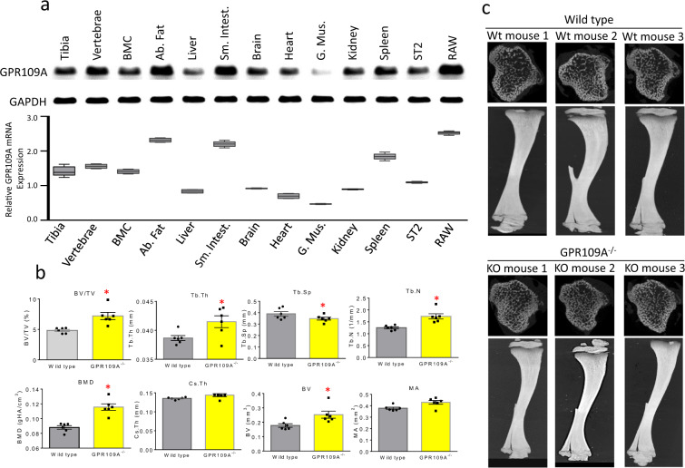
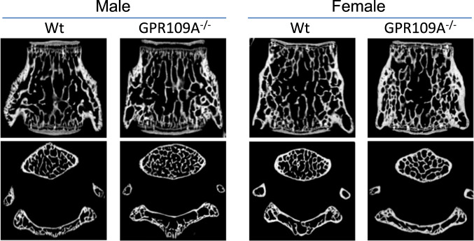
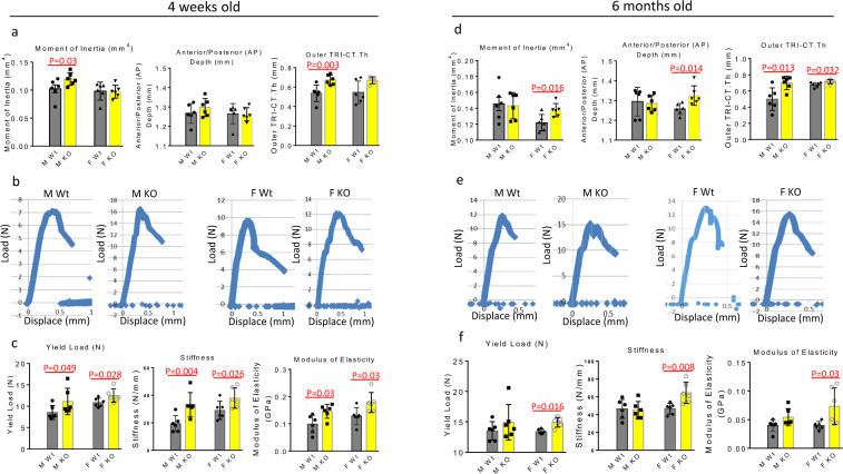
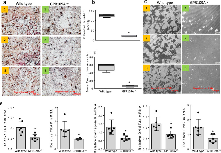
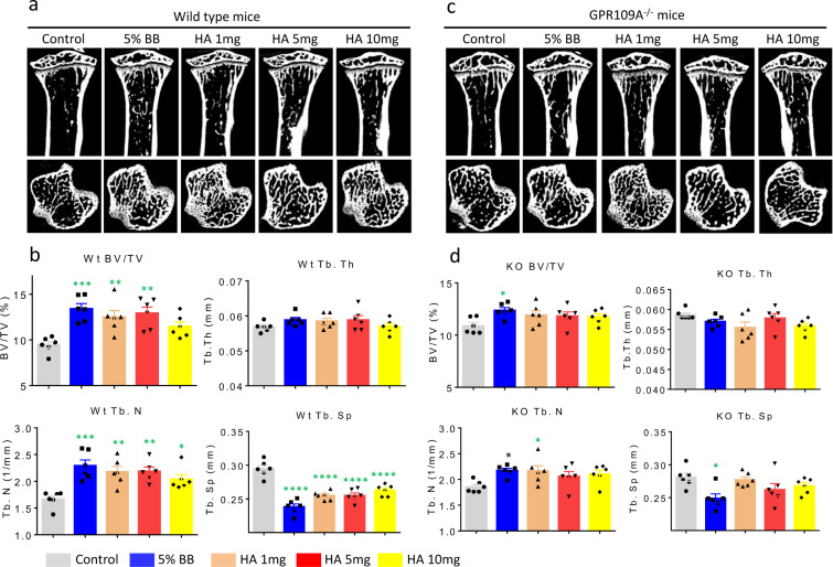
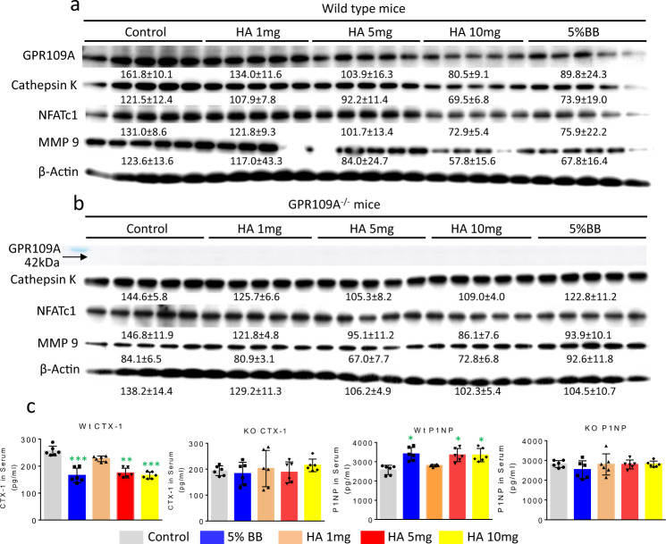
The G protein-coupled receptor 109 A (GPR109A) is robustly expressed in osteoclastic precursor macrophages. Previous studies suggested that GPR109A mediates effects of diet-derived phenolic acids such as hippuric acid (HA) and 3-(3-hydroxyphenyl) propionic acid (3-3-PPA) on promoting bone formation. However, the role of GPR109A in metabolic bone homeostasis and osteoclast differentiation has not been investigated. Using densitometric, bone histologic and molecular signaling analytic methods, we uncovered that bone mass and strength were significantly higher in tibia and spine of standard rodent diet weaned 4-week-old and 6-month-old GPR109A gene deletion (GPR109A-/-) mice, compared to their wild type controls. Osteoclast numbers in bone and in ex vivo bone marrow cell cultures were significantly decreased in GPR109A-/- mice compared to wild type controls. In accordance with these data, CTX-1 in bone marrow plasma and gene expression of bone resorption markers (TNFα, TRAP, Cathepsin K) were significantly decreased in GPR109A-/- mice, while on the other hand, P1NP was increased in serum from both male and female GPR109A-/- mice compared to their respective controls. GPR109A deletion led to suppressed Wnt/β-catenin signaling in osteoclast precursors to inhibit osteoclast differentiation and activity. Indeed, HA and 3-3-PPA substantially inhibited RANKL-induced GPR109A expression and Wnt/β-catenin signaling in osteoclast precursors and osteoclast differentiation. Resultantly, HA significantly inhibited bone resorption and increased bone mass in wild type mice, but had no additional effects on bone in GPR109A-/- mice compared with their respective untreated control mice. These results suggest an important role for GPR109A during osteoclast differentiation and bone resorption mediating effects of HA and 3-3-PPA on inhibiting bone resorption during skeletal development.


Conflict of interest statement

The authors declare no competing interests.

Figures

Fig. 1
Fig. 1. Increased bone mass phenotype in GPR109A−/− mice.
a PCR for GPR109A mRNA expression in tissues taken from a 4-week-old male mice: tibia, vertebrae, bone marrow cells (BMC), abdominal fat (Ab. Fat), liver, small intestine (Sm. Intest.), brain, heart, gastric muscles (G. Mus.), kidney, spleen and mouse-origin stromal cell line ST2 cells and macrophage cell line RAW246.7 cells (RAW), and control GAPDH mRNA expression. Box and whiskers graph under gel is real-time PCR results for relative GPR109A mRNA expression in those tissues and cells. b Micro-CT measured parameters from 4-week-old male wild type and GPR109A−/− mouse groups. BV/TV, bone volume/total tissue volume; Tb.Th, trabecular thickness; Tb.Sp, trabecular separation; Tb.N, trabecular number; BMD, actual bone mineral density; Cs.Th, cortical thickness; BV, actual bone volume; MA, medullary area; BA/TA, bone area/total area. Data are expressed as mean ± SD (n = 6 per group). *p < 0.05 by t-test. c Representative micro-CT images of the proximal tibia from three samples from each group of mice.
Fig. 2
Fig. 2. Micro-CT on L5 vertebrae from male and female GPR109A−/− and wild type mice.
Representative micro-CT images (sagittal view above, axial view below) of the L5 vertebrae from one sample from 4-weeks-old male and female of each group of GPR109A−/− and wild type (Wt) mice, white color shows trabecular bone.
Fig. 3
Fig. 3. Biomechanical testing of mouse femurs using three-point bending.
a Three different Micro-CT parameters of femurs from 4 weeks old and, d 6 months old mouse before three-point bending tests. b Represented association curves between inverse load (y axis) and inverse displace (X axis) from three-point bending tests of femurs from 4 weeks old and, e 6 months old mice. c Bone strength parameters after three-point bending tests of 4 weeks old and, f 6 months old mouse femurs. Male wild type (M Wt), male GPR109A−/− (M KO), female wild type (F Wt), female GPR109A−/− (F KO) mice. Red p = numbers mean significantly different versus respective wild type mice by t-test, n = 6, mean ± SD.
Fig. 4
Fig. 4. Increased bone mass in GPR109A−/− mice is associated with decreased osteoclast numbers.
a Representative sagittal views of quantitative pQCT analysis of one slice of the proximal tibial from three male samples from each group of mice. Bar in the middle shows color changes from black to white indicating lower to higher bone density. b Bone histology on cryosectioned tibia close to growth plate for TRAPase staining, images of magnification 40x from epifluorescent microscope (model BH-2, Olympus) showing three representative male samples from each group. Black arrows show pink TRAPase positively stained osteoclastic cells on bone surface. c Osteoclast and d osteoblast numbers counted per bone from wild type (Wt) and GPR109A−/− mice. e Bone marrow plasma resorption marker CTX-1 and, f serum P1NP levels by ELISA. Left two bars are from male wild type and GPR109A−/− mice and right two bars from age-matched (4-weeks-old) female wild type and GPR109A−/− mice, *p < 0.05 versus respective wild type mice by t-test, n = 6, mean ± SD.
Fig. 5
Fig. 5. Decreased osteoclastogenesis in GPR109A−/− mice in ex vivo non adherent bone marrow cell culture.
a Bone marrow cells were isolated from 4-week-old male wild type and GPR109A−/− mice and suspended for 48 h, non-adherent bone marrow cells were re-cultured in the presence of 50 ng/ml RANKL for 5 days. Representative pictures showing osteoclast morphology from 3 different individual wild type or GPR109A−/− mice after TRAPase staining. b Osteoclast number per well with triplicates for samples from each mouse. c Non-adherent bone marrow cells from wild type and GPR109A−/− mice were cultured for osteoclast resorptive activity in corning osteo assay plates. Pictures showing resorption pits (white spots/areas) without Von Kossa staining. d Percentage of bone resorption area per well with triplicates for samples from each mouse. e Total RNA was isolated from non-adherent bone marrow cells from wild type and GPR109A−/− mice, real time RT-PCR shows relative NFkB, TRAP, Cathepsin K, DNMT3a, Ezh2 mRNA expression. Data are shown as the means ± SD of n = 6, gene expression was relative to housekeeping gene GAPDH. *p < 0.05 versus wild type control by t-test.
Fig. 6
Fig. 6. GPR109A-mediated inhibition of RANKL-induced osteoclastogenic signaling by HA and 3-3-PPA.
Non-adherent bone marrow cells were isolated from 4-week old male wild type mice, cells were treated with HA or 3-3-PPA (HA, 60 µg/dL; 3-3-PPA, 100 µg/dL) in the presence or absence of 50 ng/ml RANKL for 48 h. a Western blot shows RANKL activated NFκB, NFATc1, MMP9 and Cathepsin K protein expression. Both HA and 3-3-PPA inhibited RANKL-induced protein expression. b Western blot shows inhibition of β-catenin and GSK3α/β and GPR109A expression by HA and 3-3-PPA in the presence of 50 ng/ml RANKL. Numbers under blots are mean ± SD band intensities relative to loading controls.
Fig. 7
Fig. 7. BB diet and HA promote bone development in wild type mice but not in GPR109A−/− mice.
Four-week-old male wild type and GPR109A−/− mice were fed with %5 BB diet and three different doses of HA supplemental diets for 6 weeks. a Representative micro-CT images (sagittal view above, axial view below) of the proximal tibial from one sample from each group of wild type mice, white color lines and dots show bone. b Micro-CT measured parameters from with or without BB or HA treatments of wild type mouse groups. BV/TV, bone volume / total tissue volume; Tb.Th, trabecular thickness; Tb.N, trabecular number; Tb.Sp, trabecular separation. c Representative micro-CT images (sagittal view above, axial view below) of the proximal tibial from one sample from each group of GPR109A−/− mice, white color shows bone. d Micro-CT measured parameters from with or without BB or HA treatments of GPR109A−/− mouse groups. Data are expressed as mean ± SD (n = 6 per group); *p < 0.05, **p < 0.01, ***p < 0.001, ****p < 0.0001 as determined by one-way ANOVA followed by Student-Newman-Keuls posthoc analysis for multiple pairwise comparisons to control.
Fig. 8
Fig. 8. HA and 5% BB diet inhibit GPR109A expression and bone resorptive signals in ten weeks old wild type but not in GPR109A−/− male mice.
Proteins were isolated from aspirated femur bone marrow cells, 5 per group from 4-weeks-old wild type or GPR109A−/− mice with or without BB or HA treatments for six weeks. a Western blot shows HA and BB diet inhibited GPR109A protein expression, and osteoclast bone resorptive markers Cathepsin K, NFATc1 and MMP9 protein expression in samples from wild type mice. b Western blot shows HA and BB diet did not change osteoclast bone resorptive markers Cathepsin K, NFATc1 and MMP9 protein expression in samples from GPR109A−/− mice compared with samples from untreated GPR109A−/− mice. c CTX-1 and P1NP levels in serum from wild type (Wt) and GPR109−/− (KO) mice. Data are expressed as mean ± SD (n = 6 per group); *p < 0.05, **p < 0.01, ***p < 0.001, as determined by one-way ANOVA followed by Student-Newman-Keuls posthoc analysis for multiple pairwise comparisons to control.

Similar articles

Cited by

References

    1. Tunaru S, et al. PUMA-G and HM74 are receptors for nicotinic acid and mediate its anti-lipolytic effect. Nat. Med. 2003;9:352–355. doi: 10.1038/nm824. - DOI - PubMed
    1. Wise A, et al. Molecular identification of high and low affinity receptors for nicotinic acid. J. Biol. Chem. 2003;278:9869–9874. doi: 10.1074/jbc.M210695200. - DOI - PubMed
    1. Miyamoto J, et al. Ketone body receptor GPR43 regulates lipid metabolism under ketogenic conditions. Proc. Natl Acad. Sci. USA. 2019;116:23813–23821. doi: 10.1073/pnas.1912573116. - DOI - PMC - PubMed
    1. Lukasova M, et al. Nicotinic acid inhibits progression of atherosclerosis in mice through its receptor GPR109A expressed by immune cells. J. Clin. Invest. 2011;121:1163–1173. doi: 10.1172/JCI41651. - DOI - PMC - PubMed
    1. Wanders D, Judd RL. Future of GPR109A agonists in the treatment of dyslipidaemia. Diabetes Obes. Metab. 2011;13:685–691. doi: 10.1111/j.1463-1326.2011.01400.x. - DOI - PubMed

Publication types