. 2021 Feb;40(7):1375-1389.
doi: 10.1038/s41388-020-01617-0. Epub 2021 Jan 8.

PRMT1 enhances oncogenic arginine methylation of NONO in colorectal cancer

Affiliations

PRMT1 enhances oncogenic arginine methylation of NONO in colorectal cancer

Xin-Ke Yin et al. Oncogene. 2021 Feb.

Abstract

Arginine methylation is an important posttranslational modification catalyzed by protein arginine methyltransferases (PRMTs). However, the role of PRMTs in colorectal cancer (CRC) progression is not well understood. Here we report that non-POU domain-containing octamer-binding protein (NONO) is overexpressed in CRC tissue and is a potential marker for poor prognosis in CRC patients. NONO silencing resulted in decreased proliferation, migration, and invasion of CRC cells, whereas overexpression had the opposite effect. In a xenograft model, tumors derived from NONO-deficient CRC cells were smaller than those derived from wild-type (WT) cells, and PRMT1 inhibition blocked CRC xenograft progression. A mass spectrometry analysis indicated that NONO is a substrate of PRMT1. R251 of NONO was asymmetrically dimethylated by PRMT1 in vitro and in vivo. Compared to NONO WT cells, NONO R251K mutant-expressing CRC cells showed reduced proliferation, migration, and invasion, and PRMT1 knockdown or pharmacological inhibition abrogated the malignant phenotype associated with NONO asymmetric dimethylation in both KRAS WT and mutant CRC cells. Compared to adjacent normal tissue, PRMT1 was highly expressed in the CRC zone in clinical specimens, which was correlated with poor overall survival in patients with locally advanced CRC. These results demonstrate that PRMT1-mediated methylation of NONO at R251 promotes CRC growth and metastasis, and suggest that PRMT1 inhibition may be an effective therapeutic strategy for CRC treatment regardless of KRAS mutation status.


Conflict of interest statement

The authors declare that they have no conflict of interest.

Figures
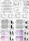
Fig. 1
Fig. 1. NONO is overexpressed in CRC tissue and promotes cell proliferation, migration, and invasion.
a NONO protein is highly expressed in CRC tissue. NONO protein level was analyzed in 28 paired colorectal tumor (T) and adjacent normal (N) tissue samples by western blotting. Glyceraldehyde 3-phosphate dehydrogenase (GAPDH) was used as a loading control. b NONO mRNA level is higher in T than in N. NONO mRNA level was examined by qPCR in 29 paired tissue samples. U6 was used as an internal control (right panel). NONO mRNA expression level in TCGA N (n = 41) and T (n = 286) CRC samples (left panel). c, d Higher NONO protein level is correlated with shorter overall survival in CRC patients. The 93 patients were divided into NONO low (n = 48) and high (n = 45) subgroups (c) and subjected to Kaplan–Meier survival analysis (d). Scale bar, 1200 μm. e NONO overexpression promotes CRC cell growth. KM12 (1 × 104) and HCT8 (2 × 104) CRC cells were seeded on day 0 and counted on days 2 and 4. f NONO KO leads to a decrease in tumor size. KM12 WT or NONO KO cells (1 × 106) were injected into nude mice (n = 5) and tumors were weighed on day 21. g–j NONO overexpression enhances CRC cell migration and invasion. The wound-healing assay was performed using KM12 (8 × 104) or HCT8 (1 × 105) cells (g, i). The transwell assay was performed using KM12 (4 × 104) or HCT8 (8 × 104) cells for 24 h (h, j). Scale bar, 400 μm. EV empty vector (pCDH-CMV)-transfected cells, NONO-OE NONO-overexpressing cells (transfected with pCDH-CMV-Flag-NONO). *P < 0.05, **P < 0.01, ***P < 0.001.
Fig. 2
Fig. 2. PRMT1 mediates NONO arginine methylation in CRC cells.
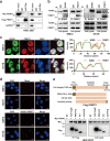
a Compared to adjacent normal (N) tissue, PRMT1 mRNA level was higher in CRC tissue (T), as determined by qPCR (n = 29 paired tissues). There were no differences in PRMT3, PRMT4, and PRMT5 mRNA levels between groups. b PRMT1 protein is highly expressed in CRC tissue. PRMT5 protein expression was similar between T and N tissues, as determined by western blotting (n = 28 paired tissues; left panel). Protein band intensity was quantified using ImageJ software (right panel). c NONO and the aDMA modification colocalized in the nucleus. Immunofluorescence analysis was performed using anti-NONO and -ADMA antibodies (right panel) in KM12 and HCT8 cells, and colocalization was analyzed using ZEN v3.0 software (Zeiss, Oberkochen, Germany) (left panel). Scale bar, 10 μm. d, e PRMT1 silencing reduces NONO aDMA level. Endogenous NONO was immunoprecipitated (IP) from control and PRMT1-deficient KM12 and HCT8 cells and analyzed by western blotting (d). NONO methylation was detected using an anti-ADMA antibody. For Duolink PLA (e), NONO aDMA level was detected using anti-NONO and -ADMA antibodies (left panel). At least 100 nuclei were counted in each group (right panel). **P < 0.01, ***P < 0.001; ns no significance.
Fig. 3
Fig. 3. NONO interacts with PRMT1 in CRC cells.
a Myc-NONO and Flag-PRMT1 interact in HEK 293T cells. pCDH-CMV-Myc-NONO and pCDH-CMV-Flag-PRMT1 were cotransfected into HEK 293T cells for 24 h before coIP analysis. b NONO interacts with PRMT1 in situ. CoIP was performed in KM12 and HCT8 cells using anti-NONO and -PRMT1 antibodies. Arrows indicate NONO or PRMT1, and circles indicate the heavy chain. c NONO and PRMT1 colocalize in the nucleus. Immunofluorescence analysis was performed using anti-NONO and -PRMT1 antibodies in KM12 and HCT8 cells (right panel). NONO and PRMT1 colocalization was analyzed using ZEN v3.0 software (left panel). Scale bar, 10 μm. d NONO interacts with PRMT1. Duolink PLA was performed with indicated antibodies. Blank, no primary antibody added in Duolink PLA. Scale bar, 10 μm. e NONO binds to the catalytic domain of PRMT1. NONO truncations are illustrated schematically at the top. HEK 293T cells were cotransfected with Myc-NONO and Flag-PRMT1 truncations for 24 h before CoIP analysis. aa amino acid, CD catalytic domain, POD post domain, PRD pre domain.
Fig. 4
Fig. 4. PRMT1-mediated methylation at R251 is required for the oncogenic function of NONO.
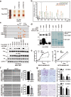
a Flag-NONO protein were immunoprecipitated from control or PRMT1-silenced KM12 cells, separated by SDS–PAGE, and subjected to silver staining. b LC–MS/MS analysis of the NONO R251 methylation site. The fragmentation pattern of the typical NONO peptide EREQPPR is shown. Fragment ions are shown as b and y ions; ++ represents the loss of doubly charged ions. c R251K mutation abolishes PRMT1-mediated aDMA modification of NONO. NONO R-to-K mutants are illustrated schematically at the top. WT or R-to-K mutant NONO protein was immunoprecipitated with Flag beads and then immunoblotted with anti-ADMA antibody. d NONO is methylated by PRMT1 at R251, as determined with the in vitro methylation assay. GST-tagged GAR, NONO, and R251K were incubated with purified GST-PRMT1 in the presence or absence of 0.6 μM SAM. Proteins were separated on SDS–PAGE and subjected to western blotting analysis and Coomassie blue staining. GAR, glycine- and arginine-rich N-terminal region of fibrillarin. e NONO R251K mutation reduces cell proliferation. KM12 (1 × 104) and HCT8 (2 × 104) cells were seeded on day 0 and counted on days 2 and 4. NONO R251K mutation inhibited KM12 cell migration in the wound-healing assay (f) and invasion in the transwell assay (g). For experiments shown in eg, KM12 cells were transfected with pCDH-CMV-Flag-NONO (WT) or pCDH-CMV-Flag-NONO-R251K (R251K mutant) for 24 h before the indicated assay. Scale bar, 400 μm. *P < 0.05, **P < 0.01, ***P < 0.001.
Fig. 5
Fig. 5. PRMT1 has an oncogenic function that is correlated with poor outcome in CRC.
a PRMT1 promotes CRC cell proliferation. KM12 (1 × 104) and HCT8 (1.8 × 104 or 2 × 104) cells were seeded on day 0 and counted every 2 days. PRMT1 knockdown inhibited and PRMT1 overexpression promoted, KM12 cell migration in the wound-healing assay (b, d) and invasion in the transwell assay (c, e). KM12 cells were transfected with pCDH-CMV-Flag-PRMT1 (PRMT1-OE) or empty vector (EV) for 24 h before the indicated assay. EV empty vector (pCDH-CMV) transfected cells, NONO-OE NONO-overexpressing cells (transfected with pCDH-CMV-Flag-NONO). Scale bar, 400 μm. f Elevated PRMT1 expression is correlated with shorter overall survival in CRC patients. The 97 patients were divided into PRMT1 low (n = 61) and high (n = 36) expression subgroups. *P < 0.05, **P < 0.01, ***P < 0.001.
Fig. 6
Fig. 6. PRMT1 inhibition reduces NONO arginine methylation and suppresses CRC progression.
a Treatment with the PRMT1 inhibitor AMI-1 reduced NONO aDMA level. KM12 and HCT8 cells treated with AMI-1 (1.2 and 0.6 mM) or DMSO for 48 h were subjected to Duolink PLA with anti-ADMA antibody. b AMI-1 treatment reduced tumor xenograft weight (n = 6 mice per group). c Treatment with the PRMT1 inhibitors MS023 and C7280948 decrease aDMA NONO level. KM12 and HCT8 cells were treated with 10 μM MS023 and 40 μM C7280948, respectively, for 48 h, and subjected to NONO IP and western blotting analysis with anti-ADMA antibody. d Treatment with MS023 or C7280948 inhibits CRC cell proliferation. KM12 WT/NONO-KO (2 × 104) and HCT8 WT/NONO-KO (4 × 104) cells were seeded on day 0 and counted on days 2 and 4 after treatment with the indicated inhibitor. KM12 and HCT8 WT/NONO-KO cells treated with MS023 and C7280948 were subjected to the wound-healing assay (e) and transwell assay (f), respectively. For (df), MS023, C7280948, or DMSO was added at the time the cells were seeded. Scale bar, 400 μm. *P < 0.05, **P < 0.01, ***P < 0.001.
Fig. 7
Fig. 7. Model of PRMT1-mediated NONO arginine methylation in CRC progression.
In CRC cells, PRMT1-mediated methylation of NONO at R251 promotes tumor cell proliferation and metastasis. Malignant progression induced by NONO arginine methylation may be abrogated by treatment with small-molecule inhibitors of PRMT1 (AMI-1, MS023, and C7280948).

References

    1. Bray F, Ferlay J, Soerjomataram I, Siegel RL, Torre LA, Jemal A. Global cancer statistics 2018: GLOBOCAN estimates of incidence and mortality worldwide for 36 cancers in 185 countries. CA Cancer J Clin. 2018;68:394–424. - PubMed
    1. Sauer R, Liersch T, Merkel S, Fietkau R, Hohenberger W, Hess C, et al. Preoperative versus postoperative chemoradiotherapy for locally advanced rectal cancer: results of the German CAO/ARO/AIO-94 randomized phase III trial after a median follow-up of 11 years. J Clin Oncol. 2012;30:1926–33. - PubMed
    1. Dienstmann R, Salazar R, Tabernero J. Personalizing colon cancer adjuvant therapy: selecting optimal treatments for individual patients. J Clin Oncol. 2015;33:1787–96. - PubMed
    1. Lièvre A, Bachet JB, Boige V, Cayre A, Le Corre D, Buc E, et al. KRAS mutations as an independent prognostic factor in patients with advanced colorectal cancer treated with cetuximab. J Clin Oncol. 2008;26:374–9. - PubMed
    1. Van Cutsem E, Köhne CH, Hitre E, Zaluski J, Chang Chien CR, Makhson A, et al. Cetuximab and chemotherapy as initial treatment for metastatic colorectal cancer. N. Engl J Med. 2009;360:1408–17. - PubMed

Publication types