. 2021 Jan 8;11(1):116.
doi: 10.1038/s41598-020-80619-0.

Fully automated preoperative segmentation of temporal bone structures from clinical CT scans

Affiliations

Fully automated preoperative segmentation of temporal bone structures from clinical CT scans

C A Neves et al. Sci Rep. .

Abstract

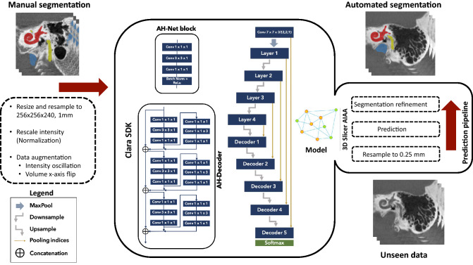
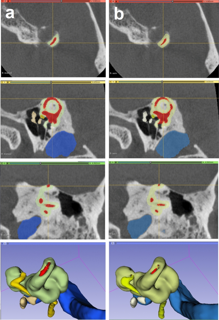
Middle- and inner-ear surgery is a vital treatment option in hearing loss, infections, and tumors of the lateral skull base. Segmentation of otologic structures from computed tomography (CT) has many potential applications for improving surgical planning but can be an arduous and time-consuming task. We propose an end-to-end solution for the automated segmentation of temporal bone CT using convolutional neural networks (CNN). Using 150 manually segmented CT scans, a comparison of 3 CNN models (AH-Net, U-Net, ResNet) was conducted to compare Dice coefficient, Hausdorff distance, and speed of segmentation of the inner ear, ossicles, facial nerve and sigmoid sinus. Using AH-Net, the Dice coefficient was 0.91 for the inner ear; 0.85 for the ossicles; 0.75 for the facial nerve; and 0.86 for the sigmoid sinus. The average Hausdorff distance was 0.25, 0.21, 0.24 and 0.45 mm, respectively. Blinded experts assessed the accuracy of both techniques, and there was no statistical difference between the ratings for the two methods (p = 0.93). Objective and subjective assessment confirm good correlation between automated segmentation of otologic structures and manual segmentation performed by a specialist. This end-to-end automated segmentation pipeline can help to advance the systematic application of augmented reality, simulation, and automation in otologic procedures.


Conflict of interest statement

The authors declare no competing interests.

Figures

Figure 1
Figure 1
Manual segmentation of temporal bone structures as seen in 3D Slicer: inner ear (red), ossicles (ivory); facial nerve (yellow), sigmoid sinus (blue). Crosshair is centered over the round window niche.
Figure 2
Figure 2
Auto-segmentation pipeline. Manual segmentation of temporal bone structures from computed tomography were used to train a deep learning model using a 3D convolutional neural network. Automated segmentation of the structures was then performed on a testing set in the 3D Slicer platform.
Figure 3
Figure 3
Training Dice similarity coefficient (a) and loss (b) for the inner ear training. The DSC graph demonstrates the improvement and eventual optimization of the model and the minimization of the loss function.
Figure 4
Figure 4
Round window niche (Crosshair) from a manual labeled scan (left—a) side by side with the auto-segmented CT dataset (right—b). From top to bottom, windows from 3D Slicer showing the axial, sagittal, coronal and 3D rendered view of the middle ear. Inner ear (red), ossicles (ivory); facial nerve (yellow), sigmoid sinus (blue) and otic capsule (green).
Figure 5
Figure 5
Superior semicircular canal dehiscence as demonstrated from manual (a) and auto-segmented (b) CT dataset seen in 3D Slicer. Note the lack of bone covering the balance canal (at the crosshair). Understanding the location and size of such a defect can facilitate surgical planning. Inner ear (red), ossicles (ivory); facial nerve (yellow), sigmoid sinus (blue) and otic capsule (green).

References

    1. Meng J, Li S, Zhang F, Li Q, Qin Z. Cochlear size and shape variability and implications in cochlear implantation surgery. Otol. Neurotol. 2016;37:1307–1313. doi: 10.1097/MAO.0000000000001189. - DOI - PubMed
    1. Locketz GD, et al. Anatomy-specific virtual reality simulation in temporal bone dissection: Perceived utility and impact on surgeon confidence. Otolaryngol. Neck Surg. 2017;156:1142–1149. doi: 10.1177/0194599817691474. - DOI - PubMed
    1. Barber SR, et al. Augmented reality, surgical navigation, and 3D printing for transcanal endoscopic approach to the petrous apex. OTO Open. 2018;2:2473974. doi: 10.1177/2473974X18804492. - DOI - PMC - PubMed
    1. Neves CA, et al. Application of holographic augmented reality for external approaches to the frontal sinus. Int. Forum Allergy Rhinol. 2020;10:920–925. doi: 10.1002/alr.22546. - DOI - PubMed
    1. Gare BM, et al. Multi-atlas segmentation of the facial nerve from clinical CT for virtual reality simulators. Int. J. Comput. Assist. Radiol. Surg. 2020;15:259–267. doi: 10.1007/s11548-019-02091-0. - DOI - PubMed

Publication types