Review
. 2021 Mar;22(3):137-151.
doi: 10.1038/s41583-020-00415-7. Epub 2021 Jan 8.

Neurexins: molecular codes for shaping neuronal synapses

Affiliations
Review

Neurexins: molecular codes for shaping neuronal synapses

Andrea M Gomez et al. Nat Rev Neurosci. 2021 Mar.

Abstract

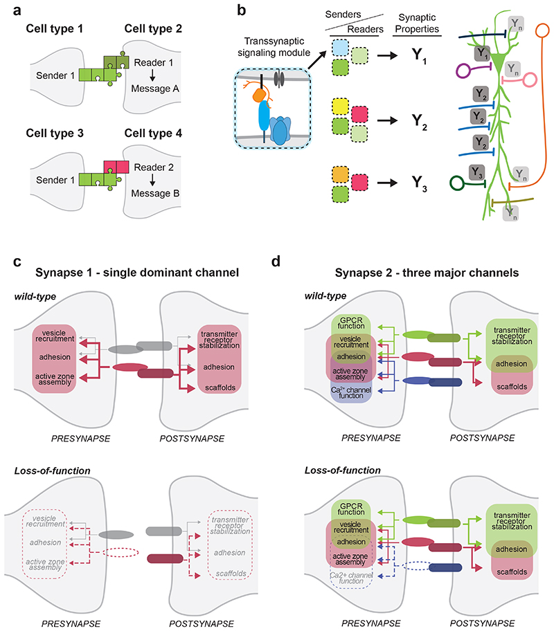
The function of neuronal circuits relies on the properties of individual neuronal cells and their synapses. We propose that a substantial degree of synapse formation and function is instructed by molecular codes resulting from transcriptional programmes. Recent studies on the Neurexin protein family and its ligands provide fundamental insight into how synapses are assembled and remodelled, how synaptic properties are specified and how single gene mutations associated with neurodevelopmental and psychiatric disorders might modify the operation of neuronal circuits and behaviour. In this Review, we first summarize insights into Neurexin function obtained from various model organisms. We then discuss the mechanisms and logic of the cell type-specific regulation of Neurexin isoforms, in particular at the level of alternative mRNA splicing. Finally, we propose a conceptual framework for how combinations of synaptic protein isoforms act as 'senders' and 'readers' to instruct synapse formation and the acquisition of cell type-specific and synapse-specific functional properties.


Figures

Figure 1
Figure 1. Synaptogenic function of Neurexins.
a) Illustration of bi-directional synapse-organizing activity of Neurexins. Presentation of Neurexin proteins or their ligands on synthetic surfaces in vitro and overexpression of Neurexin ligands in vitro and in vivo drives assembly of pre- and postsynaptic structures, respectively. b) Neurexin isoforms interact with a large array of structurally unrelated extracellular binding partners. Only a selection of ligands is displayed in this simplified schematic. Depending on cellular context, several ligands can be co-expressed at single synapses or can be differentially expressed across neuronal cell populations. c) Example for loss-of-function phenotype resulting from loss of Neurexin (nrx-1 mutant) at the neuromuscular junction of Drosophila larvae (adapted from Li et al. ), synaptic release sites are marked in orange). d) Illustration of contribution of Neurexin-Neuroligin adhesion system to growth of axonal arborisations of the motoneurons that innervate the abdominal pleural muscles of adult Drosophila (adapted from Constance et al. ).
Figure 2
Figure 2. Molecular and structural features of Neurexin isoform diversity.
a) Schematic illustrating the alternatively spliced segments of mouse Neurexin genes. Mouse Neurexin transcript isoforms are generated from three genes (Nrxn1 = 1.1Mb, Nrxn2 = 0.1Mb, Nrxn3 = 1.8Mb; note that given the big differences in gene sizes, the exons and introns are not drawn to scale), each containing up to three alternative promoters (α, β and γ) and exhibiting extensive alternative splicing at six alternatively spliced segments (AS1-6). Individual segments can contain single alternative cassette exons (e.g. AS4, AS6) or consist of complex combinations of alternative splice donor and acceptor sites (e.g. AS1, AS2, AS3, AS5). Numbers (2,3,4,8,30) depict counts of potential splice variations generated at each segment. Alternative exons are illustrated in color, constitutive exons in grey, alternative donor or acceptor sites are striped. b) Alternative promoters and alternative mRNA splicing result in Neurexin protein isoforms that share transmembrane domain (TMD) and cytoplasmic sequences but differ in their extracellular protein sequences. The extracellular sequences are composed of three major elements: Laminin-Neurexin-Sex hormone-binding globulin domains (LNS), epidermal growth factor-like domains (EGF), and attachment sites for heparan sulfates (HS). The largest Neurexin proteins are the NRXNα forms composed of six LNS domains (LNS 1-6), three interposed EGF domains (EGF A-C) and the HS attachment sites. Interaction surfaces for ligands are marked with purple lines. The NRXNβ forms contain a single LNS domain and HS attachment sites whereas the NRXNγ is the smallest form lacking LNS and EGF domains. c) Mapping of alternatively spliced segments (AS2, AS3, AS4), ligand-binding domains, and sequence variants on a prototypical LNS domain: (left) ribbon diagram and positions of alternatively spliced segments, (middle) view of a 90-degree rotation and surface representation LNS domain with mapped alternatively spliced segments, (right) surface representation as in middle panel highlighting the position of ligand-binding domains and the naturally occurring R498W variant in NRXN3α which has been linked to behavioral alterations in mice . d) Illustration of approximate sizes and hypothetical conformation of adhesion molecule complexes in the synaptic cleft: β-Neurexin (orange, left panel) with LRRTM2 (green), and α-Neurexin (orange, right panel) with Neurexophilin (NXPH 1, pink) and Neuroligin (NLGN, blue). Structural models of the extracellular domains were drawn with ChimeraX 1.0 from the following Protein Data Bank IDs: 3POY , 3B3Q , 6PNP , 5Z8Y . The position of stalk, transmembrane and cytoplasmic sequences is indicated as dashed lines. Diagrams at the bottom display positions within these structures where alternative splicing at AS1, AS6 and AS5 in NRXN β (left) and NRXN α (right) modifies the flexibility of the extracellular domains in the synaptic cleft
Figure 3
Figure 3. Control of synapse specification by alternative splicing programs.
a) Schematic illustrating the control of alternative splicing by RNA motifs and trans-acting factors (coloured spheres X1, X2, X3…). The displayed alternatively spliced segment contains an alternative splice donor site in the upstream exon (depicted as grey and blue boxes on left), followed by a cassette exon (purple box) and a downstream constitutive exon (grey box, right). Exons can contain exonic splicing enhancers (ESE) and exonic splicing silencers (ESS). The intronic between the exon boxes contain RNA motifs that act as intronic splicing enhancers (ISE) and silencers (ISS). GU marks the 5’ splice site, A-Py-AG marks the branchpoint and Polypyrimidine tract followed by the terminal AG sequence in the intron. Motifs recruit trans-acting RNA-binding proteins (depicted as colored spheres) which either promote or suppress usage of individual splice donor acceptor sites, resulting in inclusion or skipping of the alternative cassette exon (isoform 1 and isoform 2). Usage of the alternative donor site in the first exon, results in a third transcript isoform (isoform3). b) Intersection of Nrxn transcriptional programs and the combinatorial action of splicing regulators. The illustration depicts two hypothetical cell types (cell type 1 and cell type 2) that produce different Neurexin transcript repertoires. Cell type-specific transcription from promoters/enhancers (arrowheads) drives the differential transcription of the primary transcripts from the Nrxn genes (e.g. Cell type 1 transcribes high levels of NRXN1α whereas Cell type 2 transcribes high levels of NRXN3α). In addition, each expresses a specific battery of splicing regulators (Cell type 1: X1,X2, X3,…; Cell type 2: X1, X3, X5,…). The intersection of these splicing regulators (colored spheres) with the primary Nrxn transcripts then produces the cell type-specific Neurexin isoforms (e.g. β Nrxn AS4-AS5- in cell type 1 and β Nrxn AS4+AS5+ in cell type 2) ,, .
Figure 4
Figure 4. Examples of synaptic interaction modules nucleated by Neurexin proteins.
Simplified model illustrating trans-synaptic interaction modules assembled around presynaptic Neurexin protein isoforms. Amongst other components, CA3-CA1 Schaffer collateral synapses in the mouse hippocampus contain trans-synaptic NRXNAS4--NLGN1 and NRXN-LRRTM complexes which recruit postsynaptic NMDA and AMPA-type glutamate receptors. Note that LRRTM proteins bind α and β Neurexins – but for simplicity only the interaction with α is depicted here. Cerebellar parallel fiber synapses largely rely on a single trans-synaptic module consisting of NRXNAS4+ isoforms, extracellular CBLN1 proteins and the postsynaptic receptor GLUD2. CBLN1 is secreted from lysosome-like carrier vesicles . Note that CBLN1 interacts with α and β Neurexins – but for simplicity only the interaction with β is depicted here. GABAergic basket cell synapses in the mouse hippocampus contain modules of NRXNAS4+ and NRXNAS2- isoforms linking to postsynaptic NLGN2 or α/β Dystroglycan proteins, respectively. Red dots within synaptic vesicles indicate the neurotransmitter glutamate, blue dots represent GABA. One major contribution of Neurexins at synapses is the incorporation of functional voltage-gated calcium channels (VGCC) at synapses, facilitating the calcium-dependent release of synaptic vesicles ,, . The lower row displays illustrations of individual trans-synaptic modules present at the respective synapses.
Figure 5
Figure 5. Context-dependent functions of synaptic interaction modules.
a) Model for the action of synaptic interaction modules. Cell types express specific complements of “senders” (light green) and “readers” (dark green and red), such as Neurexin splice isoforms and corresponding binding partners. The impact on synapse assembly and functional specification (“message”) depends on the cell type-specific abundance of corresponding senders and readers (dark green and red). Cell type 1 and cell type 3 both express the same sender but because their downstream synaptic partners cell types 2 and 4 express different readers (reader 1 and 2, respectively), the messages will be different (message A and message B, respectively). Thus, the same presynaptic sender can convey divergent messages at different synapses. b) Across the surface of an individual neuron (here an illustration of a pyramidal cell), the combinations of axonal senders and dendritic readers create trans-synaptic modules (depicted as blue, green, light green boxes etc.) which represent a molecular code. This code sets synapse-specific properties (Y1, Y2, Y3, .. Yn; the blue labels represent glutamatergic synapses, the red labels GABAergic synapses) and, thereby, input integration in the postsynaptic cell. c) Synapses across the central nervous system employ various numbers of trans-synaptic modules that can be viewed as trans-synaptic communication channels (displayed in different colors). Some synapses, rely on a single dominant channel (here depicted in red), which has a major contribution to synapse assembly, stability and functional properties (“Synapse 1” - an example for this would be parallel fiber synapses in the cerebellum which rely on the NRXN-CBLN1-GLUD2 module ,, ). Loss of a single presynaptic sender (illustrated in the lower panel, e.g. CBLN1) results in disruption of the trans-synaptic module and substantial disruption of synapse formation and function, despite the presence of an additional, minor channel (depicted in grey). d) Other synapses (“Synapse 2”) contain multiple prominent trans-synaptic modules (here depicted in red, green, and purple) – likely to afford a larger dynamic range of plasticity. These modules drive overlapping elements of synaptic differentiation. For example, the green module drives bi-directional adhesion, presynaptic vesicle recruitment, presynaptic GPCR function, and post-synaptic stabilization of neurotransmitter receptors, whereas the purple module controls adhesion, active zone assembly and calcium channel function. Loss of a single presynaptic sender (the purple module) results in a loss of presynaptic calcium channel function but active zone assembly and adhesion are maintained by the overlapping red and green modules at this synapse. See references for examples on Ca2+ channel function ,, .

References

    1. Lu W, Bushong EA, Shih TP, Ellisman MH, Nicoll RA. The cell-autonomous role of excitatory synaptic transmission in the regulation of neuronal structure and function. Neuron. 2013;78:433–439. - PMC - PubMed
    1. Sigler A, et al. Formation and Maintenance of Functional Spines in the Absence of Presynaptic Glutamate Release. Neuron. 2017;94:304–311.:e304. - PMC - PubMed
    1. Sando R, et al. Assembly of Excitatory Synapses in the Absence of Glutamatergic Neurotransmission. Neuron. 2017;94:312–321.:e313. - PMC - PubMed
    1. Hobert O. Terminal Selectors of Neuronal Identity. Curr Top Dev Biol. 2016;116:455–475. - PubMed
    1. Hassan BA, Hiesinger PR. Beyond Molecular Codes: Simple Rules to Wire Complex Brains. Cell. 2015;163:285–291. - PMC - PubMed

Publication types

Substances