

Inflammatory response in lungs and extrapulmonary sites detected by [18F] fluorodeoxyglucose PET/CT in convalescing COVID-19 patients tested negative for coronavirus
- PMID: 33420914
- PMCID: PMC7794623
- DOI: 10.1007/s00259-020-05083-4
Inflammatory response in lungs and extrapulmonary sites detected by [18F] fluorodeoxyglucose PET/CT in convalescing COVID-19 patients tested negative for coronavirus
Abstract
Background: The severe acute respiratory syndrome coronavirus 2 (SARS-CoV-2) has resulted in an ongoing global pandemic of coronavirus disease 2019 (COVID-19). The challenges associated with imaging infected patients have resulted, to date, in a paucity of metabolic imaging studies of patients with severe COVID-19 infection. Furthermore, it remains unclear if any abnormal metabolic events are taking place in patients who have recovered from COVID-19.
Purpose: To use [18F] fluorodeoxyglucose ([18F] FDG) positron emission tomography/computed tomography (PET/CT) to measure metabolic activity in inflamed organs of patients convalescing post severe COVID-19 infection.
Materials and methods: A prospective study was performed in seven convalescing patients who were recovering from severe COVID-19 infection in February 2020. Prior to [18F] FDG PET/CT, all patients had received two consecutive negative results of real-time reverse transcriptase polymerase chain reaction (RT-PCR) for SARS-CoV-2 nucleic acid. Clinical intake including symptoms, treatment, laboratory test results, and follow-up was performed. The PET/CT images of COVID-19 patients were compared to a control group of patients that were matched for age and sex.
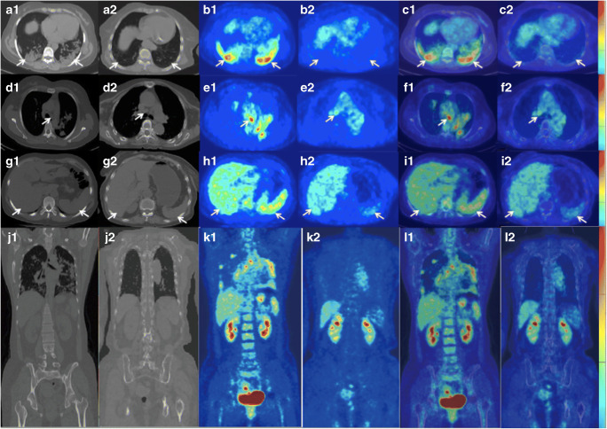
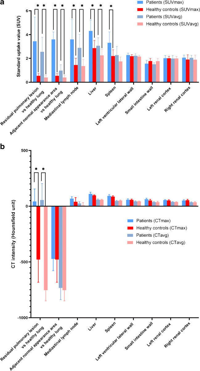
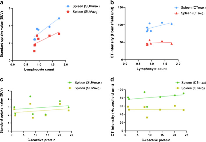
Results: Residual pulmonary lesions were present in all patients and maximum standard uptake value (SUVmax), average standard uptake value (SUVavg), maximum CT intensity (CTmax), and average CT intensity (CTavg) were all significantly greater than in the control group (p < 0.01 for all). In addition, SUVmax and SUVavg were significantly greater in the mediastinal lymph node and liver, and SUVmax was significantly greater in the spleen, of COVID-19 patients compared with controls (p < 0.05 for all). For the spleen, SUVmax (r2 = 0.863, p = 0.003) and SUVavg (r2 = 0.797, p = 0.007) were significantly correlated with blood lymphocyte count, and which was below the normal range in five of the seven (71.4%) patients convalescing post severe COVID-19 infection.
Conclusion: [18F] FDG PET/CT quantitative analysis has shown that significant inflammation remained in lungs, mediastinal lymph nodes, spleen, and liver after two consecutive negative RT-PCR tests in patients convalescing post severe COVID-19 infection.
Keywords: Coronavirus disease 2019; PET/CT; Severe acute respiratory syndrome coronavirus 2; [18F] Fluorodeoxyglucose.
Conflict of interest statement
The authors declare that they have no conflict of interest.
Figures
Similar articles
-
FDG-PET/CT of COVID-19 and Other Lung Infections.Semin Nucl Med. 2022 Jan;52(1):61-70. doi: 10.1053/j.semnuclmed.2021.06.017. Epub 2021 Jun 22. Semin Nucl Med. 2022. PMID: 34246449 Free PMC article. Review.
-
The imaging quantification of multiple organs by dynamic 18F-FDG PET/CT in discharged COVID-19 patients: A prospective pilot study.Int J Med Sci. 2022 Sep 6;19(10):1539-1547. doi: 10.7150/ijms.73801. eCollection 2022. Int J Med Sci. 2022. PMID: 36185330 Free PMC article.
-
Vasculitis changes in COVID-19 survivors with persistent symptoms: an [18F]FDG-PET/CT study.Eur J Nucl Med Mol Imaging. 2021 May;48(5):1460-1466. doi: 10.1007/s00259-020-05084-3. Epub 2020 Oct 30. Eur J Nucl Med Mol Imaging. 2021. PMID: 33123760 Free PMC article.
-
Incidental Findings Suggestive of COVID-19 Pneumonia in Oncologic Patients Undergoing 18F-FDG PET/CT Studies: Association Between Metabolic and Structural Lung Changes.J Nucl Med. 2022 Feb;63(2):274-279. doi: 10.2967/jnumed.121.261915. Epub 2021 Jun 4. J Nucl Med. 2022. PMID: 34088776 Free PMC article.
-
A systematic review and meta-analysis of 18F-fluoro-d-deoxyglucose positron emission tomography interpretation methods in vascular graft and endograft infection.J Vasc Surg. 2020 Dec;72(6):2174-2185.e2. doi: 10.1016/j.jvs.2020.05.065. Epub 2020 Jul 6. J Vasc Surg. 2020. PMID: 32645420
Cited by
-
Mapping COVID-19 with nuclear imaging: from infection to functional sequelae.Am J Nucl Med Mol Imaging. 2021 Feb 15;11(1):59-63. eCollection 2021. Am J Nucl Med Mol Imaging. 2021. PMID: 33688456 Free PMC article.
-
New Onset of Acute and Chronic Hepatic Diseases Post-COVID-19 Infection: A Systematic Review.Biomedicines. 2024 Sep 10;12(9):2065. doi: 10.3390/biomedicines12092065. Biomedicines. 2024. PMID: 39335578 Free PMC article. Review.
-
Transient immune hepatitis as post-coronavirus disease complication: A case report.World J Clin Cases. 2021 Jun 6;9(16):4032-4039. doi: 10.12998/wjcc.v9.i16.4032. World J Clin Cases. 2021. PMID: 34141763 Free PMC article.
-
[18F]FDG PET/CT in Patients Affected by SARS-CoV-2 and Lymphoproliferative Disorders and Treated with Tocilizumab.J Pers Med. 2022 Nov 4;12(11):1839. doi: 10.3390/jpm12111839. J Pers Med. 2022. PMID: 36579547 Free PMC article.
-
FDG-PET/CT of COVID-19 and Other Lung Infections.Semin Nucl Med. 2022 Jan;52(1):61-70. doi: 10.1053/j.semnuclmed.2021.06.017. Epub 2021 Jun 22. Semin Nucl Med. 2022. PMID: 34246449 Free PMC article. Review.
References
MeSH terms
Substances
LinkOut - more resources
Full Text Sources
Other Literature Sources
Medical
Miscellaneous
