Review
. 2021 Mar;31(2):228-243.
doi: 10.1111/jon.12819. Epub 2021 Jan 9.

COVID-19: Neuroimaging Features of a Pandemic

Affiliations
Review

COVID-19: Neuroimaging Features of a Pandemic

Theodoros Ladopoulos et al. J Neuroimaging. 2021 Mar.

Abstract

Background and purpose: The ongoing Coronavirus Disease 2019 (COVID-19) pandemic is caused by the novel severe acute respiratory syndrome coronavirus 2 (SARS-CoV-2). COVID-19 is occasionally associated with manifold diseases of the central nervous system (CNS). We sought to present the neuroimaging features of such CNS involvement. In addition, we sought to identify typical neuroimaging patterns that could indicate possible COVID-19-associated neurological manifestations.

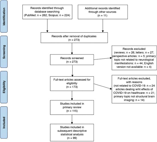
Methods: In this systematic literature review, typical neuroimaging features of cerebrovascular diseases and inflammatory processes associated with COVID-19 were analyzed. Reports presenting individual patient data were included in further quantitative analysis with descriptive statistics.

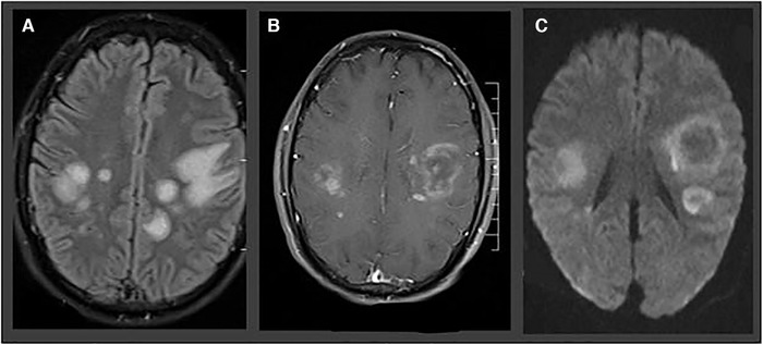
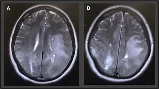
Results: We identified 115 studies reporting a total of 954 COVID-19 patients with associated neurological manifestations and neuroimaging alterations. A total of 95 (82.6%) of the identified studies were single case reports or case series, whereas 660 (69.2%) of the reported cases included individual information and were thus included in descriptive statistical analysis. Ischemia with neuroimaging patterns of large vessel occlusion event was revealed in 59.9% of ischemic stroke patients, whereas 69.2% of patients with intracerebral hemorrhage exhibited bleeding in a location that was not associated with hypertension. Callosal and/or juxtacortical location was identified in 58.7% of cerebral microbleed positive images. Features of hemorrhagic necrotizing encephalitis were detected in 28.8% of patients with meningo-/encephalitis.

Conclusions: Manifold CNS involvement is increasingly reported in COVID-19 patients. Typical and atypical neuroimaging features have been observed in some disease entities, so that familiarity with these imaging patterns appears reasonable and may assist clinicians in the differential diagnosis of COVID-19 CNS manifestations.

Keywords: COVID-19; CT; MRI; SARS-CoV-2; neuroimaging.


Figures

Fig 1
Fig 1
“Preferred Reporting Items for Systematic Reviews and Meta‐Analyses”—flow diagram of the study. n = number.
Fig 2
Fig 2
(A) CT angiography demonstrates left internal carotid artery occlusion (large vessel occlusion stroke) in a 79‐year‐old female patient. (B) Despite thrombolysis in cerebral infarction scale 2b recanalization, complete infarction of the left media and anterior cerebral artery was visible on follow‐up diffusion‐weighted imaging sequence.
Fig 3
Fig 3
Atypical ICH in a 57‐year‐old woman with prolonged encephalopathy and hypoxic respiratory failure. Polymerase chain reaction for SARS‐CoV‐2 was positive in the nasopharyngeal swab. (A) Brain CT performed in the acute setting revealed a right frontal lobe hemorrhage with perifocal edema. (B) T2* image demonstrating the right frontal lobar intracerebral hemorrhage.
Fig 4
Fig 4
Two‐dimensional time‐of‐flight MR venography (A) with atypical internal cerebral vein thrombosis in a 30‐year‐old female SARS‐CoV‐2‐positive patient, presenting with severe headache, vomiting, and bilateral papilledema. Axial 2‐dimensional fluid‐attenuated inversion recovery revealed bilateral thalamic edema due to venous congestion in the same patient (B).
Fig 5
Fig 5
Acute necrotizing hemorrhagic encephalopathy in a 32‐year‐old man presenting with speech impairment, disorientation, and epileptic seizures. MRIs show predominantly subcortical fluid‐attenuated inversion recovery hyperintensities (A), with ring enhancement in T1‐weighted sequence (B) and ring‐shaped diffusion‐restriction (C).
Fig 6
Fig 6
A 54‐year‐old man with no previous medical history admitted with headache, disorientation, nausea, vomiting, and blurred vision. T2‐weighted images show pronounced acute disseminated encephalomyelitis like leukoencephalopathy in the parietal and frontal lobes involving u‐fibers as well as cortical areas on both sides.

References

    1. Renu K, Prasanna PL, Valsala Gopalakrishnan A. Coronaviruses pathogenesis, comorbidities and multi‐organ damage ‐ a review. Life Sci 2020;255:117839. - PMC - PubMed
    1. Zhu N, Zhang D, Wang W, et al. A novel Coronavirus from patients with pneumonia in China, 2019. N Engl J Med 2020;382:727‐33. - PMC - PubMed
    1. Gupta A, Madhavan MV, Sehgal K, et al. Extrapulmonary manifestations of COVID‐19. Nat Med 2020;26:1017‐32. - PMC - PubMed
    1. Tsivgoulis G, Palaiodimou L, Katsanos AH, et al. Neurological manifestations and implications of COVID‐19 pandemic. Ther Adv Neurol Disord 2020;13:1756286420932036. - PMC - PubMed
    1. Mao L, Jin H, Wang M, et al. Neurologic manifestations of hospitalized patients with Coronavirus disease 2019 in Wuhan, China. JAMA Neurol 2020;77:683‐90. - PMC - PubMed