. 2021 May;36(5):1191-1202.
doi: 10.1002/mds.28477. Epub 2021 Jan 9.

Visual Dysfunction Predicts Cognitive Impairment and White Matter Degeneration in Parkinson's Disease

Affiliations

Visual Dysfunction Predicts Cognitive Impairment and White Matter Degeneration in Parkinson's Disease

Angeliki Zarkali et al. Mov Disord. 2021 May.

Abstract

Background: Visual dysfunction predicts dementia in Parkinson's disease (PD), but whether this translates to structural change is not known. The objectives of this study were to identify longitudinal white matter changes in patients with Parkinson's disease and low visual function and also in those who developed mild cognitive impairment.

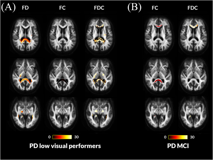
Methods: We used fixel-based analysis to examine longitudinal white matter change in PD. Diffusion MRI and clinical assessments were performed in 77 patients at baseline (22 low visual function/55 intact vision and 13 PD-mild cognitive impairment/51 normal cognition) and 25 controls and again after 18 months. We compared microstructural changes in fiber density, macrostructural changes in fiber bundle cross-section and combined fiber density and cross-section, across white matter, adjusting for age, sex, and intracranial volume.

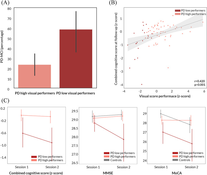
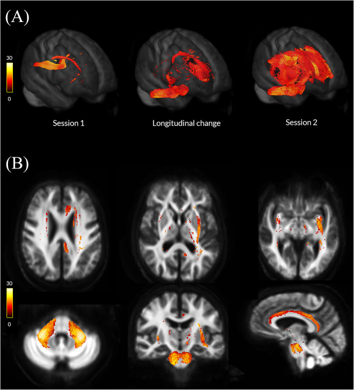
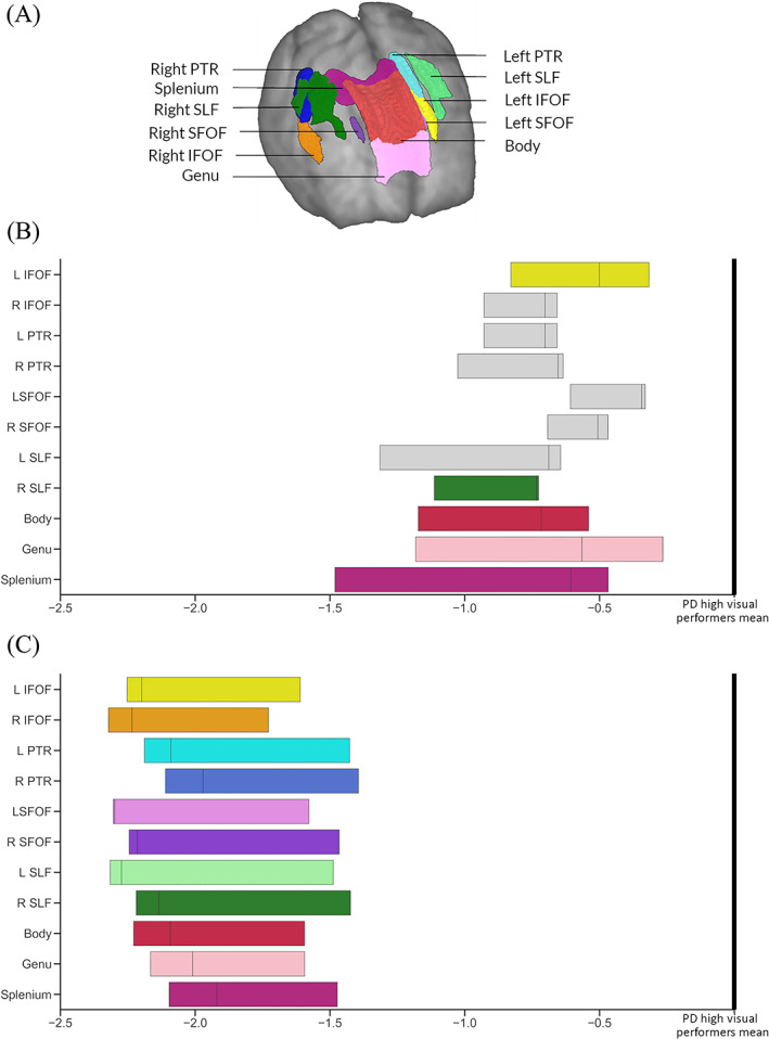
Results: Patients with PD and visual dysfunction showed worse cognitive performance at follow-up and were more likely to develop mild cognitive impairment compared with those with normal vision (P = 0.008). Parkinson's with poor visual function showed diffuse microstructural and macrostructural changes at baseline, whereas those with mild cognitive impairment showed fewer baseline changes. At follow-up, Parkinson's with low visual function showed widespread macrostructural changes, involving the fronto-occipital fasciculi, external capsules, and middle cerebellar peduncles bilaterally. No longitudinal change was seen in those with mild cognitive impairment at baseline or converters, even when the 2 groups were combined.

Conclusion: Parkinson's patients with poor visual function show increased white matter damage over time, providing further evidence for visual function as a marker of imminent cognitive decline. © 2021 The Authors. Movement Disorders published by Wiley Periodicals LLC on behalf of International Parkinson and Movement Disorder Society.

Keywords: Parkinson's disease; Parkinson's disease dementia; diffusion weighted imaging; fixel; white matter.


Figures

FIG. 1
FIG. 1
Longitudinal changes in cognition in patients with Parkinson's disease. (A) Percentage of patients who developed mild cognitive impairment (PD‐MCI) in PD low visual performers compared with PD high visual performers. (B) Correlation between a marker of overall visual performance at baseline and combined cognitive performance at 18‐month follow‐up in patients with Parkinson's disease (95% confidence interval). Visual performance is presented as the summed z score of the 2 computer‐based visual tasks (cats and dogs task and biological motion task: Z = Z biolmotion + Z cats&dogs). Cognitive performance is presented as combined cognitive score (z scored against the performance of age‐matched controls). (C) Longitudinal change in combined cognitive score, MMSE and MoCA in PD low visual performers, PD high visual performers, and controls. Error bars represent 95% confidence intervals. MMSE, Mini–Mental State Examination; MoCA, Montreal Cognitive Assessment. [Color figure can be viewed at wileyonlinelibrary.com]
FIG. 2
FIG. 2
Macrostructural white matter changes in patients with Parkinson's disease and low visual performance over time. (A) Changes in white matter macrostructure (as seen by reduction in fiber cross‐section [FC]) in PD low visual performers compared with PD high visual performers (FWE‐corrected P < 0.05) in session 1 (baseline, left), at longitudinal change (difference between the 2 images, middle), and in session 2 (18 months follow‐up, right). Results are presented as streamlines and colored by percentage reduction. (B) Statistically significant (FWE‐corrected P < 0.05) longitudinal reductions in fiber cross‐section (FC) in PD low visual performers compared with PD high visual performers. Results are colored by percentage reduction. [Color figure can be viewed at wileyonlinelibrary.com]
FIG. 3
FIG. 3
Fiber tract–specific reductions at baseline in PD low visual performers compared with PD high visual performers and PD with mild cognitive impairment compared with PD with normal cognition from whole‐brain fixel‐based analysis. (A) PD low visual performers showed widespread microstructural (changes in fiber density [FD]) compared with PD high visual performers, with reductions within the genu, body, and splenium of the corpus callosum, the right internal capsule, the cingulum bilaterally, tapetum bilaterally, posterior thalamic radiations bilaterally, right hippocampus, and the right corticospinal tract. Macrostructural changes (changes in fiber density) were also seen within the splenium of the corpus callosum, the right cingulum, and bilateral posterior thalamic radiations. Changes in the combined FDC metric were seen within the genu, body, and splenium of the corpus callosum, the right internal capsule, the cingulum bilaterally, tapetum bilaterally, posterior thalamic radiations bilaterally, right hippocampus, and the right corticospinal tract; these represent impaired overall ability to relay information in these tracts in PD low visual performers. (B) Patients with Parkinson's disease who developed mild cognitive impairment (MCI) showed macrostructural changes (changes in fiber cross‐section [FC]) compared with Parkinson's disease with stable vision within the genu and splenium of the corpus callosum, posterior thalamic radiations bilaterally and the right hippocampus. Changes in the combined FDC metric are seen in the genu and the right hippocampus; this represents impaired overall ability to relay information in these tracts in PD‐MCI compared with PD with normal cognition (NC). No changes were seen in the FD metric for this patient group. Results are displayed as streamlines; these correspond to fixels that significantly differed between PD low and high visual performers (FWE‐corrected P < 0.05). Streamlines are colored by percentage reduction (color bars). More details on the baseline changes seen in these groups are seen in Figure S2. [Color figure can be viewed at wileyonlinelibrary.com]
FIG. 4
FIG. 4
Significant tracts in Parkinson's low performers; tract of interest analysis. (A) Anatomical representation of all analyzed tracts. PTR, posterior thalamic and optic radiations; SLF, superior longitudinal fasciculi; IFOF, Inferior fronto‐occipital fasciculi (segmentation includes the inferior longitudinal fasciculus); and SLF, superior fronto‐occipital fasciculi. (B) Baseline visit. Reduction (mean, 95% CI) in fiber cross‐section (FC) visualized as percentage reduction from the mean of patients with Parkinson's disease with high visual performance. Tracts with significantly reduced FC (FDR‐corrected P < 0.05) are shown in color, whereas tracts with no significant changes in FDC are plotted in gray. L, left; R, right; C, visit 2 (18‐month follow‐up). Reduction (mean, 95% CI) in fiber cross‐section (FC) visualized as percentage reduction from the mean of patients with Parkinson's disease with high visual performance at follow‐up. All 11 of the selected tracts showed significantly reduced FC (FDR‐corrected P < 0.05). L, left; R, right. [Color figure can be viewed at wileyonlinelibrary.com]

References

    1. Williams‐Gray CH, Mason SL, Evans JR, et al. The CamPaIGN study of Parkinson's disease: 10‐year outlook in an incident population‐based cohort. J Neurol Neurosurg Psychiatry 2013;84(11):1258–1264. - PubMed
    1. Hely MA, Reid WGJ, Adena MA, et al. The Sydney multicenter study of Parkinson's disease: the inevitability of dementia at 20 years. Mov Disord 2008;23(6):837–844. - PubMed
    1. Anang JBM, Gagnon J‐F, Bertrand J‐A, et al. Predictors of dementia in Parkinson disease: a prospective cohort study. Neurology 2014;83(14):1253–1260. - PMC - PubMed
    1. Hamedani AG, Abraham DS, Maguire MG, Willis AW. Visual impairment is more common in Parkinson's disease and is a risk factor for poor health outcomes. Mov Disord 2020;35:1542–1549. - PMC - PubMed
    1. Leyland L‐A, Bremner FD, Mahmood R, et al. Visual tests predict dementia risk in Parkinson disease. Neurol Clin Pract 2019;10:29–39. - PMC - PubMed

Publication types

MeSH terms

LinkOut - more resources