. 2021 Apr;19(4):1018-1028.
doi: 10.1111/jth.15241. Epub 2021 Feb 9.

Anti-β2-glycoprotein I and anti-prothrombin antibodies cause lupus anticoagulant through different mechanisms of action

Affiliations

Anti-β2-glycoprotein I and anti-prothrombin antibodies cause lupus anticoagulant through different mechanisms of action

Tessa Noordermeer et al. J Thromb Haemost. 2021 Apr.

Abstract

Background: The presence of lupus anticoagulant (LA) is an independent risk factor for thrombosis. This laboratory phenomenon is detected as a phospholipid-dependent prolongation of the clotting time and is caused by autoantibodies against beta2-glycoprotein I (β2GPI) or prothrombin. How these autoantibodies cause LA is unclear.

Objective: To elucidate how anti-β2GPI and anti-prothrombin antibodies cause the LA phenomenon.

Methods: The effects of monoclonal anti-β2GPI and anti-prothrombin antibodies on coagulation were analyzed in plasma and with purified coagulation factors.

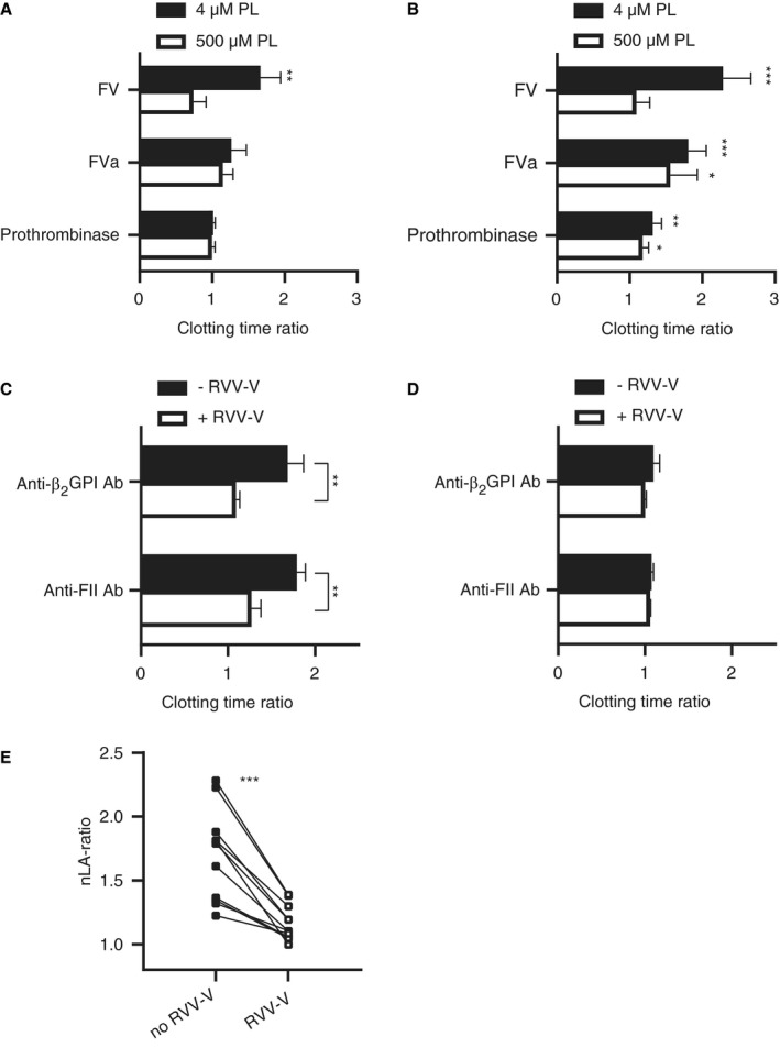
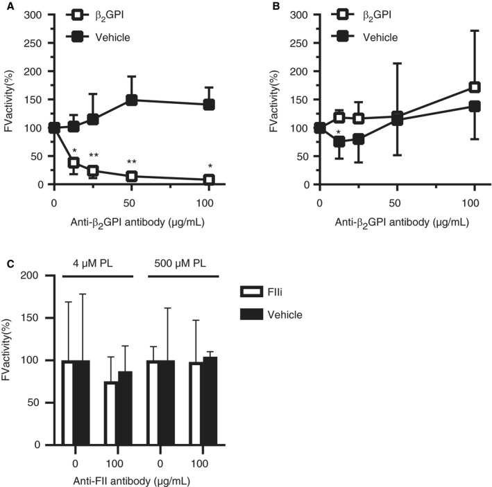
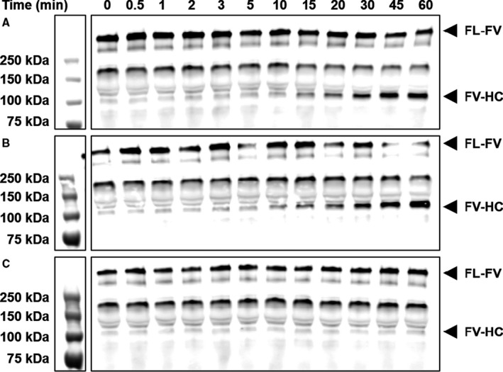
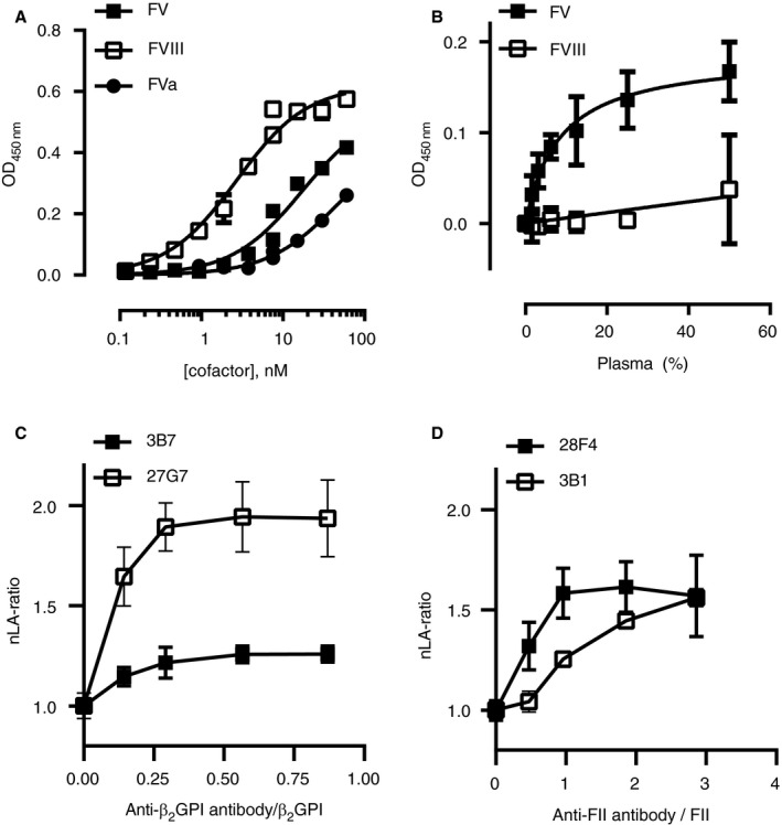
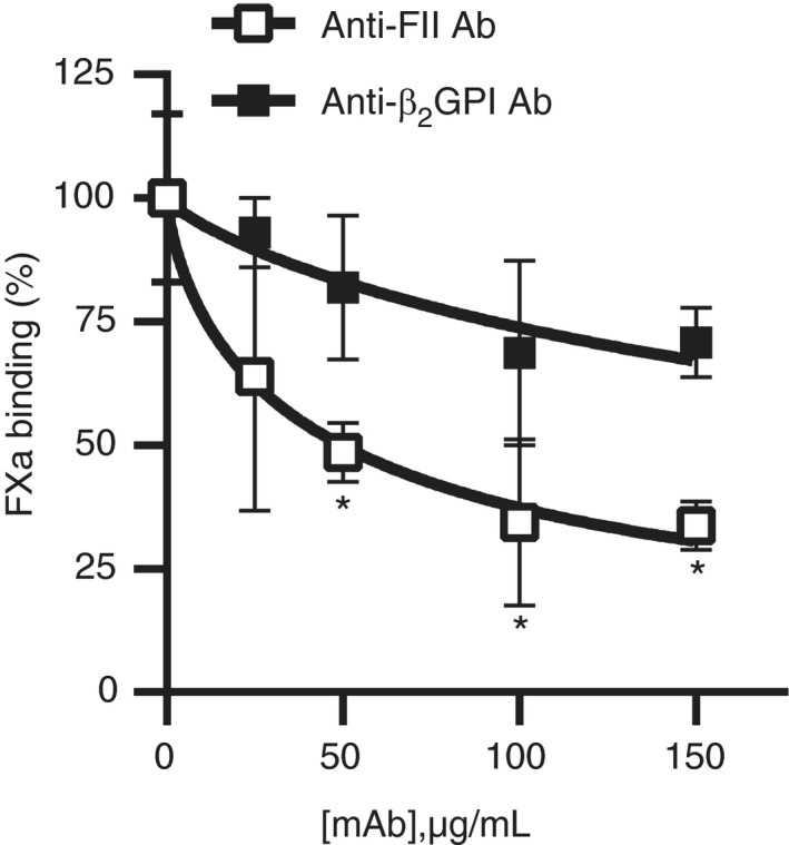
Results: Detection of LA caused by anti-β2GPI or anti-prothrombin antibodies required the presence of the procofactor factor V (FV) in plasma. LA effect disappeared when FV was replaced by activated FV (FVa), both in a model system and in patient plasma, although differences between anti-β2GPI and anti-prothrombin antibodies were observed. Further exploration of the effects of the antibodies on coagulation showed that the anti-β2GPI antibody attenuated FV activation by activated faxtor X (FXa), whereas the anti-prothrombin antibody did not. Binding studies showed that β2GPI--antibody complexes directly interacted with FV with high affinity. Anti-prothrombin complexes caused the LA phenomenon through competition for phospholipid binding sites with coagulation factors as reduced FXa binding to lipospheres was observed with flow cytometry in the presence of these antibodies.

Conclusion: Anti-β2GPI and anti-prothrombin antibodies cause LA through different mechanisms of action: While anti-β2GPI antibodies interfere with FV activation by FXa through a direct interaction with FV, anti-prothrombin antibodies compete with FXa for phospholipid binding sites. These data provide leads for understanding the paradoxical association between thrombosis and a prolonged clotting time in the antiphospholipid syndrome.

Keywords: antiphospholipid antibodies; beta2-glycoprotein I; factor V; lupus anticoagulant; prothrombin.


Conflict of interest statement

There are no conflicts of interest reported by any of the authors.

Figures

Figure 1
Figure 1
Lupus anticoagulant induced by anti‐beta2‐glycoprotein I (β2GPI) and anti‐prothrombin antibodies requires factor V (FV). A, B, FV‐depleted plasma was reconstituted with FV, activated FV (FVa), or preformed prothrombinase complex in the presence or absence of monoclonal anti‐β2GPI (3B7) (A) or anti‐prothrombin (anti‐FII; 28F4; B) antibodies. Clotting was initiated with activated factor X (FXa) and CaCl2 at 4 µM or 500 µM phospholipids. Clotting time ratios at low (4 µM) and high (500 µM) phospholipids were calculated between clotting times with and without monoclonal antibodies (mAbs; n = 4–5). Significant differences were determined between clotting times with and without mAbs using an unpaired Student’s t‐test. C, D, Pooled normal plasma was supplemented with the FV‐activating snake venom enzyme RVV‐V in the presence or absence of mAbs. Clotting times were obtained using dilute Russell's viper venom time (dRVVT) screen (C) and confirm (D) reagents and ratios were calculated between clotting times with and without mAbs (n = 3). Significant differences were determined between clotting time ratios with and without RVV‐V using an unpaired Student’s t‐test. E, Similar experiments were performed with mixed lupus anticoagulant (LA)‐positive patients plasma (n = 11) positive for anti‐β2GPI (n = 9) or anti‐prothrombin antibodies (n = 11). Results are shown as individual LA ratios of patients’ plasma incubated with or without RVV‐V and were calculated as described in the Method section. Connected lines represent paired data. Significant differences were determined using a paired sample t‐test. *P‐value<0.05, **P‐value<0.01, ***P‐value<0.005
Figure 2
Figure 2
Antibody‐beta2‐glycoprotein I (β2GPI) but not antibody‐prothrombin complexes attenuate activated factor X (FXa)‐dependent factor V (FV) activation. A, B, FV was activated with FXa at either 4 (A) or 500 (B) µM phospholipids, with or without β2GPI and a monoclonal anti‐β2GPI antibody (3B7). C, FV was activated with FXa at either 4 or 500 µM phospholipids, with or without prothrombin active site mutant (FIIi) or a monoclonal anti‐prothrombin antibody (28F4). Data are expressed as relative FV activity compared to the conditions without antibodies (n = 3–5). Significant differences were determined between samples with and without β2GPI or FIIi using a Mann‐Whitney test. *P‐value<0.05, **P‐value<0.01
Figure 3
Figure 3
Antibody‐beta2‐glycoprotein I (β2GPI) complexes delay activated factor X (FXa)‐dependent factor V (FV) activation over time. 10 nM FV was activated with 0.1 nM FXa in the presence of phospholipids and CaCl2 and anti‐β2GPI antibody (3B7; A), β2GPI (B), or both (C) at 37°C. Samples were taken at the indicated time points and subjected to SDS‐PAGE and western blot
Figure 4
Figure 4
Antibody‐beta2‐glycoprotein I (β2GPI) complexes bind to factor V (FV) with high affinity and have an indirect effect on lupus anticoagulant. 3B7 was immobilized on a microtiter plate and saturated with β2GPI. A, Plates were incubated with purified FV, factor VIII (FVIII), or activated FV (FVa) at the indicated concentrations and binding was assessed with polyclonal antibodies (n = 2). B, Plates were incubated with several dilutions of pooled normal plasma and binding of FV or FVIII was assessed (n = 3). Data were analyzed with non‐linear regression assuming a 1:1 interaction. C, D, Normalized lupus anticoagulant (nLA) ratios were obtained with dilute Russell's viper venom time (dRVVT) screen and confirm reagents in pooled normal plasma supplemented with increasing concentrations of monoclonal anti‐β2GPI (3B7 or 27G7; C) or anti‐prothrombin (28F4 or 3B1; D) antibodies (25, 50, 100, and 150 µg/mL; n = 3). Antibody concentrations were expressed relative to the concentrations of β2GPI or prothrombin in plasma
Figure 5
Figure 5
Antibody‐prothrombin complexes compete with coagulation factors on phospholipid binding sites. Biotinylated activated factor X (FXa) was incubated with lipospheres and competition with antibody‐beta2‐glycoprotein I (β2GPI) or antibody‐prothrombin complexes was assessed with flow cytometry. Significant differences were determined between baseline FXa binding at 0 µg/mL and other concentrations of monoclonal antibodies using an unpaired Student’s t‐test (n = 3). *P‐value<0.05

References

    1. Keeling D, Mackie I, Moore GW, Greer IA, Greaves M. Guidelines on the investigation and management of antiphospholipid syndrome. Br J Haematol. 2012;157(1):47‐58. - PubMed
    1. Limper M, de Leeuw K, Lely AT, et al. Diagnosing and treating antiphospholipid syndrome: A consensus paper. Neth J Med. 2019;77(3):98‐108. - PubMed
    1. Miyakis S, Lockshin MD, Atsumi T, et al. International consensus statement on an update of the classification criteria for definite antiphospholipid syndrome (APS). J Thromb Haemost. 2006;4(2):295‐306. - PubMed
    1. Pengo V, Banzato A, Denas G, et al. Correct laboratory approach to APS diagnosis and monitoring. Autoimmun Rev. 2013;12(8):832‐834. - PubMed
    1. Lockshin MD, Kim M, Laskin CA, et al. Lupus anticoagulant, but not anticardiolipin antibody, predicts adverse pregnancy outcome in patients with antiphospholipid antibodies. Arthritis Rheum. 2012;64(7):2311‐2318. - PMC - PubMed

Publication types