. 2021 Apr;232(4):642-655.
doi: 10.1016/j.jamcollsurg.2020.11.031. Epub 2021 Jan 7.

Effect of Mitochondrial Antioxidant (Mito-TEMPO) on Burn-Induced Cardiac Dysfunction

Affiliations

Effect of Mitochondrial Antioxidant (Mito-TEMPO) on Burn-Induced Cardiac Dysfunction

Jake J Wen et al. J Am Coll Surg. 2021 Apr.

Abstract

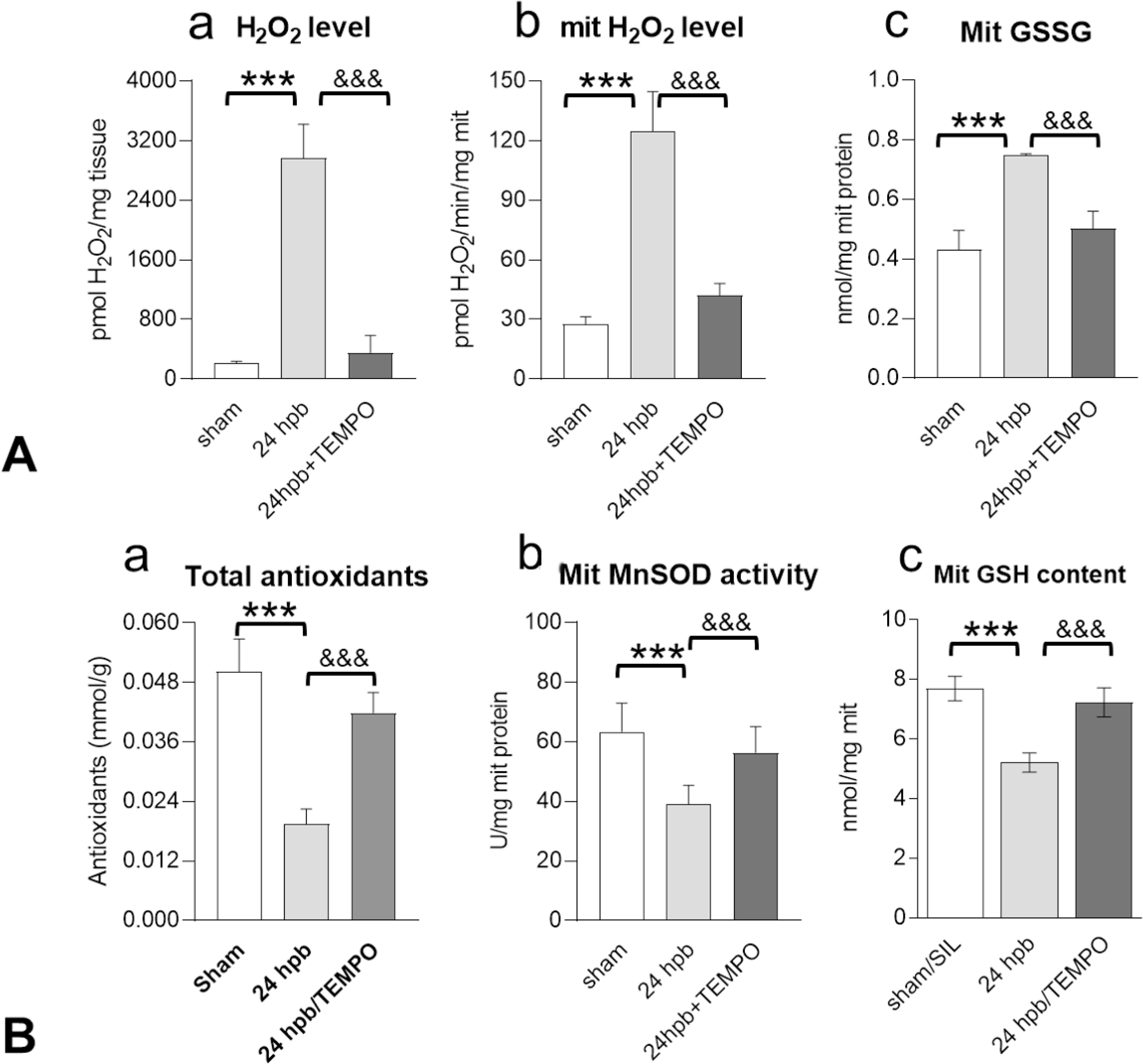
Background: Imbalance of oxidants/antioxidants results in heart failure, contributing to mortality after burn injury. Cardiac mitochondria are a prime source of reactive oxygen species (ROS), and a mitochondrial-specific antioxidant may improve burn-induced cardiomyopathy. We hypothesize that the mitochondrial-specific antioxidant, Triphenylphosphonium chloride (Mito-TEMPO), could protect cardiac function after burn.

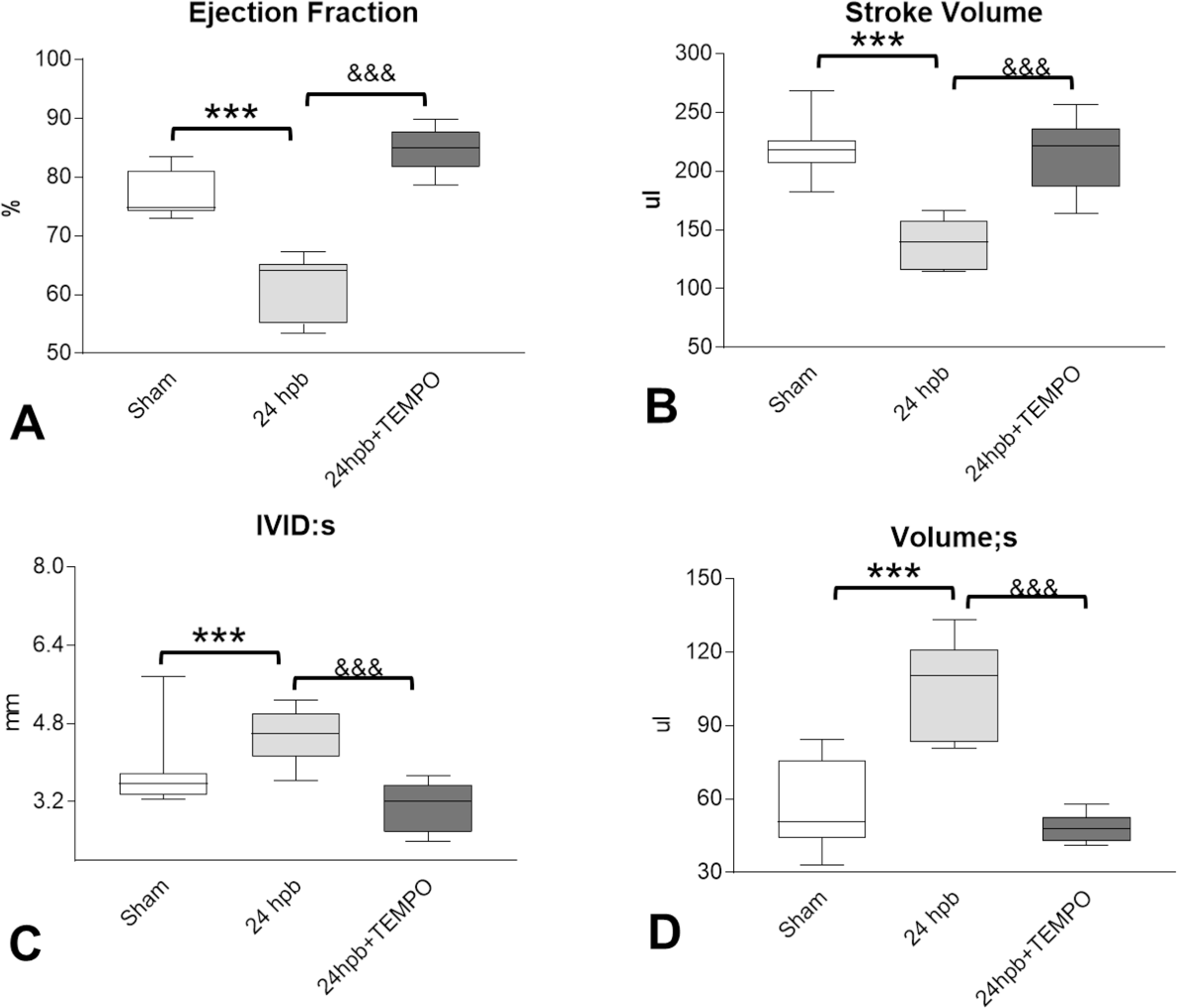
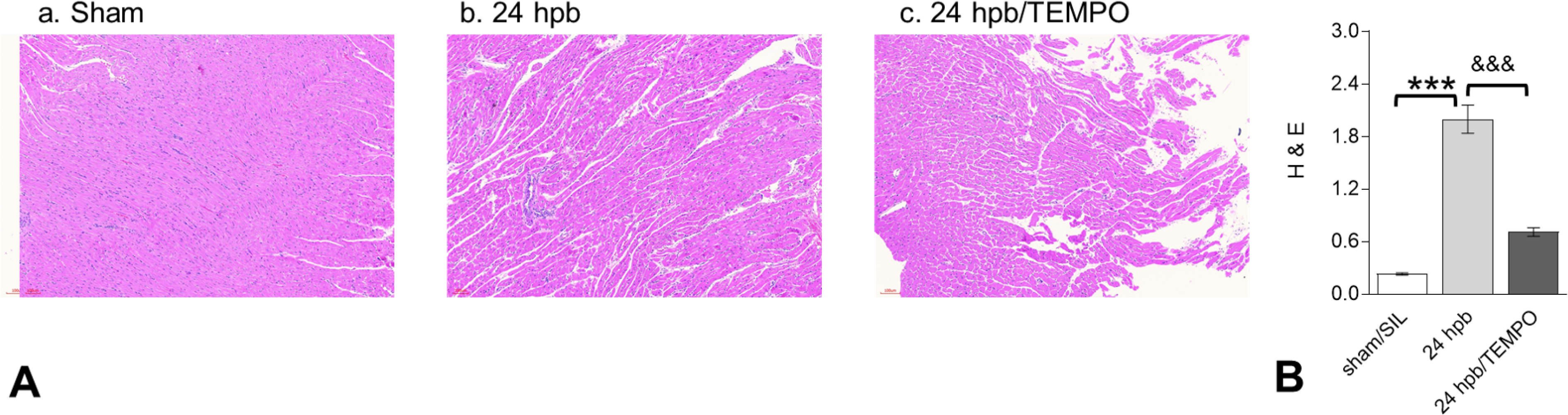
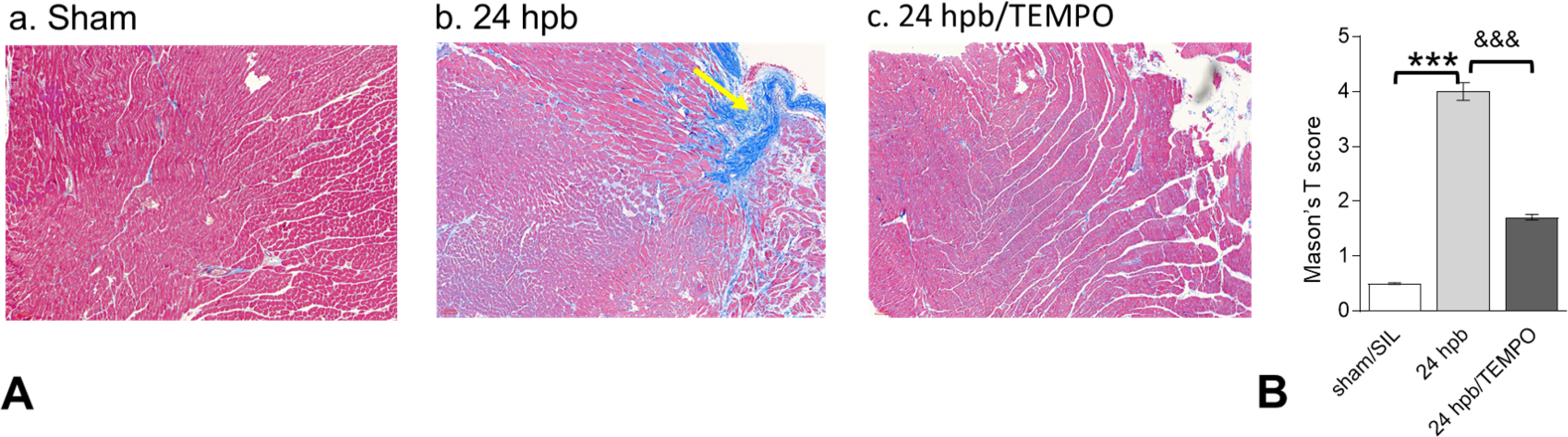
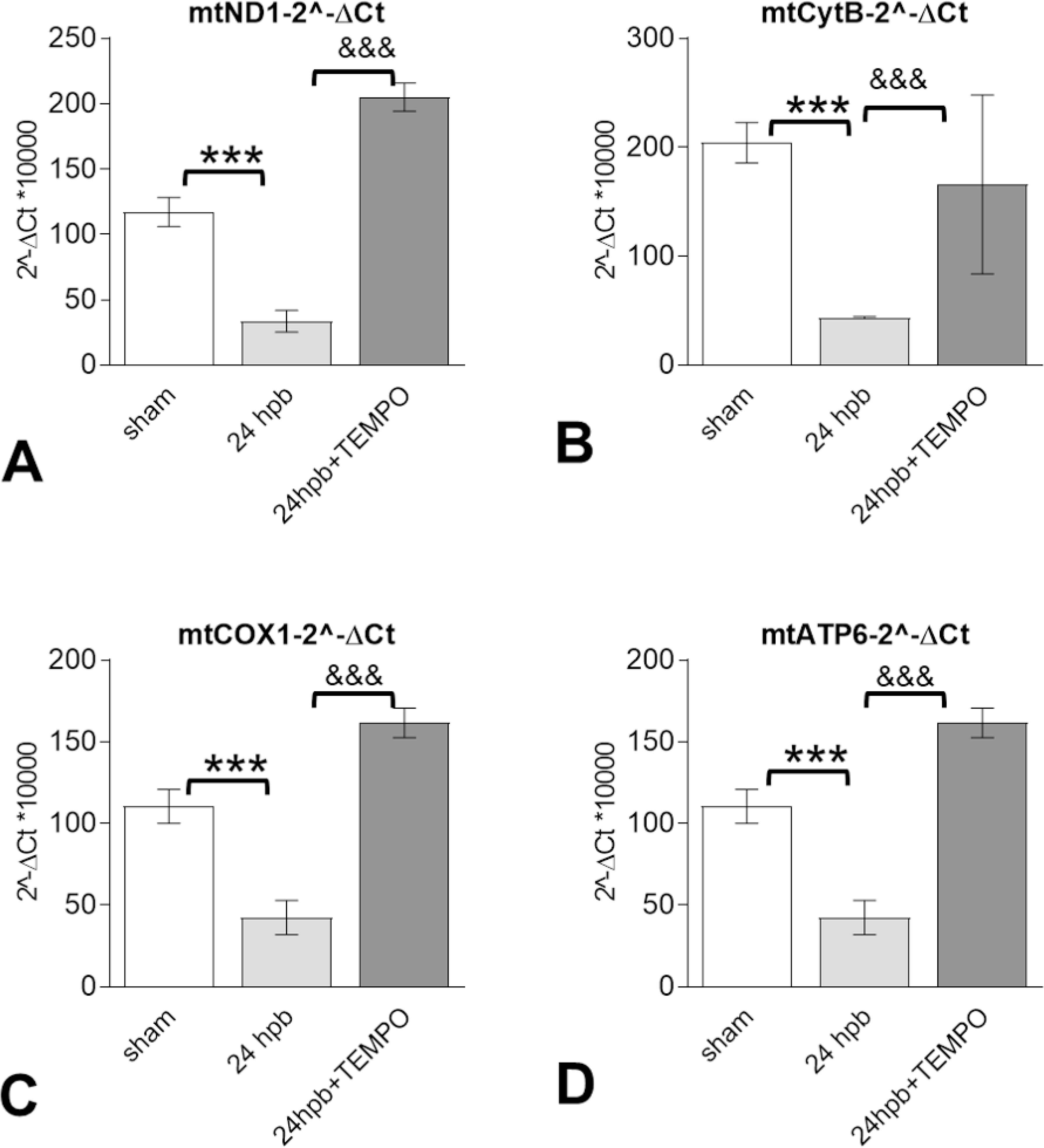
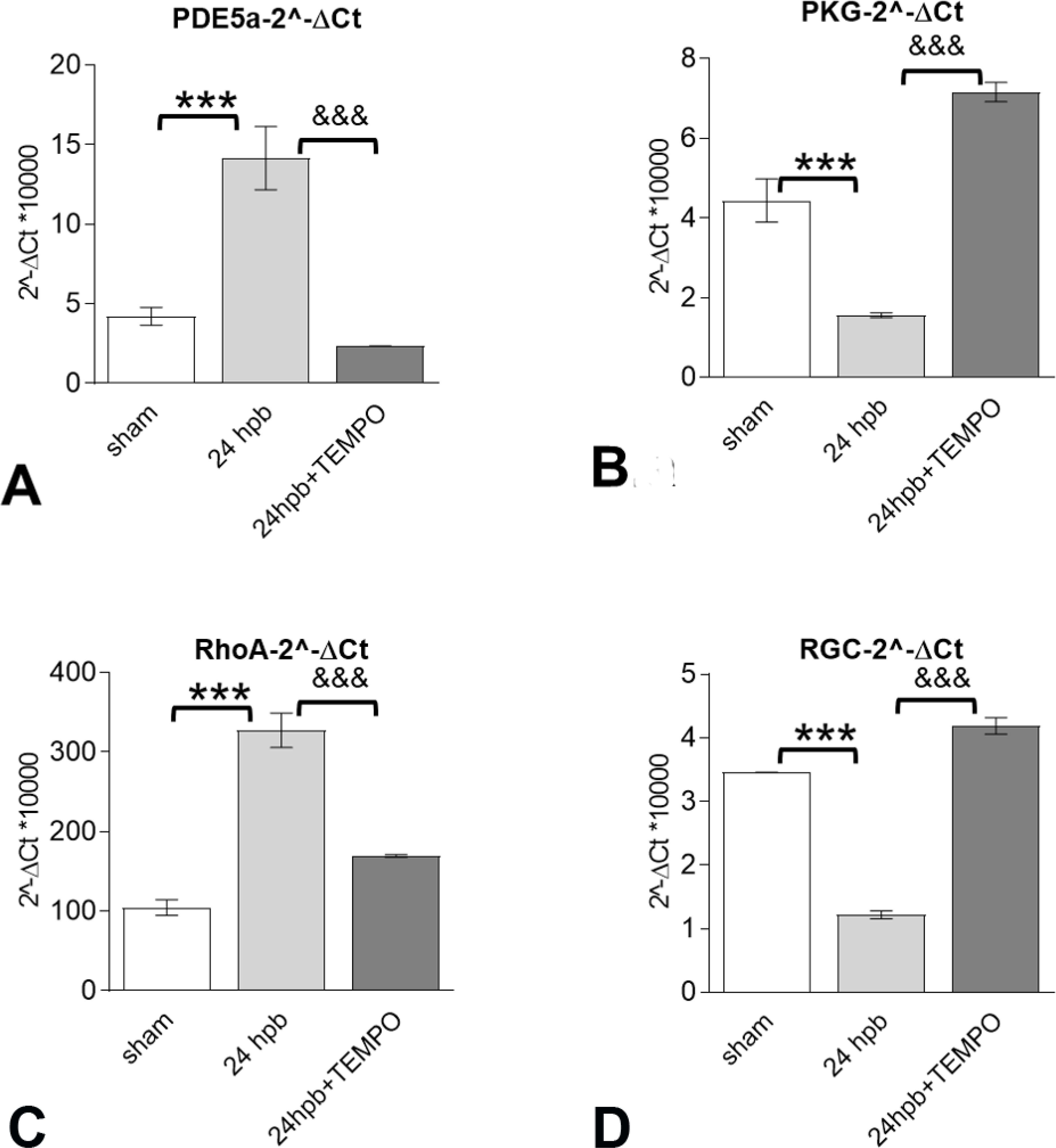
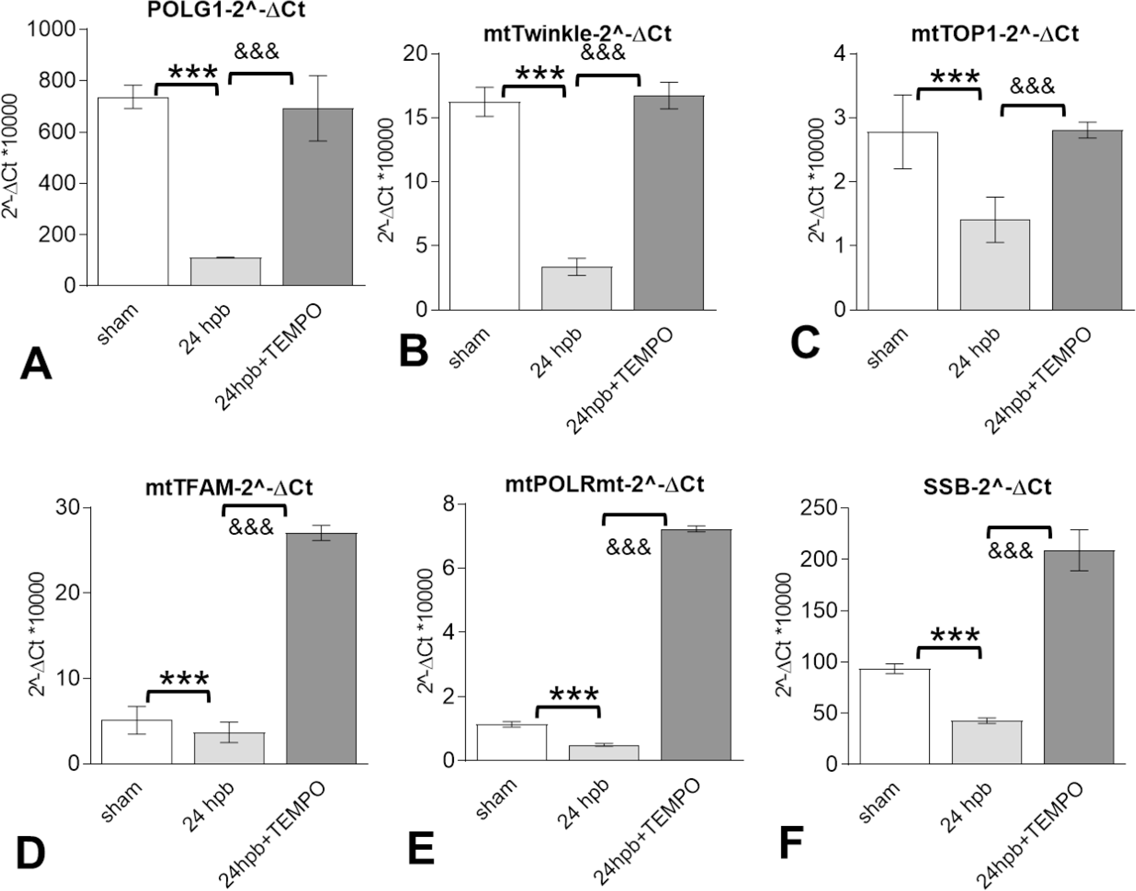
Study design: Male rats had a 60% total body surface area (TBSA) scald burn injury and were treated with/without Mito-TEMPO (7 mg/kg-1, intraperitoneal) and harvested at 24 hours post-burn. Echocardiography (ECHO) was used for measurement of heart function. Masson Trichrome and hematoxylin and eosin (H & E) staining were used for cardiac fibrosis and immune response. Qualitative polymerase chain reaction (qPCR) was used for mitochondrial DNA replication and gene expression.

Results: Burn-induced cardiac dysfunction, fibrosis, and mitochondrial damage were assessed by measurement of mitochondrial function, DNA replication, and DNA-encoded electron transport chain-related gene expression. Mito-TEMPO partially improved the abnormal parameters. Burn-induced cardiac dysfunction was associated with crosstalk between the NFE2L2-ARE pathway, PDE5A-PKG pathway, PARP1-POLG-mtDNA replication pathway, and mitochondrial SIRT signaling.

Conclusions: Mito-TEMPO reversed burn-induced cardiac dysfunction by rescuing cardiac mitochondrial dysfunction. Mitochondria-targeted antioxidants may be an effective therapy for burn-induced cardiac dysfunction.


Figures

Figure 1.
Figure 1.
Effect of Mito-TEMPO on the balance of cardiac oxidants and antioxidants after burn injury. Shown are levels of (A) cardiac total H2O2 (a), cardiac mitochondrial H2O2 (b) and cardiac mitochondrial GSSG content after burn injury (± Mito-TEMPO). Shown are levels of (B) total cardiac antioxidant capacity (B.a), cardiac mitochondrial MnSOD activity (B.b) and cardiac mitochondrial GSH content (B.c) after burn injury (± Mito-TEMPO). In all figures, data are plotted as mean value ± SD. Significance is shown as * (24 hpb vs. sham) or # (24 hpb vs. 24 hpb/TEMPO), and presented as *, #p<0.05, **, ##p<0.01, ***, ### p<0.001 (n = ≥6 per group).
Figure 2.
Figure 2.
Effect of Mito-TEMPO on burn-induced cardiac dysfunction. Shown are (A) ejection fraction (EF), (B) stroke volume (SV), (C) Left ventricular internal diameter end systole (LVID;s), (D) left ventricular systolic volume (LV Vol;s.). Data are presented as mean value ± SD, shown as **, && p < 0.01, ***, &&& p < 0.001 (n = ≥ 6 per group).
Figure 3.
Figure 3.. Effect of Mito-TEMPO on burn-induced myocardial inflammation.
(A) Shown are representative H&E stained images of the left ventricle (magnification: 20×. Pink: muscle/cytoplasm/keratin, blue: nuclear) (panels A.a to A.c). (B) Shown are the density measurements from panel A. In all figures, data are plotted as mean value ± SEM (n ≥ 6 per group). Significance is shown as * (24 hpb vs. matched control) or & (24 hpb/untreated vs. 24 hpb/TEMPO), and presented as ***,&&& p < 0.001 (n = ≥ 6 per group).
Figure 4.
Figure 4.. Effect of Mito-TEMPO on burn-induced cardiac fibrogenesis.
(A) Shown are representative images of the left ventricle with Masson’s trichrome staining (panels a–c). (B) Shown are the relative density measurements from panel A. In all figures, data are plotted as mean value ± SEM (n ≥ 6 per group). Significance is shown as * (24 hpb vs. matched control) or & (24 hpb/untreated vs. 24 hpb/TEMPO), and presented as ***,&&& p < 0.001 (n = ≥ 6 per group).
Figure 5.
Figure 5.. Effects of Mito-TEMPO on mitochondrial DNA-encoded gene expression after burn injury.
Shown are (A) representatives of the mitochondrial DNA encoded complex I genes (mtND1), (B) mitochondrial DNA encoded complex III gene (CYTB) (C) mitochondrial DNA encoded complex IV gene (COXI), and (D) mitochondrial DNA encoded complex V genes (ATP6). In all figures, data are presented as mean value SD. Significance is shown as * (24 hpb vs. sham control) or & (24 hpb vs. 24 hpb/TEMPO), and presented as ***, &&& p < 0.001 (n = 6 per group).
Figure 6.
Figure 6.. The effects of Mito-TEMPO on burn-induced cardiac dysfunction occur through the PDE5A-PKG pathway.
Shown are (A) Myocardial levels of PDE5A mRNA and Myocardial levels of PKG (B) as well as PKG-down-regulated genes including RhoA (C) and RGC (D) mRNA levels. Results were normalized to GAPDH and β-actin mRNAs, and represent as 2 −ΔCt x 1000. In all figures, data are plotted as mean value ± SD. Significance is shown as * (24 hpb vs. control) or & (24 hpb vs. 24 hpb/TEMPO), and presented as ***, &&& p < 0.001 (n = ≥ 6 per group).
Figure 7.
Figure 7.. The benefits of Mito-TEMPO on burn-induced cardiac dysfunction were through regulation of mitochondrial DNA replication-related genes.
Shown are mitochondrial DNA replication-related genes included (A) mitochondrial DNA polymerase gamma 1 (POLG1), (B) mitochondrial twinkle, (C) mitochondrial topoisomerase 1 (TOP1), (D) mitochondrial transcription factor A (TFAM), (E) mitochondrial DNA directed RNA polymerase (mtPOLR), (F) mitochondrial single stranded DNA binding protein (mtSSB). Results were normalized to GAPDH and β-actin mRNAs and represent fold change after burn injury (±MitoTEMPO), as compared to sham. In all figures, data are plotted as mean value ± SD. Significance is shown as * (24 hpb vs. control) or & (24 hpb vs. 24 hpb/TEMPO), and presented as ***, &&& p < 0.001 (n = ≥ 6 per group).
Figure 8.
Figure 8.. The benefit of Mito-TEMPO on burn-induced cardiac dysfunction via Nrf2-ARE-ROS axis.
Shown are target genes of Nrf2-ARE-ROS axis including: (A)Nrf2 (A), HO-1 (B), NQO1 (C), GCLM (D), PGC1-alpha (E), PARP1 and SIRT3 (G). Results were normalized to rat GAPDH and β-actin mRNAs and represent fold change after a burn (±TEMPO), as compared to that noted in matched normal controls. In all figures, data are plotted as mean value ± SD. Significance is shown as * (24 hpb vs. control) or & (24 hpb vs. 24 hpb/TEMPO), and presented as ***, &&& p < 0.001 (n = ≥ 6 per group).

Comment in

  • Invited Commentary.
    Gaines BA. Gaines BA. J Am Coll Surg. 2021 Apr;232(4):655. doi: 10.1016/j.jamcollsurg.2020.12.029. J Am Coll Surg. 2021. PMID: 33771323 No abstract available.

References

    1. Klein MB, Goverman J, Hayden DL, et al. Benchmarking outcomes in the critically injured burn patient. Ann Surg. 2014. May;259(5):833–41. - PMC - PubMed
    1. Miller SF, Bessey PQ, Schurr MJ, et al. National Burn Repository 2005: a ten-year review. J Burn Care Res. 2006. Jul-Aug;27(4):411–36. - PubMed
    1. Kallinen O, Maisniemi K, Bohling T, et al. Multiple organ failure as a cause of death in patients with severe burns. J Burn Care Res. 2012. Mar-Apr;33(2):206–11. - PubMed
    1. Jeschke MG, Gauglitz GG, Kulp GA, et al. Long-term persistance of the pathophysiologic response to severe burn injury. PLoS One. 2011;6(7):e21245. - PMC - PubMed
    1. Guillory AN, Clayton RP, Herndon DN, Finnerty CC. Cardiovascular Dysfunction Following Burn Injury: What We Have Learned from Rat and Mouse Models. Int J Mol Sci. 2016. Jan 2;17(1). - PMC - PubMed

MeSH terms