. 2021 Jan 9;40(1):25.
doi: 10.1186/s13046-020-01823-4.

The EGFR-HSF1 axis accelerates the tumorigenesis of pancreatic cancer

Affiliations

The EGFR-HSF1 axis accelerates the tumorigenesis of pancreatic cancer

Weikun Qian et al. J Exp Clin Cancer Res. .

Abstract

Background: Pancreatic ductal adenocarcinoma (PDAC) is one of the most malignant diseases because of its non-symptomatic tumorigenesis. We previous found heat shock factor 1 (HSF1) was critical for PDAC progression and the aim of this study was to clarified the mechanisms on early activation of HSF1 and its role in the pancreatic cancer tumorigenesis.

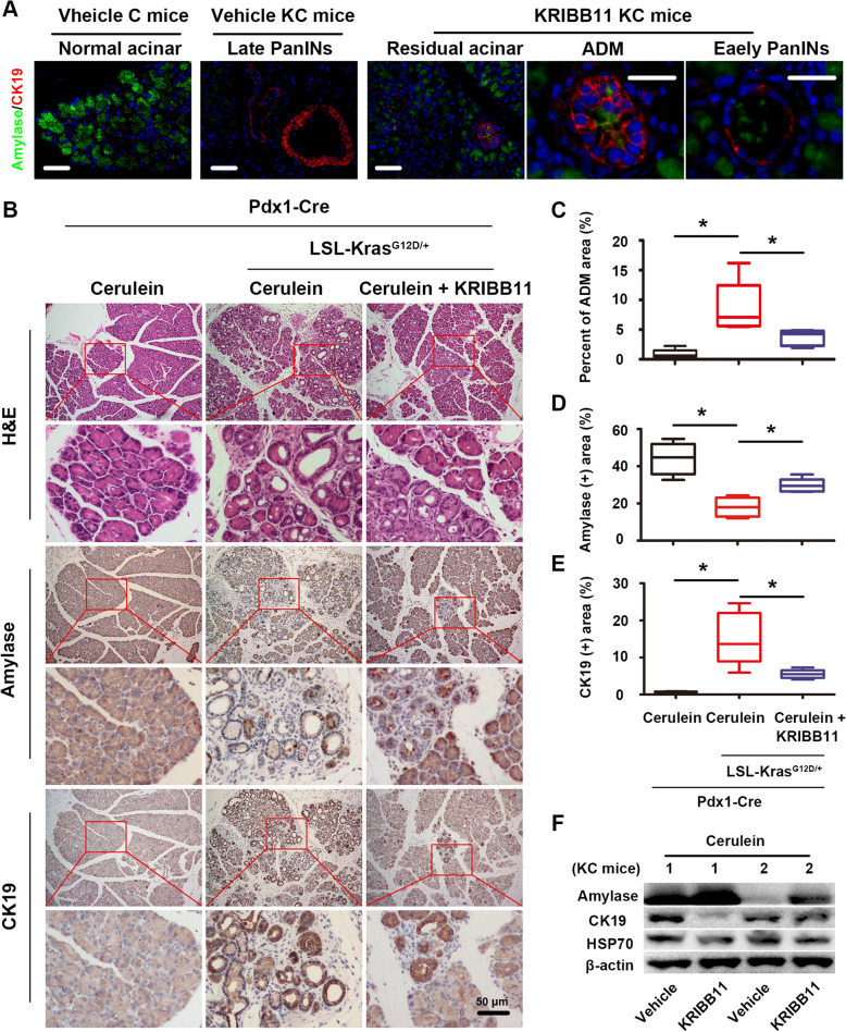
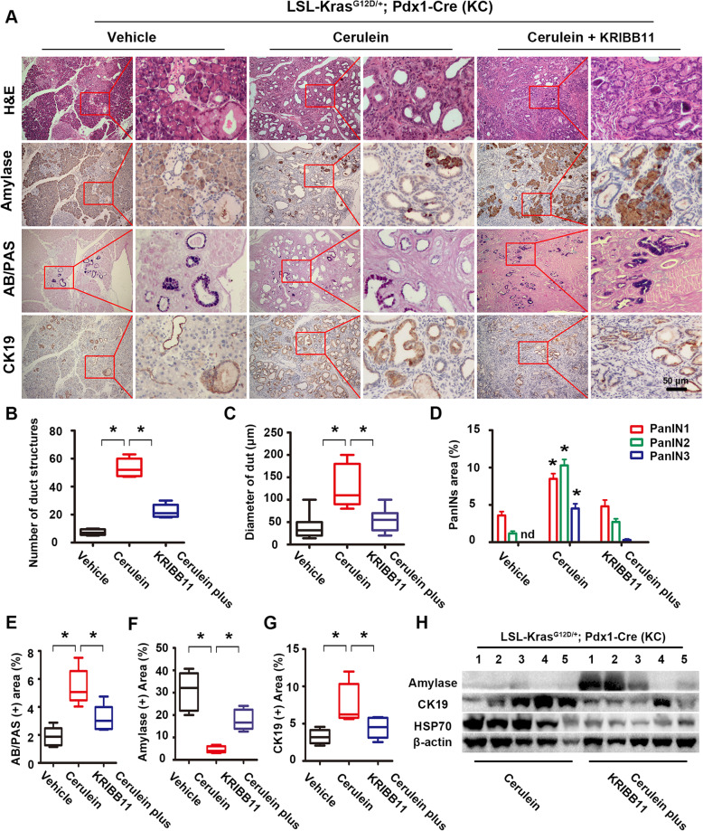
Methods: The expression and location of HSF1 on human or mice pancreatic tissues were examined by immunohistochemically staining. We mainly used pancreatic acinar cell 3-dimensional (3D) culture and a spontaneous pancreatic precancerous lesion mouse model called LSL-KrasG12D/+; Pdx1-Cre (KC) (and pancreatitis models derived from KC mice) to explore the pro-tumorigenesis mechanisms of the HSF1 in vitro and in vivo. Bioinformatics and molecular experiments were used to explore the underlying mechanisms between HSF1 and epidermal growth factor receptor (EGFR).

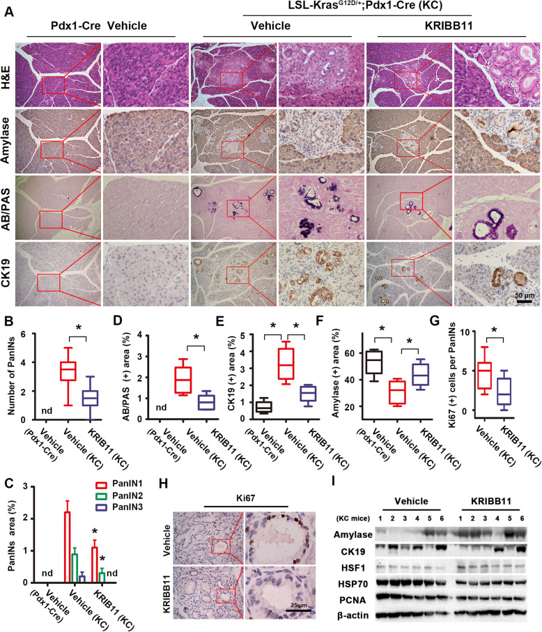
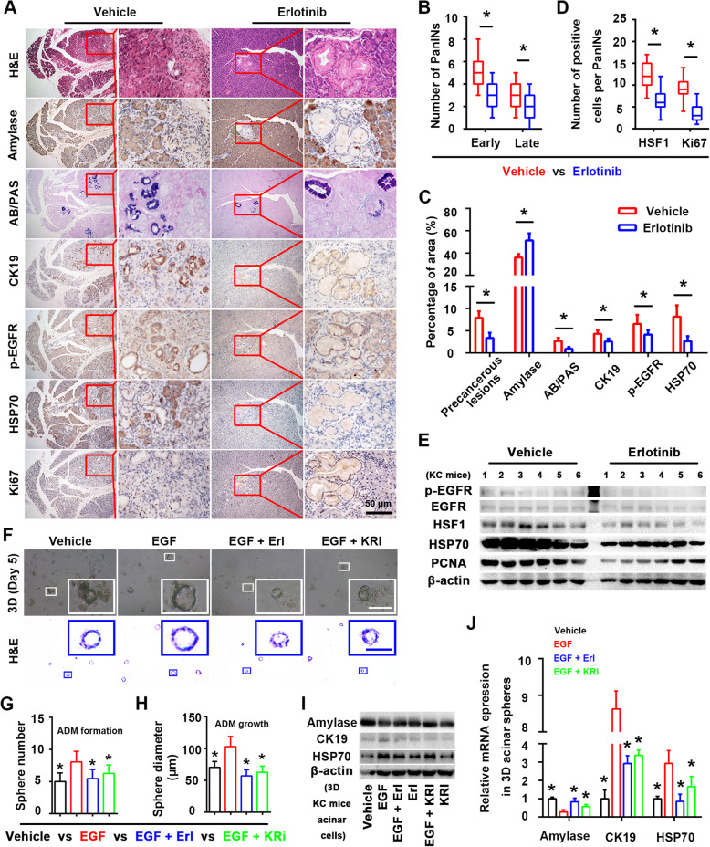
Results: In this study, we found that pharmacological inhibition of HSF1 slowed pancreatic cancer initiation and suppressed the pancreatitis-induced formation of pancreatic precancerous lesion. Next, bioinformatics analysis revealed the closely linked between HSF1 and EGFR pathway and we also confirmed their parallel activation in pancreatic precancerous lesions. Besides, the pharmacological inhibition of EGFR suppressed the initiation of pancreatic cancer and the activation of HSF1 in vivo. Indeed, we demonstrated that the EGFR activation that mediated pancreatic cancer tumorigenesis was partly HSF1-dependent in vitro.

Conclusion: Hence, we concluded that the EGFR-HSF1 axis promoted the initiation of pancreatic cancer.

Keywords: Epidermal growth factor receptor; Heat shock factor 1; Pancreatic ductal adenocarcinoma; Transgenic mice; Tumorigenesis.


Conflict of interest statement

The authors declare that they have no competing financial and non-financial interest.

Figures

Fig. 1
Fig. 1
Abnormal HSF1 activation is an early molecular event in pancreatic cancer tumorigenesis. a-b Representative IHC staining of HSF1 positive cells and Masson staining of collagen I positive fibrosis in all stages of in all stages of the tumorigenesis of human pancreatic cancer (normal acinar and ADM (a), early PanINs, late PanINs, invasive and infiltrative PDAC tissues (b). c Representative histopathology (H&E staining) and the IHC staining of HSF1-positive cells in all stages of the tumorigenesis of murine (KC mice) pancreatic cancer. d The relative HSF1 expression in pancreatic cancer tissues of mutant KRAS samples (KRASMUT) and non-mutant KRAS samples (KRASWT) samples according to TCGA datasets. e The relative HSF1 expression in normal pancreas tissues and pancreatic cancer tissues according to TCGA & GTEx datasets. f The relative HSF1 protein expression in pancreatic ductal progenitor cell line hTERT-HPNE and pancreatic cancer cell lines BxPC-3, CFPAC-1, PANC-1, MIAPaCa-2, and AsPC-1. g Representative IHC staining of HSF1-positive cells in pancreatic acinar cells from C mice and KC mice pancreatic cancer. h-i Representative histopathology (H&E staining) and IHC staining of HSF1-positive cells in the acute pancreatitis tissues (h) and chronic pancreatitis tissues (i) of KC mice. Scale bars = 10/25 μm. * P < 0.05, ns P > 0.05
Fig. 2
Fig. 2
Pharmacological inhibition of HSF1 suppressed the initiation of pancreatic cancer. a Representative histopathology (H&E staining), low-grade PanINs lesions (AB/PAS staining) and the IHC staining of amylase (acinar marker), CK19 (ductal marker) in the pancreas of vehicle C mice, vehicle KC mice and KC mice treated with KRIBB11 (HSF1 inhibitor). b-f Quantification of the PanINs numbers (b) and the percentage of the PanINs area (c), the AB/PAS-positive blue area (d), the CK19-positive ductal area (e) and the amylase-positive acinar area (f) per 400× field in the pancreas of vehicle C mice, vehicle KC mice and KC mice treated with KRIBB11. g-h IHC staining and its quantification of Ki67-positive proliferative cells in PanINs structures per 400× field in the pancreas of vehicle KC mice and KC mice treated with KRIBB11. i Western blotting assay were performed to evaluate the effect of KRIBB11 on the expression of amylase, CK19, HSF1 and HSP70 and PCNA in KC mice pancreases. Scale bars = 25/50 μm. nd = not detected, * P < 0.05
Fig. 3
Fig. 3
Pharmacological inhibition of HSF1 suppressed the formation of ADM in vivo. a Double-label IF staining showed the expression of amylase and CK19 in the pancreas of vehicle C mice, vehicle KC mice and KC mice treated with KRIBB11. b Representative histopathology (H&E staining) and the IHC staining of amylase (acinar marker), CK19 (ductal marker) in the pancreas of C mice treated with short-term cerulein, KC mice treated with short-term cerulein and KC mice treated with short-term cerulein plus KRIBB11. c-e Quantification of the PanINs area (c), the amylase-positive acinar area (d) and the CK19-positive ductal area (e) per 400× field in the pancreas of C mice treated with short-term cerulein, KC mice treated with short-term cerulein and KC mice treated with short-term cerulein plus KRIBB11. f Western blotting assay were performed to evaluate the effect of KRIBB11 on the expression of amylase, CK19 and HSP70 in KC mice (short-term cerulein) acute pancreatitis tissues. Scale bars = 50 μm. * P < 0.05
Fig. 4
Fig. 4
Pharmacological inhibition of HSF1 suppressed the formation of PanINs. a Representative histopathology (H&E staining), low-grade PanINs lesions (AB/PAS staining) and the IHC staining of amylase (acinar marker), CK19 (ductal marker) in the pancreas of vehicle KC mice, KC mice treated with long-term cerulein and KC mice treated with long-term cerulein plus KRIBB11. b-c Quantification of the number (b) and the diameter (c) of ductal-like structures per 400× field in the pancreas of vehicle KC mice, KC mice treated with long-term cerulein and KC mice treated with long-term cerulein plus KRIBB11. d-g Quantification of the percentage of the PanINs area (d), the AB/PAS-positive blue area (e), the amylase-positive acinar area (f) and the CK19-positive ductal area (g) per 400× field in the pancreas of vehicle KC mice, KC mice treated with long-term cerulein and KC mice treated with long-term cerulein plus KRIBB11. h Western blotting assay were performed to evaluate the effect of KRIBB11 on the expression of amylase, CK19 and HSP70 in KC mice (long-term cerulein) chronic pancreatitis tissues. Scale bars = 50 μm. nd = not detected, * P < 0.05
Fig. 5
Fig. 5
HSF1 was potential downstream molecule of EGFR in pancreatic cancer tumorigenesis. a The possible related biological pathways of HSF1 and its related proteins in pancreatic cancer. b IHC staining of EGFR in all stages of the tumorigenesis of pancreatic cancer in KC mice. c The relative EGFR expression in normal pancreas tissues and pancreatic cancer tissues according to TCGA & GTEx datasets. d The relationship between EGFR and the OS of pancreatic cancer according to TCGA database. e The forest graph showed HR and its 95% CI of EGFR in pancreatic cancer patients’ OS according to TCGA database. (f) GSEA analysis of biological pathways related to pancreatic cancer in EGFR high expression group vs EGFR low expression group according to TCGA database. g-h The relationship between HSF1 and EGFR singling pathway related molecules (EGFR (g)/MEK (h)) in pancreatic cancer. i-j The relationship between EGFR and HSF1 target genes (HSPA4 (i)/HSP90AA1(j)) in pancreatic cancer. k IHC staining of EGFR, HSF1, HSP90 and Ki67-positive areas in early/late PanINs of KC mice. l GSEA analysis of HSF1 binding motifs/signature (target genes gene sets, represented by RGAANNTTC_V$HSF1_01 and HSF1_01) in EGFR high expression group vs EGFR low expression group according to a TCGA datasets of pancreatic cancer. Scale bars = 25/50 μm. * P < 0.05
Fig. 6
Fig. 6
EGFR-HSF1 axis is momentous in pancreatic cancer initiation. a Representative histopathology (H&E staining), low-grade PanINs lesions (AB/PAS staining) and the IHC staining of amylase (acinar marker), CK19 (ductal marker), p-EGFR, HSP70 and Ki67 in the pancreas of vehicle KC mice and KC mice treated with erlotinib. b Quantification of the number of early and late PanINs per 400× field in the pancreas of vehicle KC mice and KC mice treated with erlotinib. c Quantification of the percentage of the precancerous lesion area, the AB/PAS-positive blue area, the amylase-positive acinar area, the CK19-positive ductal area, the p-EGFR-positive area and HSP70-positive area per 400× field in the pancreas of vehicle KC mice and KC mice treated with erlotinib. d Quantification of the number of HSF1- and Ki67-positive cells per 400× field in the pancreas of vehicle KC mice and KC mice treated with erlotinib. e Western blotting assay were performed to evaluate the effect of erlotinib on the expression of p-EGFR, EGFR, HSF1, HSP70 and PCNA in KC mice pancreas tissues. f Representative bright field/H&E staining images of 3D acinar/ductal-like spheres in the vehicle, EGF, EGF plus erlotinib (Erl) and EGF plus KRIBB11 (KRI) groups. g-h Quantification of the sphere number (g) and diameter (h) per 100× field in the vehicle, EGF, EGF plus erlotinib and EGF plus KRIBB11 groups (the EGF group was a control). i Western blotting was performed to evaluate the expression of acinar marker amylase, ductal marker CK19 and HSF1 target gene HSP70 among vehicle, EGF, EGF plus erlotinib, erlotinib, EGF plus KRIBB11 and KRIBB11 group in 3D acinar/ductal-like spheres. j Relative amylase, CK19 and HSP70 mRNA levels of 3D acinar/ductal-like spheres among the vehicle, EGF, EGF pluserlotinib and EGF plus KRIBB11 groups (the EGF group was a control). Scale bars = 50 μm. * P < 0.05

References

    1. Siegel RL, Miller KD, Jemal A. Cancer statistics, 2019. CA Cancer J Clin. 2019;69:7–34. doi: 10.3322/caac.21551. - DOI - PubMed
    1. Kamisawa T, Wood LD, Itoi T, Takaori K. Pancreatic cancer. Lancet. 2016;388:73–85. doi: 10.1016/S0140-6736(16)00141-0. - DOI - PubMed
    1. Awasthi N, Kronenberger D, Stefaniak A, Hassan MS, von Holzen U, Schwarz MA, Schwarz RE. Dual inhibition of the PI3K and MAPK pathways enhances nab-paclitaxel/gemcitabine chemotherapy response in preclinical models of pancreatic cancer. Cancer Lett. 2019;459:41–49. doi: 10.1016/j.canlet.2019.05.037. - DOI - PubMed
    1. Vihervaara A, Sistonen L. HSF1 at a glance. J Cell Sci. 2014;127:261–266. doi: 10.1242/jcs.132605. - DOI - PubMed
    1. Brandvold KR, Morimoto RI. The Chemical Biology of Molecular Chaperones--Implications for Modulation of Proteostasis. J Mol Biol. 2015;427:2931–2947. doi: 10.1016/j.jmb.2015.05.010. - DOI - PMC - PubMed