Review
. 2021 Jan 9;11(1):13.
doi: 10.1186/s13578-020-00522-z.

Structure, kinetic properties and biological function of mechanosensitive Piezo channels

Affiliations
Review

Structure, kinetic properties and biological function of mechanosensitive Piezo channels

Xiang-Zhi Fang et al. Cell Biosci. .

Abstract

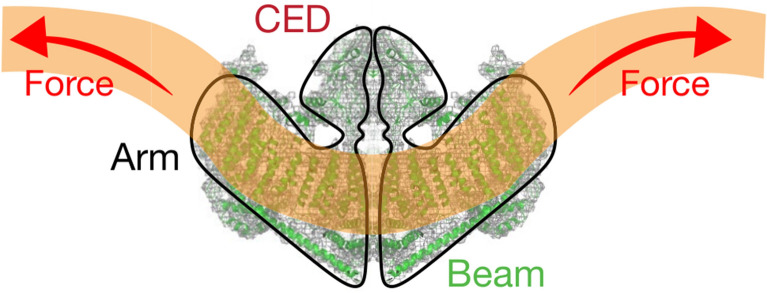
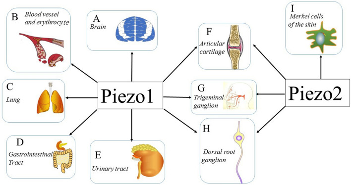
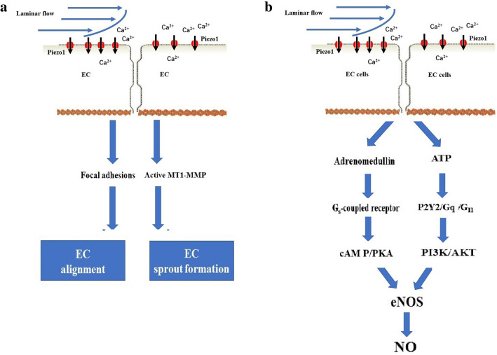
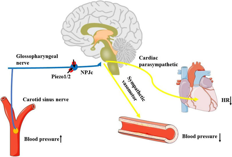
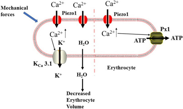
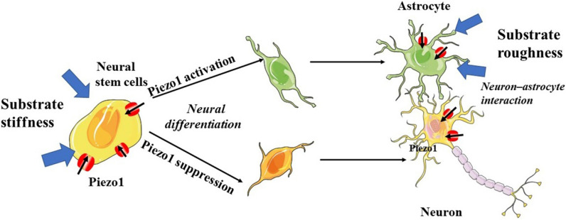
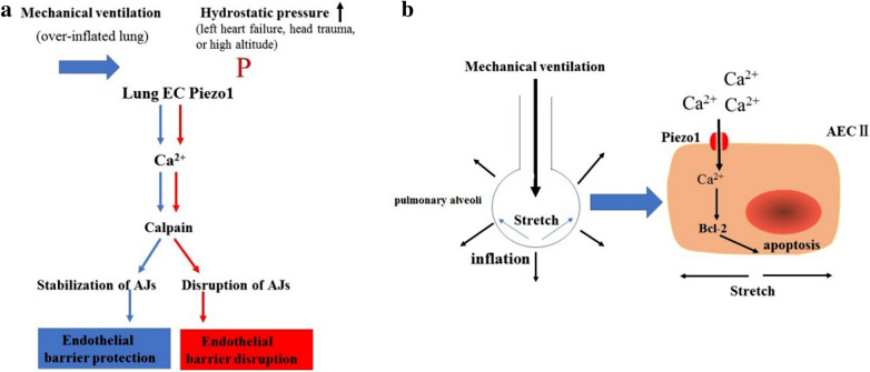
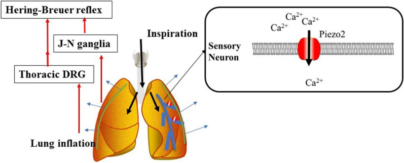
Mechanotransduction couples mechanical stimulation with ion flux, which is critical for normal biological processes involved in neuronal cell development, pain sensation, and red blood cell volume regulation. Although they are key mechanotransducers, mechanosensitive ion channels in mammals have remained difficult to identify. In 2010, Coste and colleagues revealed a novel family of mechanically activated cation channels in eukaryotes, consisting of Piezo1 and Piezo2 channels. These have been proposed as the long-sought-after mechanosensitive cation channels in mammals. Piezo1 and Piezo2 exhibit a unique propeller-shaped architecture and have been implicated in mechanotransduction in various critical processes, including touch sensation, balance, and cardiovascular regulation. Furthermore, several mutations in Piezo channels have been shown to cause multiple hereditary human disorders, such as autosomal recessive congenital lymphatic dysplasia. Notably, mutations that cause dehydrated hereditary xerocytosis alter the rate of Piezo channel inactivation, indicating the critical role of their kinetics in normal physiology. Given the importance of Piezo channels in understanding the mechanotransduction process, this review focuses on their structural details, kinetic properties and potential function as mechanosensors. We also briefly review the hereditary diseases caused by mutations in Piezo genes, which is key for understanding the function of these proteins.

Keywords: Function; Ion channel; Mechanotransduction; Piezo.


Conflict of interest statement

The authors declare that they have no competing interests.

Figures

Fig. 1
Fig. 1
Cryo-EM structure of the mPiezo1 channel. (adapted from Zhao et al. [35]). a Multiple views of the sharpened map of the trimeric channel with the major domains labeled, with the three subunits colored red, green and blue. b Cartoon model in which the three subunits are colored red, green and blue. In the middle panel, the front subunit has been omitted to provide a better view of the curvature of the TMs
Fig. 2
Fig. 2
A 38-TM topology model and key functional sites in mPiezo1. (adapted from Zhao et al. [35]). a A model showing one subunit with individual THUs and featured structural components. Residues L1342 and L1345 in the beam are indicated by red spheres. b A 38-TM topology model color-coded to match the cartoon model in A
Fig. 3
Fig. 3
Structure of the central pore module. (adapted from Zhao et al. [35]). a Ribbon diagram of the ion-conduction pore comprising the OH, CED, IH, and CTD from three color-coded subunits. The central solvent-accessible pathway is marked with dotted mesh generated by the program HOLE. b Pore module presenting the surface electrostatic potentials showing negative (red) and positive (blue) potential. Extracellular and intracellular fenestrations are marked by cyan and green dashed lines, respectively. The lateral portal is marked by yellow dashed lines
Fig. 4
Fig. 4
Model of the lever-like mechanotransduction model. The curved blades can act as a mechanosensor, while the beam structure, with residues Ll1342 and Ll1345 acting as a pivot, can act as a lever-like apparatus. Coupling of the distal blades and central pore through the lever-like apparatus converts mechanical force into cation conduction. a Proposed model of the force-induced gating of Piezo channels. The blue and orange models represent the channel in its closed and open states, respectively. Red dashed lines indicate possible ion-conduction pathways. Adapted from Ge et al. [34]. b A lever-like mechano-gating model in Piezo1. The blue and red dashed arrows indicate input and output forces, respectively
Fig. 5
Fig. 5
Model of the membrane doming mechanisms. Changes in membrane curvature lead to a gating force applied to the Piezo1 channel
Fig. 6
Fig. 6
Expression and function of Piezo channels Multiple tissues and cells express Piezo channels, and each of those shown is discussed in this review. a–e demonstrate the vital role of the Piezo1 channel in the CNS, blood vessels, erythrocytes, lungs, gastrointestinal tract and urinary tract. f–h illustrate the expression of both the Piezo1 channel and Piezo2 channel in articular cartilage, trigeminal ganglia, and dorsal root ganglia. shows that the Piezo2 channel is expressed in Merkel cells, which are involved in sensing light touch
Fig. 7
Fig. 7
Role of the Piezo1 channel in vascular development and tone. a In blood vessels, shear stress (laminar flow: blue arrow) triggered Piezo1-mediated Ca2+ influx and thereby facilitated endothelial cell (EC) alignment via the regulation of focal adhesions and EC sprout formation via the activation of MT1-MMP signaling. b In blood vessels, shear stress (laminar flow: blue arrow) activated the Piezo1 channel in ECs and subsequently mediated vascular tone. Specifically, shear stress led to Piezo1-dependent adrenomedullin release in ECs, which then activated the Gs-coupled endothelial adrenomedullin receptor. The subsequent increase in cAMP levels promoted the phosphorylation of endothelial NO synthase (eNOS) and caused NO production and vasodilation. Additionally, shear stress activated the Piezo1 channel in ECs and subsequently mediated the release of ATP in part by pannexin channels. Extracellular ATP, in turn, stimulated Gq/G11-coupled purinergic P2Y2 receptors, resulting in the phosphorylation of eNOS via PI3K/AKT signaling and increased NO formation
Fig. 8
Fig. 8
Overview of the Piezo1/2 channel-regulated baroreceptor reflex. Both Piezo1 and Piezo2 channels were highly expressed in the nodose-petrosal-jugular-ganglion complex (NPJc), which contains the cell bodies of baroreceptor neurons. Shear stress from high blood pressure was transformed into an electronic signal through Piezo1/2 channel activation, which was relayed to the medullary cardiovascular center. Subsequently, the efferent impulse contacted its effector organs (heart and blood vessels), decreasing the subject’s blood pressure and heart rate
Fig. 9
Fig. 9
Role of Piezo1 in erythrocytes. The left side of the figure depicts the Piezo1 channel regulating erythrocyte volume. In overhydrated red blood cells (RBCs), Piezo1-mediated Ca2+ influx activates K+ efflux through the Gardos channel (KCa3.1), which in turn leads to water loss and RBC dehydration. The right side of the figure shows the working model for how Piezo1 channel activation regulates the release of ATP from erythrocytes. In erythrocytes, shear can activate the Piezo1 channel and induce Ca2+ influx, which triggers significant ATP release that is dependent on pannexin-1 (Px1)
Fig. 10
Fig. 10
Role of the Piezo1 channel in regulating CNS processing. Mechanical properties of the neural progenitor cell environment activate Piezo1 channels in neural stem cells, astrocytes and neurons, thereby leading to neuronal differentiation, the development of neurite morphology and neuron–astrocyte interactions
Fig. 11
Fig. 11
Role of the Piezo1 channel in lung vascular endothelial cells (ECs) and alveolar type II epithelial cells (AEC-IIs). a Shear stress (mechanical ventilation: blue arrow) triggered Piezo1-mediated Ca2+ influx and thereby stabilized adherens junctions (AJs) of ECs through calpain. Piezo1 activation by hydrostatic pressure (P, red arrow) in lung ECs caused disruption of the AJs. b Alveoli exposed to cyclic stretch due to mechanical ventilation. Mechanical stretching of the alveoli induced Piezo1 channel activation of AEC-IIs, which resulted in the apoptosis of AEC-IIs via Ca2+ influx
Fig. 12
Fig. 12
Overview of the Piezo2-regulated Hering-Breuer inflation reflex. Piezo2 channels are expressed in airway vagal sensory neurons. Shear stress from lung inflation triggers the Piezo2 channel and activates the Hering-Breuer reflex through the jugular-nodose ganglia complex and the thoracic dorsal root ganglia

Similar articles

Cited by

References

    1. Fernandez-Sanchez ME, Brunet T, Röper JC, Farge E. Mechanotransduction’s impact on animal development, evolution, and tumorigenesis. Annu Rev Cell Dev Biol. 2015;31:373–97. doi: 10.1146/annurev-cellbio-102314-112441. - DOI - PubMed
    1. Chighizola M, Dini T, Lenardi C, Milani P, Podestà A, Schulte C. Mechanotransduction in neuronal cell development and functioning. Biophys Rev. 2019;11(5):701–2. doi: 10.1007/s12551-019-00587-2. - DOI - PMC - PubMed
    1. Costigan M, Scholz J, Woolf CJ. Neuropathic pain: A maladaptive response of the nervous system to damage. Annu Rev Neurosci. 2009;32:1–32. doi: 10.1146/annurev.neuro.051508.135531. - DOI - PMC - PubMed
    1. Blount P, Sukharev SI, Moe PC, Martinac B, Kung C. Mechanosensitive channels of bacteria. Methods Enzymol. 1999;294:458–482. doi: 10.1016/S0076-6879(99)94027-2. - DOI - PubMed
    1. Morris CE. Mechanosensitive ion channels. J Membr Biol. 1990;113(2):93–107. doi: 10.1007/BF01872883. - DOI - PubMed