

Accelerated brain aging in chronic low back pain
- PMID: 33422525
- PMCID: PMC7939694
- DOI: 10.1016/j.brainres.2020.147263
Accelerated brain aging in chronic low back pain
Abstract
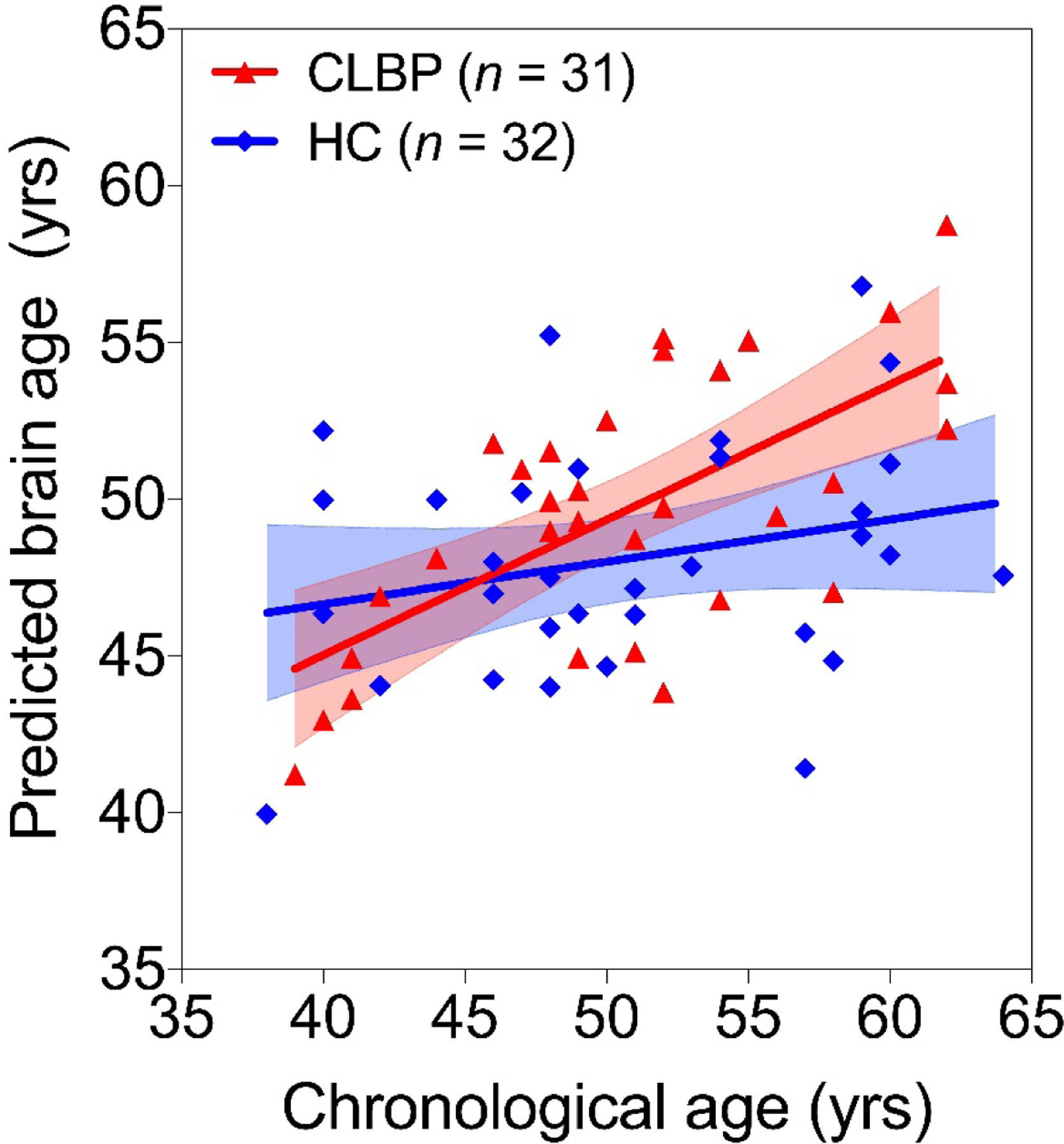
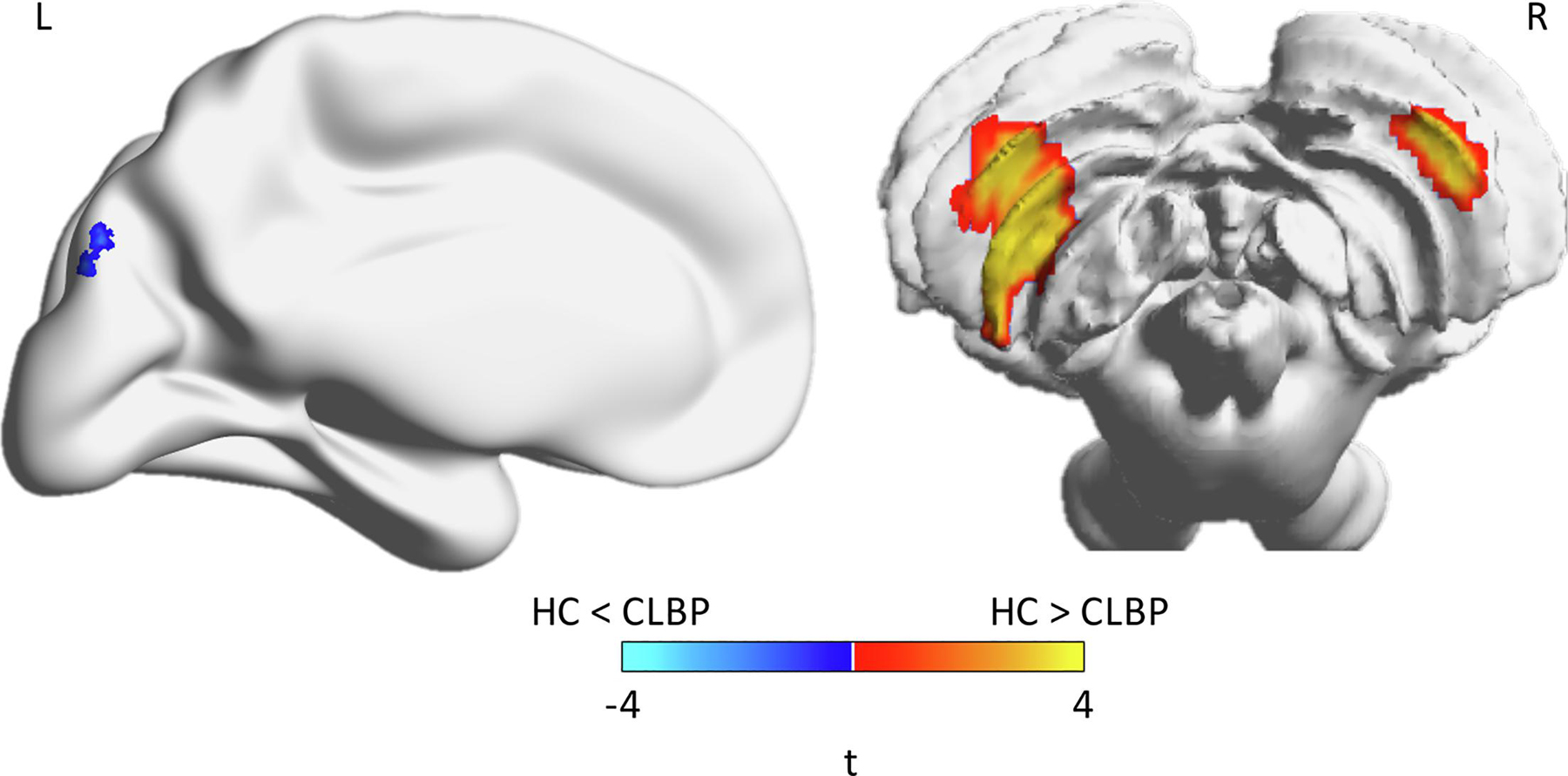
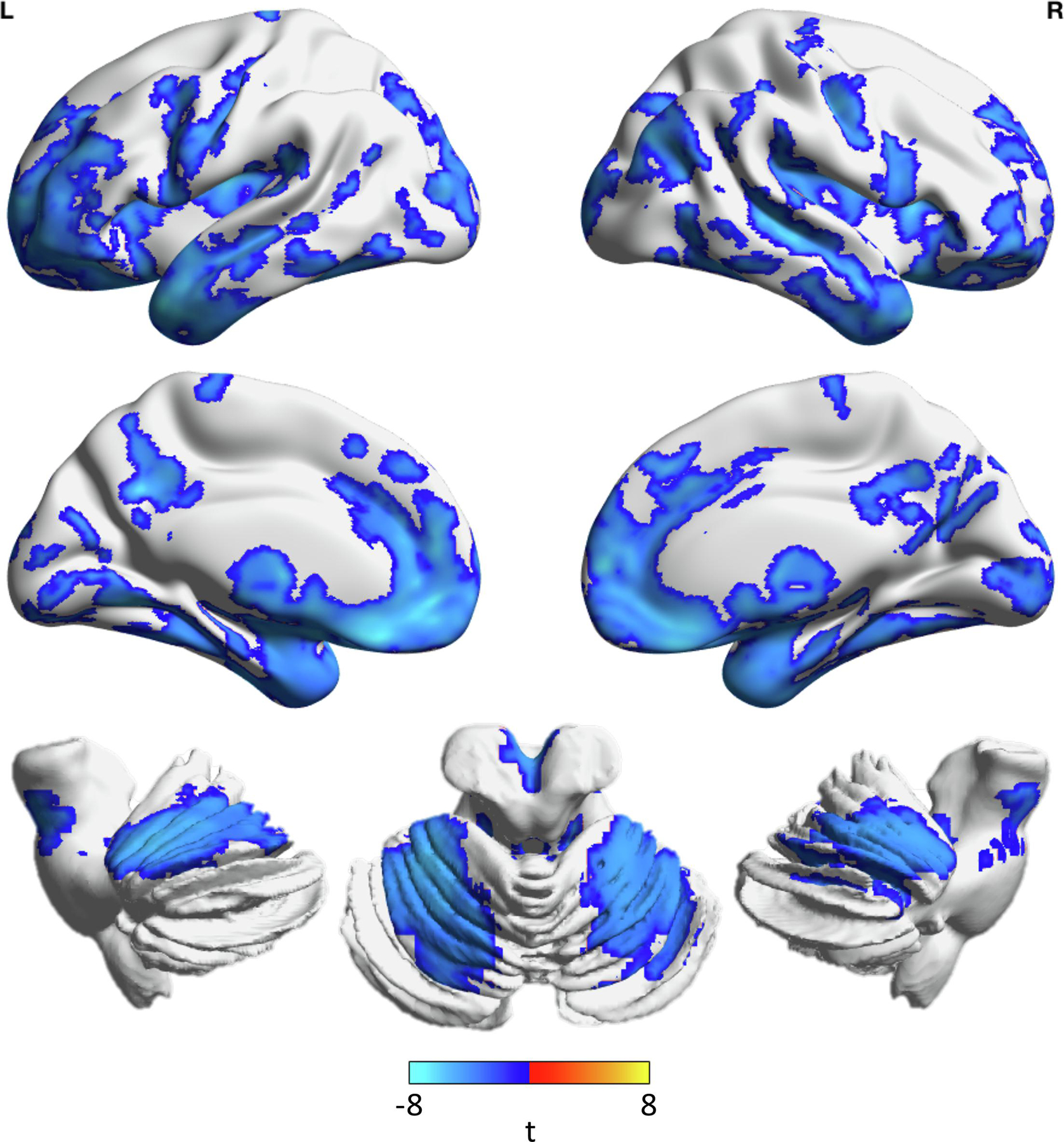
Chronic low back pain (CLBP) is a leading cause of disability and is associated with neurodegenerative changes in brain structure. These changes lead to impairments in cognitive function and are consistent with those seen in aging, suggesting an accelerated aging pattern. In this study we assessed this using machine-learning estimated brain age (BA) as a holistic metric of morphometric changes associated with aging. Structural imaging data from 31 non-depressed CLBP patients and 32 healthy controls from the Pain and Interoception Imaging Network were included. Using our previously developed algorithm, we estimated BA per individual based on grey matter density. We then conducted multivariable linear modeling for effects of group, chronological age, and their interaction on BA. We also performed two voxel-wise analyses comparing grey matter density between CLBP and control individuals and the association between gray matter density and BA. There was an interaction between CLBP and greater chronological age on BA such that the discrepancy in BA between healthy and CLBP individuals was greater for older individuals. In CLBP individuals, BA was not associated with sex, current level of pain, duration of CLBP, or mild to moderate depressive symptoms. CLBP individuals had lower cerebellar grey matter density compared to healthy individuals. Brain age was associated with lower gray matter density in numerous brain regions. CLBP was associated with greater BA, which was more profound in later life. BA as a holistic metric was sensitive to differences in gray matter density in numerous regions which eluded direct comparison between groups.
Keywords: Aging; Chronic low back pain; Machine learning; Structural neuroimaging.
Copyright © 2021 Elsevier B.V. All rights reserved.
Conflict of interest statement
Declarations of interest: none
Figures
References
Publication types
MeSH terms
Grants and funding
LinkOut - more resources
Full Text Sources
Other Literature Sources
Medical
