Review
. 2021 Mar:111:103590.
doi: 10.1016/j.mcn.2021.103590. Epub 2021 Jan 8.

Macrophage roles in peripheral nervous system injury and pathology: Allies in neuromuscular junction recovery

Affiliations
Review

Macrophage roles in peripheral nervous system injury and pathology: Allies in neuromuscular junction recovery

Rachel Rios et al. Mol Cell Neurosci. 2021 Mar.

Abstract

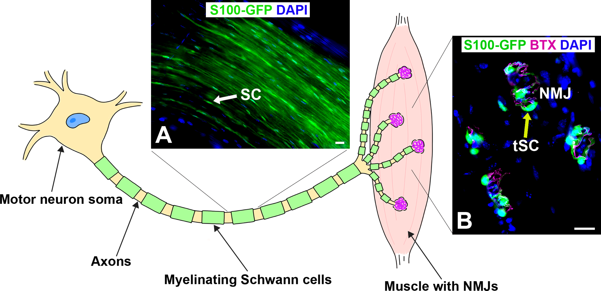
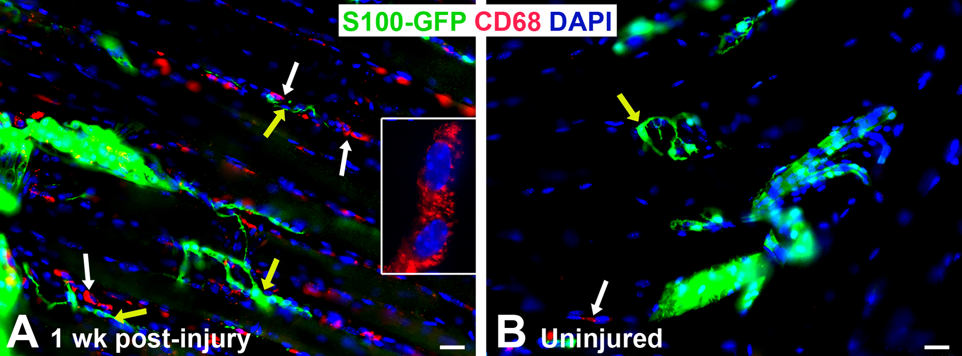
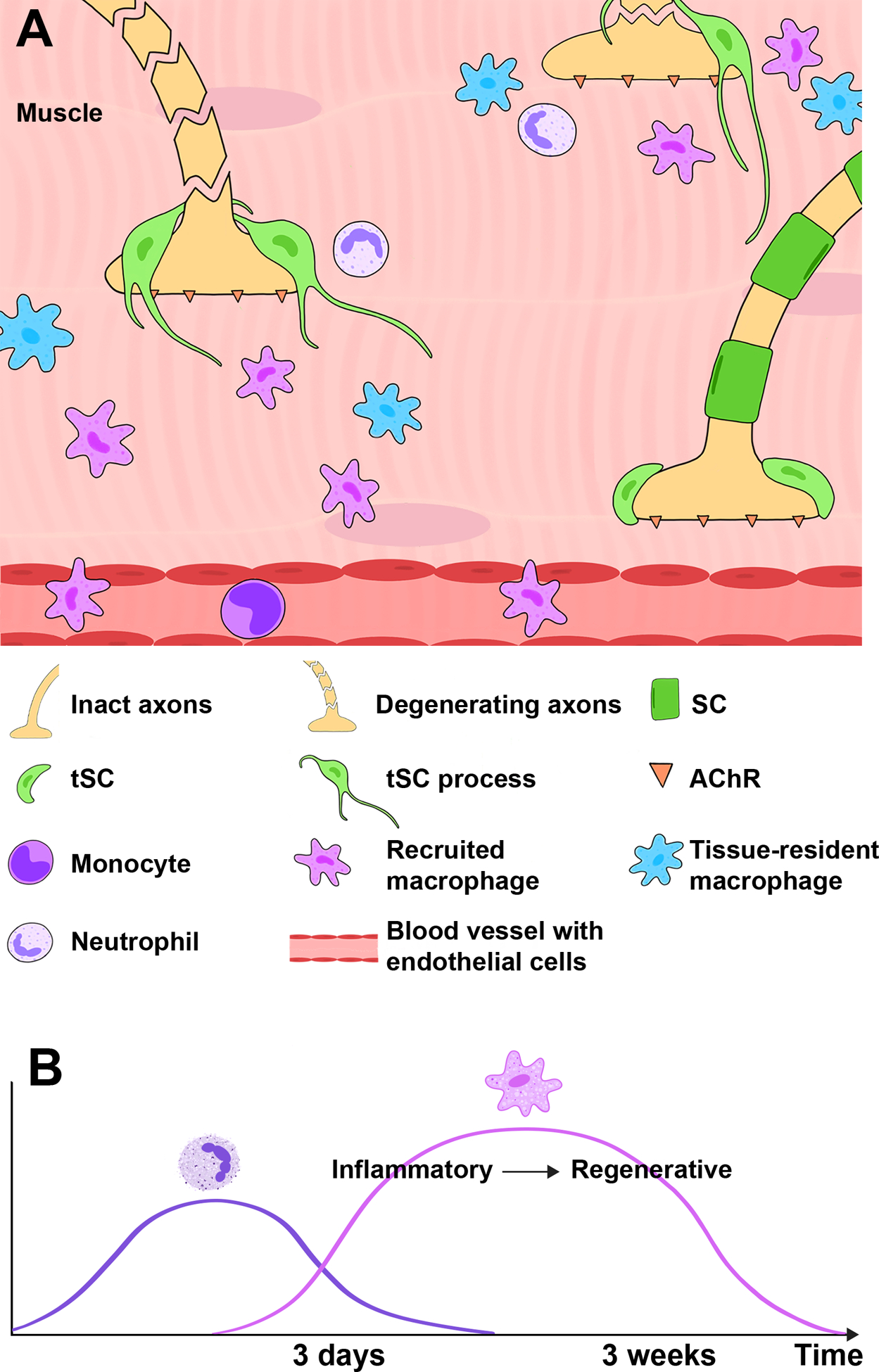
Peripheral nerve injuries remain challenging to treat despite extensive research on reparative processes at the injury site. Recent studies have emphasized the importance of immune cells, particularly macrophages, in recovery from nerve injury. Macrophage plasticity enables numerous functions at the injury site. At early time points, macrophages perform inflammatory functions, but at later time points, they adopt pro-regenerative phenotypes to support nerve regeneration. Research has largely been limited, however, to the injury site. The neuromuscular junction (NMJ), the synapse between the nerve terminal and end target muscle, has received comparatively less attention, despite the importance of NMJ reinnervation for motor recovery. Macrophages are present at the NMJ following nerve injury. Moreover, in denervating diseases, such as amyotrophic lateral sclerosis (ALS), macrophages may also play beneficial roles at the NMJ. Evidence of positive macrophages roles at the injury site after peripheral nerve injury and at the NMJ in denervating pathologies suggest that macrophages may promote NMJ reinnervation. In this review, we discuss the intersection of nerve injury and immunity, with a focus on macrophages.

Keywords: Glial cells; Macrophage; Nerve injury; Nerve regeneration; Neuromuscular junction.


Conflict of interest statement

Disclosure of Conflicts of Interest: Declarations of interest: none.

Figures

Figure 1.
Figure 1.. Illustration of motor neuron and the end target muscle with neuromuscular junctions (NMJs).
A) Representative image of myelinating Schwann cells (SC, green, arrow) along the axons within the sciatic nerve in young adult S100-GFP mice. B) Representative image of longitudinal section of extensor digitorum longus muscle with neuromuscular junctions containing non-myelinating terminal SCs (tSC, green, yellow arrow) overlaying acetylcholine receptors (BTX, magenta). S100-GFP = glial cells (green), BTX = α‐bungarotoxin, DAPI = nuclear staining (blue). Scale bar = 20 μm. N = 4 mice.
Figure 2.
Figure 2.. Macrophages are present in the extensor digitorum longus muscle after nerve injury in young adult S100-GFP mice.
A) Representative image of immunofluorescence staining showing increased CD68+ macrophages/image area (red, white arrows) around the NMJ (yellow arrows) one week (wk) following sciatic nerve transection and immediate repair, while uninjured (control) mice (B) have nearly no macrophages present (white arrow). Yellow arrow indicates NMJ with tSCs. Anti-CD68 rat Ab (#MCA1957, BioRad) = macrophages (red), S100-GFP = glial cells (green), DAPI = nuclear staining (blue). Scale bar = 20 μm. N = 4 mice/time point.
Figure 3.
Figure 3.. Diagram showing tSC injury response and the cellular immune response at the NMJ in the muscle after nerve injury.
A) After nerve cut and repair, tissue-resident macrophages and recruited neutrophils and monocytes/macrophages surround NMJs and promote NMJ reinnervation. In addition, tSCs at denervated synapses extend their processes to nearby NMJs with intact innervation. These processes serve as a guide for intact nerves to reach denervated NMJs, promoting reinnervation. B) Neutrophils are early responders, while macrophage infiltration increases at 3 days after injury. Over several weeks, macrophages progress from inflammatory to regenerative phenotypes.

References

    1. Padovano WM, et al. , Incidence of Nerve Injury After Extremity Trauma in the United States. Hand (N Y), 2020: p. 1558944720963895. - PMC - PubMed
    1. Akel BS, et al. , Health-related quality of life in children with obstetrical brachial plexus palsy. Qual Life Res, 2013. 22(9): p. 2617–24. - PubMed
    1. Bergmeister KD, et al. , Acute and long-term costs of 268 peripheral nerve injuries in the upper extremity. PLoS One, 2020. 15(4): p. e0229530. - PMC - PubMed
    1. Hong TS, et al. , Indirect Cost of Traumatic Brachial Plexus Injuries in the United States. J Bone Joint Surg Am, 2019. 101(16): p. e80. - PMC - PubMed
    1. Mehta P, et al. , Prevalence of amyotrophic lateral sclerosis - United States, 2010–2011. MMWR Suppl, 2014. 63(7): p. 1–14. - PubMed

Publication types

MeSH terms