

Volumetric distribution of perivascular space in relation to mild cognitive impairment
- PMID: 33422892
- PMCID: PMC7902350
- DOI: 10.1016/j.neurobiolaging.2020.12.010
Volumetric distribution of perivascular space in relation to mild cognitive impairment
Abstract
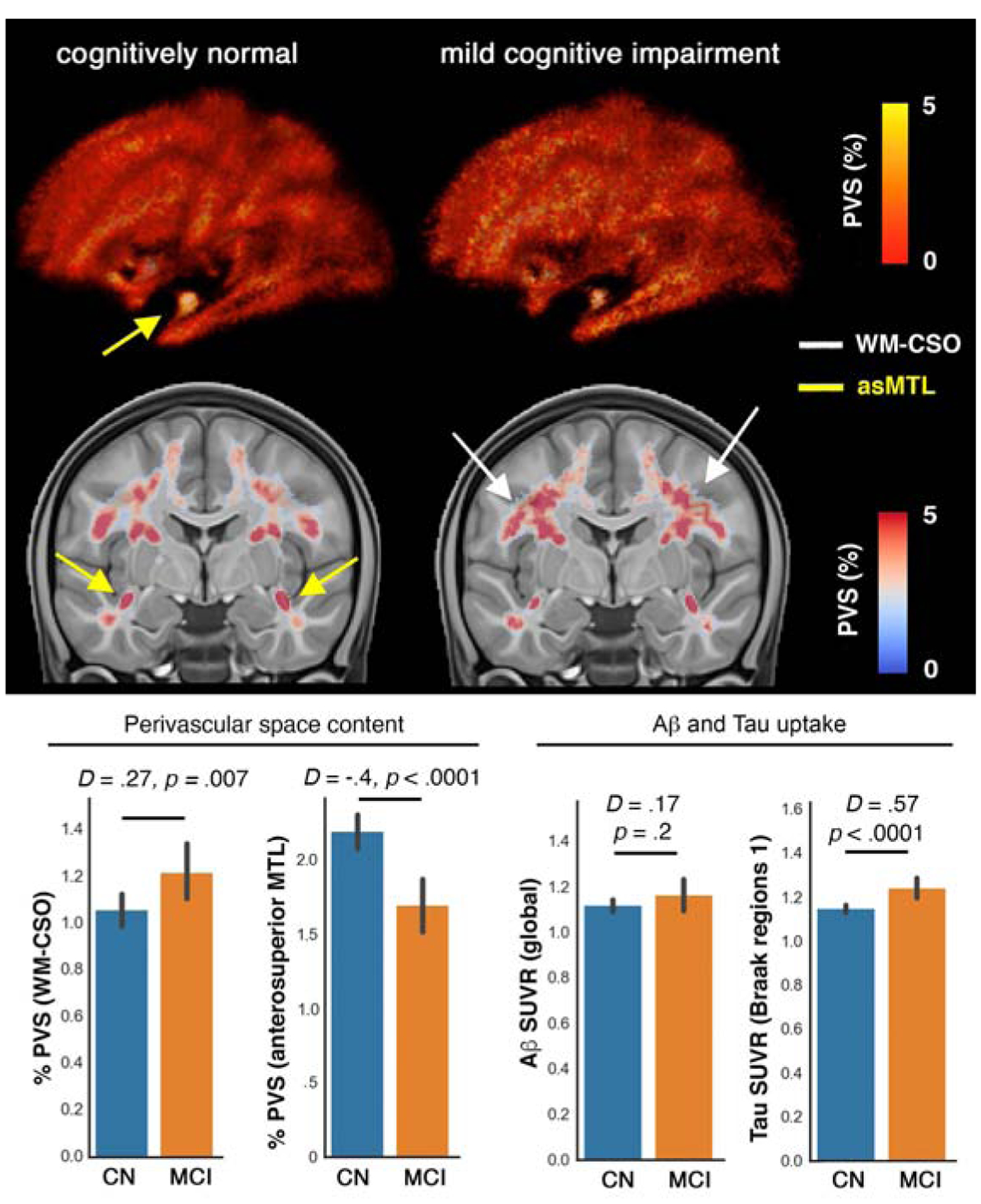
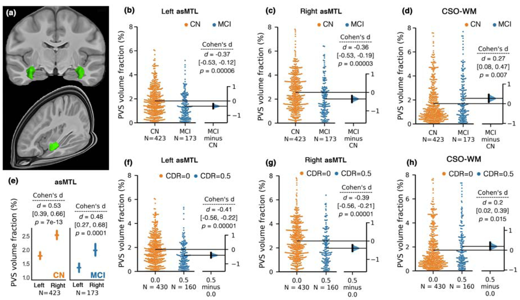
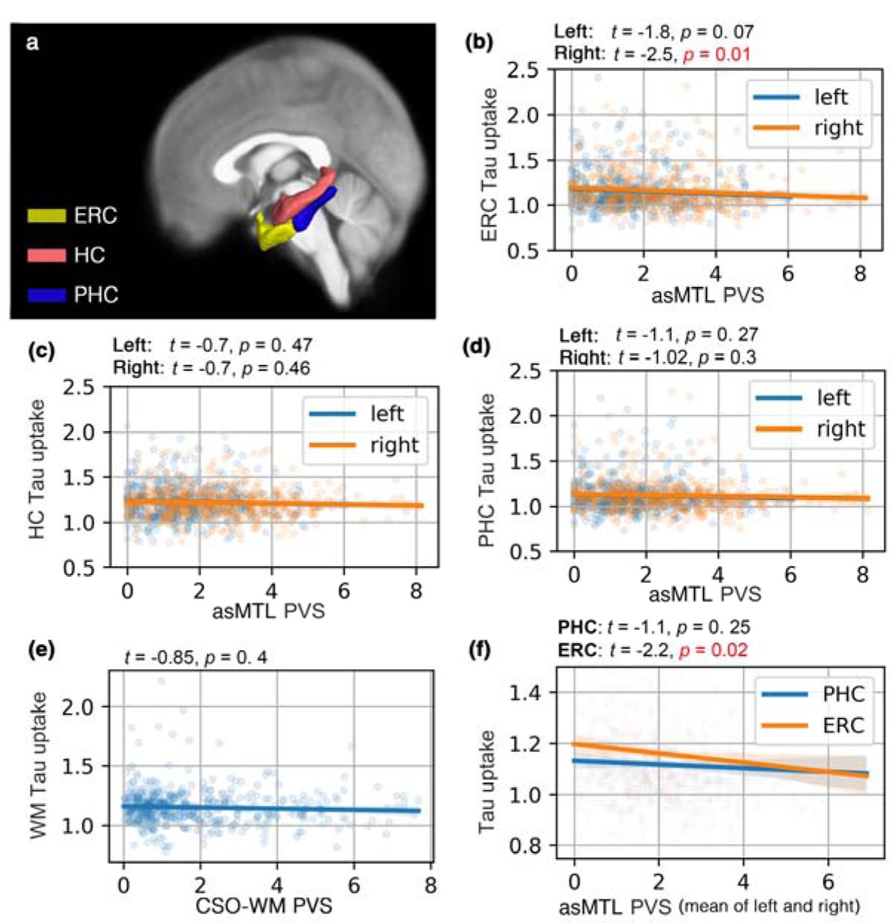
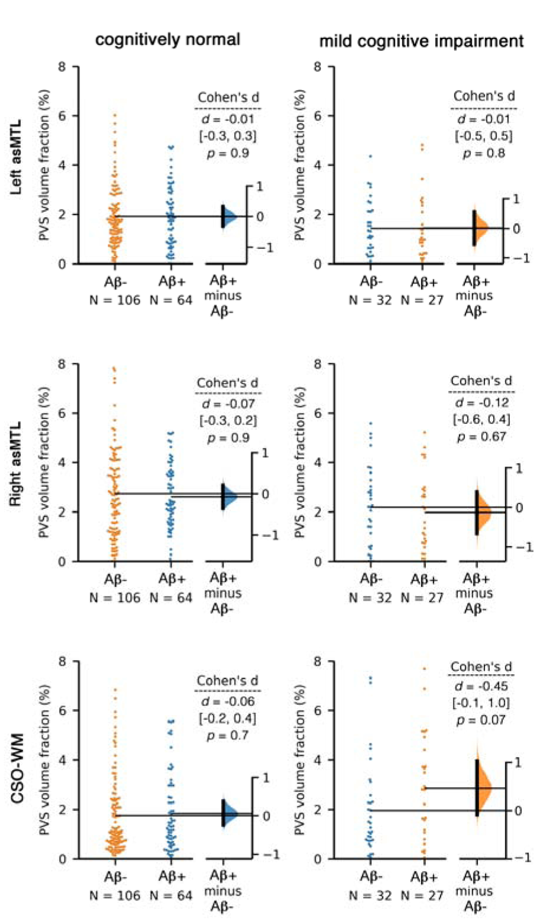
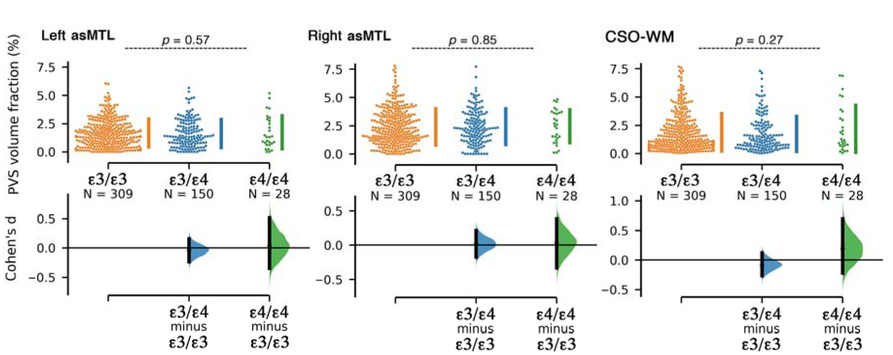
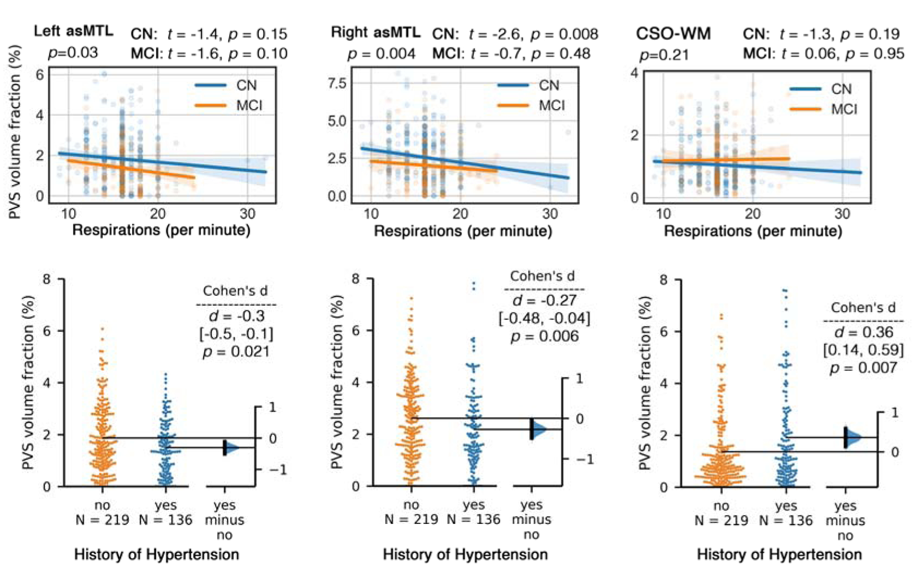
Vascular contributions to early cognitive decline are increasingly recognized, prompting further investigation into the nature of related changes in perivascular spaces (PVS). Using magnetic resonance imaging, we show that, compared to a cognitively normal sample, individuals with early cognitive dysfunction have altered PVS presence and distribution, irrespective of Amyloid-β. Surprisingly, we noted lower PVS presence in the anterosuperior medial temporal lobe (asMTL) (1.29 times lower PVS volume fraction in cognitively impaired individuals, p < 0.0001), which was associated with entorhinal neurofibrillary tau tangle deposition (beta (standard error) = -0.98 (0.4); p = 0.014), one of the hallmarks of early Alzheimer's disease pathology. We also observed higher PVS volume fraction in centrum semi-ovale of the white matter, but only in female participants (1.47 times higher PVS volume fraction in cognitively impaired individuals, p = 0.0011). We also observed PVS changes in participants with history of hypertension (higher in the white matter and lower in the asMTL). Our results suggest that anatomically specific alteration of the PVS is an early neuroimaging feature of cognitive impairment in aging adults, which is differentially manifested in female.
Keywords: Alzheimer’s disease; Clearance system; Cognitive decline; Perivascular space.
Copyright © 2020 Elsevier Inc. All rights reserved.
Conflict of interest statement
Disclosure statement
Authors have no conflict of interest to declare.
Verification
Authors have no any actual or potential conflicts of interest including any financial, personal or other relationships with other people or organizations within three years of beginning the work submitted that could inappropriately influence (bias) their work.
None of the authors has contracts relating to this research through which it or any other organization may stand to gain financially now or in the future.
None of the authors has other agreements of authors or their institutions that could be seen as involving a financial interest in this work.
Figures
Similar articles
-
Perivascular space enlargement accelerates in ageing and Alzheimer's disease pathology: evidence from a three-year longitudinal multicentre study.Alzheimers Res Ther. 2024 Oct 31;16(1):242. doi: 10.1186/s13195-024-01603-8. Alzheimers Res Ther. 2024. PMID: 39482759 Free PMC article.
-
Increased perivascular space volume in white matter and basal ganglia is associated with cognition in Parkinson's Disease.Brain Imaging Behav. 2024 Feb;18(1):57-65. doi: 10.1007/s11682-023-00811-4. Epub 2023 Oct 19. Brain Imaging Behav. 2024. PMID: 37855955 Free PMC article.
-
Associations Between GFAP, Aβ42/40 Ratio, and Perivascular Spaces and Cognitive Domains in Vascular Cognitive Impairment.Int J Mol Sci. 2025 Apr 9;26(8):3541. doi: 10.3390/ijms26083541. Int J Mol Sci. 2025. PMID: 40332019 Free PMC article.
-
Imaging brain fluid dynamics and waste clearance involving perivascular spaces in cerebral small vessel disease.Alzheimers Dement. 2025 Apr;21(4):e70212. doi: 10.1002/alz.70212. Alzheimers Dement. 2025. PMID: 40289686 Free PMC article. Review.
-
Perivascular spaces and brain waste clearance systems: relevance for neurodegenerative and cerebrovascular pathology.Neuroradiology. 2021 Oct;63(10):1581-1597. doi: 10.1007/s00234-021-02718-7. Epub 2021 May 21. Neuroradiology. 2021. PMID: 34019111 Free PMC article. Review.
Cited by
-
Perivascular spaces as a potential biomarker of Alzheimer's disease.Front Neurosci. 2022 Oct 18;16:1021131. doi: 10.3389/fnins.2022.1021131. eCollection 2022. Front Neurosci. 2022. PMID: 36330347 Free PMC article. Review.
-
Effects of one-night partial sleep deprivation on perivascular space volume fraction: Findings from the Stockholm Sleepy Brain Study.bioRxiv [Preprint]. 2024 Oct 27:2024.10.26.620382. doi: 10.1101/2024.10.26.620382. bioRxiv. 2024. Update in: Sleep Med. 2025 Jul;131:106537. doi: 10.1016/j.sleep.2025.106537. PMID: 39484474 Free PMC article. Updated. Preprint.
-
Parenchymal border macrophages regulate tau pathology and tau-mediated neurodegeneration.Life Sci Alliance. 2023 Aug 10;6(11):e202302087. doi: 10.26508/lsa.202302087. Print 2023 Nov. Life Sci Alliance. 2023. PMID: 37562846 Free PMC article.
-
Deep medullary veins: a promising neuroimaging marker for mild cognitive impairment in outpatients.BMC Neurol. 2023 Jan 5;23(1):3. doi: 10.1186/s12883-022-03037-x. BMC Neurol. 2023. PMID: 36604624 Free PMC article.
-
Neuroimaging aspects and clinical significance of giant perivascular spaces in the brain.Precis Cancer Med. 2022 Dec 30;5:31. doi: 10.21037/pcm-22-27. Precis Cancer Med. 2022. PMID: 36619900 Free PMC article. No abstract available.
References
-
- Adachi M, Hosoya T, Haku T, Yamaguchi K, 1998. Dilated Virchow-Robin spaces: MRI pathological study. Neuroradiology 40, 27–31. - PubMed
-
- Avants BB, Tustison N, Song G, 2009. Advanced Normalization Tools (ANTS). Insight J 1–35.
Publication types
MeSH terms
Substances
Grants and funding
LinkOut - more resources
Full Text Sources
Other Literature Sources
Research Materials
