. 2021 Jun;17(3):1053-1067.
doi: 10.1007/s12015-020-10113-7. Epub 2021 Jan 9.

Off-the-Shelf, Immune-Compatible Human Embryonic Stem Cells Generated Via CRISPR-Mediated Genome Editing

Affiliations

Off-the-Shelf, Immune-Compatible Human Embryonic Stem Cells Generated Via CRISPR-Mediated Genome Editing

Annie Kim et al. Stem Cell Rev Rep. 2021 Jun.

Abstract

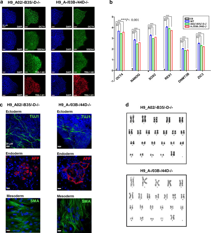
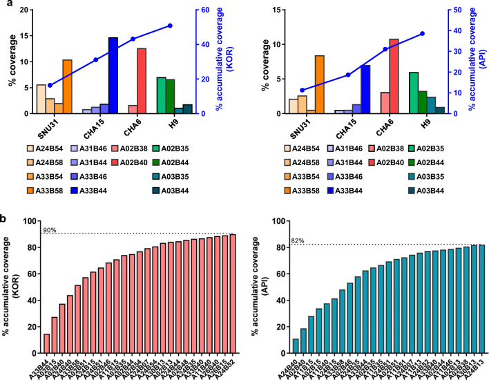
Human embryonic stem cells (hESCs) hold promise in regenerative medicine but allogeneic immune rejections caused by highly polymorphic human leukocyte antigens (HLAs) remain a barrier to their clinical applications. Here, we used a CRISPR/Cas9-mediated HLA-editing strategy to generate a variety of HLA homozygous-like hESC lines from pre-established hESC lines. We edited four pre-established HLA-heterozygous hESC lines and created a mini library of 14 HLA-edited hESC lines in which single HLA-A and HLA-B alleles and both HLA-DR alleles are disrupted. The HLA-edited hESC derivatives elicited both low T cell- and low NK cell-mediated immune responses. Our library would cover about 40% of the Asian-Pacific population. We estimate that HLA-editing of only 19 pre-established hESC lines would give rise to 46 different hESC lines to cover 90% of the Asian-Pacific population. This study offers an opportunity to generate an off-the-shelf HLA-compatible hESC bank, available for immune-compatible cell transplantation, without embryo destruction. Graphical Abstract.

Keywords: CRISPR/Cas9; HLA homozygous-like hESCs; HLA-editing; Human embryonic stem cells; Immune-compatible hESC banking.


Conflict of interest statement

J.-S.K. is a cofounder of, and holds stock in, ToolGen, Inc.

Figures

None
Graphical Abstract
Fig. 1
Fig. 1
Generation an HLA-A, B hemizygous hESC library and validation of knockout cells. (a) Schematic diagram of the HLA-DRB1 (null), HLA-A (hemizygous), and HLA-B (hemizygous) knockout strategy in H9 hESCs. The HLA-DRB1 gene was biallelically targeted by transient delivery of preassembled single-chain guide RNA (sgRNA)/Cas9 protein ribonucleoproteins (RNPs), followed by the monoallelic knockout of the HLA-A and HLA-B genes. (b) Target sites and indel patterns of the isolated mutant clones derived from H9 hESCs. The protospacer adjacent motif and the sgRNA target sequence are shown in red and blue, respectively. Deletions and insertions of nucleotides are indicated as dashes and lowercase letters, respectively. Nucleotide changes are shown in orange upper case letters. (c) Relative HLA-DRB1 mRNA levels in H9_WT, HLA-DRB1 null, and HLA-AB hemizygous hESC clones were compared. The level of HLA-DRB1 mRNA in H9_WT cells was arbitrarily set to 1. Ordinary one-way ANOVA was performed, followed by Tukey’s multiple comparison test. ****P < 0.0001. (d) Relative HLA-B mRNA levels in HLA-DRB1 null and HLA-AB hemizygous hESC clones were compared. Two-way ANOVA followed by Tukey’s multiple comparison test was performed. Cells were treated with 100 ng/ml IFN-γ for 48 h before the experiment and HLA-DRB1 and HLA-B expression was measured by RT-qPCR. The level of HLA-B mRNA in the H9 A02/03B35/44D−/− line was arbitrarily set to 1. Means ± SEM were from six independent experiments. ****p < 0.0001. (e) HLA-A*02 and HLA-A*03 expression levels in the H9_WT, A02/−B35/−D−/−, A−/03B−/44D−/−, and B2M−/−D−/− hESC clones were measured by flow cytometry. Cells were treated with 100 ng/ml IFN-γ for 48 h before the experiment. See Fig. S2D for the B2M−/− D−/− indel pattern
Fig. 2
Fig. 2
Characterization of HLA-edited hESC clones. (a) The HLA-edited hESC clones used for T cell response analysis, A02/−B35/−D−/− and A−/03B−/44D−/−, were positive for the pluripotent cell markers OCT4, SSEA4, TRA-1-60, and TRA-1-81. Scale bar: 50 μm. (b) Quantitative real-time RT-PCR was performed to detect the expression of the representative pluripotency markers, OCT4, NANOG, SOX2, REX1, DNMT3B, and ZIC3. Data are represented as means ± SEMs (n = 3). ***p < 0.001, two way ANOVA, followed by Tukey’s multiple comparison test. (c) HLA-edited hESC clones were spontaneously differentiated into cell derivatives of the three germ layers, and the expression levels of representative markers of the ectoderm (TUJ1), mesoderm (SMA), and endoderm (AFP) lineages were examined. (d) Karyotyping analysis was performed to detect gross chromosomal abnormalities
Fig. 3
Fig. 3
Genome-edited HLA-A, B hemizygous and -DRB1 null cells evade NK cell activity in vitro. (a) Cell-surface CD31 expression levels on IFN-γ-treated ECs differentiated from H9_WT, A02/−B35/−D−/−, and B2M−/−D−/− hESC lines. Data are represented as means ± SEMs (n = 5). ***p < 0.001, one-way ANOVA, followed by Tukey’s test. (b) Flow cytometric analysis of HLA-ABC expression on ECs derived from H9_WT, A02/−B35/−D−/−, and B2M−/−D−/− hESC lines. The expression of HLA-ABC was examined with or without IFN-γ treatment. Data are represented as means ± SEMs (n = 5). ***p < 0.001, one-way ANOVA, followed by Tukey’s test. (c) 7-AAD assay to measure the percentage of apoptotic target cells in EC populations differentiated from H9_WT, A02/−B35/−D−/−, B2M−/−D−/− hESC lines after incubation with HLA null-reactive NK cells (derived from 3 donors) at various effector: target (E:T) ratios (left panel). The percentage of 7-AAD+ apoptotic target cells among different target EC populations when treated at an E:T ratio of 1:1 is shown (right panel). (d) CD107a expression on CD3CD56+ NK cells was measured by flow cytometry after co-culture with various types of target cells at an E:T ratio of 1:1. Data are represented as means ± SEMs. *p < 0.05, and ** p < 0.005, followed by Mann-Whitney nonparametric test
Fig. 4
Fig. 4
T cell responses to genome-edited HLA-A, B hemizygous cells in vitro. (a) The gating strategy for analysis of the mixed lymphocyte reaction is shown. Gates were applied to exclude dead cells and target cells, to obtain CD3+ T cells and subsequently CD4+ or CD8+ T cells. Detailed analysis for CTVlow proliferating cells in response to antigen is shown by histogram. The mitotic index was calculated as the number of mitotic events/number of absolute precursors. For each type of T cell, the mitotic index and cytokine production were determined by flow cytometric analysis. (b) Mitotic indices of proliferating CD8+ and CD4+ T cells in the presence of different cell types are shown. (c) Correlation between cytokine production and proliferation in CD4+ and CD8+ T cells. *p < 0.05, **p < 0.005, ***p < 0.001, ****p < 0.0001 followed by Spearman’s nonparametric test
Fig. 5
Fig. 5
Estimation of population coverage. (a) The proportions of Korean (KOR) and Asian-Pacific Islander (API) populations matched to our small HLA hemizygous hESC library are shown. Our HLA-A, B hemizygous hESC library, made from four established hESC lines, covers >50% of the KOR population and about 40% of the API population. (b) Twenty five different hemizygous hESC lines from 16 pre-established hESCs can cover ~90% of the KOR population. Likewise, 25 different hemizygous hESC lines from a different set of 13 pre-established hESCs can cover ~82% of the API population

References

    1. Bradley JA, Bolton EM, Pedersen RA. Stem cell medicine encounters the immune system. Nature Reviews Immunology. 2002;2(11):859–871. - PubMed
    1. Edgerly CH, Weimer ET. The past, present, and future of HLA typing in transplantation. Methods in Molecular Biology. 2018;1802:1–10. - PubMed
    1. Drukker M, Katchman H, Katz G, Even-Tov Friedman S, Shezen E, Hornstein E, Mandelboim O, Reisner Y, Benvenisty N. Human embryonic stem cells and their differentiated derivatives are less susceptible to immune rejection than adult cells. Stem Cells. 2006;24(2):221–229. - PubMed
    1. Li L, Baroja ML, Majumdar A, Chadwick K. Human embryonic stem cells possess immune-privileged properties. Stem. 2004;22:448–456. - PubMed
    1. Lin G, Ou-Yang Q, Qian X, Lu G. Construction of human embryonic stem cell banks: Prospects for tissue matching. In: Fairchild PJ, editor. The immunological barriers to regenerative medicine. New York: Springer; 2013. pp. 111–128.

Publication types