. 2021 Jan 10;18(1):21.
doi: 10.1186/s12974-020-02048-0.

The role of microglia membrane potential in chemotaxis

Affiliations

The role of microglia membrane potential in chemotaxis

Laura Laprell et al. J Neuroinflammation. .

Erratum in

Abstract

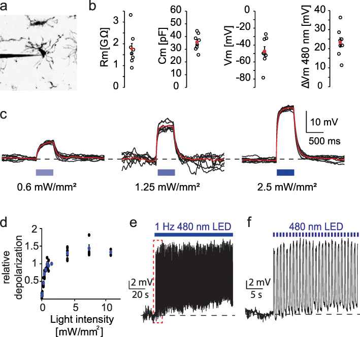
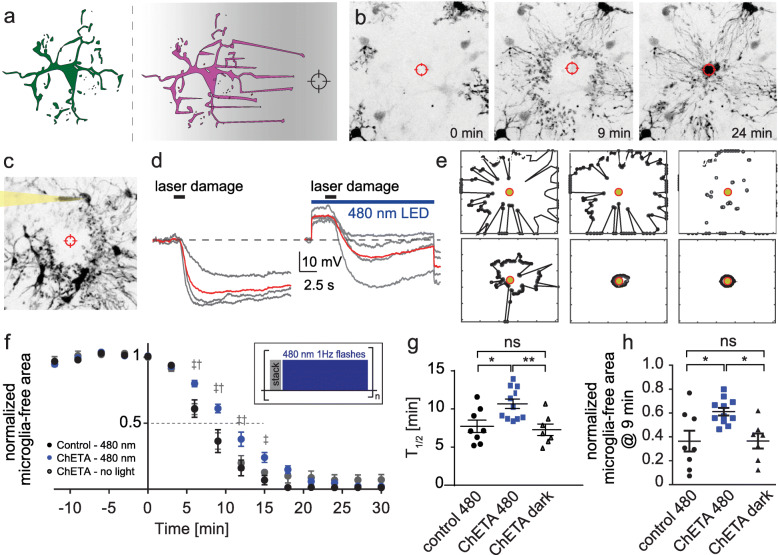
Microglia react to danger signals by rapid and targeted extension of cellular processes towards the source of the signal. This positive chemotactic response is accompanied by a hyperpolarization of the microglia membrane. Here, we show that optogenetic depolarization of microglia has little effect on baseline motility, but significantly slows down the chemotactic response. Reducing the extracellular Ca2+ concentration mimics the effect of optogenetic depolarization. As the membrane potential sets the driving force for Ca2+ entry, hyperpolarization is an integral part of rapid stimulus-response coupling in microglia. Compared to typical excitable cells such as neurons, the sign of the activating response is inverted in microglia, leading to inhibition by depolarizing channelrhodopsins.

Keywords: Calcium signaling; Channelrhodopsin; Chemotaxis; Hippocampus; Laser damage; Microglia; Optogenetics; Sterile inflammation; Two-photon microscopy.


Conflict of interest statement

The authors declare that they have no competing interests.

Figures

Fig. 1
Fig. 1
Microglia-specific ChETA expression in organotypic hippocampal slice cultures. a Schematic overview of microglia-specific expression. The microglia-driver line Cx3cr1-CreERT2 (blue) is crossed with a reporter mouse line (R26-LSL-tdTomato, red) and the ChETA mouse line (R26-LSL-ChETA, green). After injection of (Z)-4-hydroxytamoxifen, the tdTomato and ChETA are expressed in microglia. b Illustration of the Channelrhodopsin-variant ChETA activated by blue light. Scale bar 25 μm. c Immunostaining using antibodies against the reporter (tdTomato - red) and microglia (iba1 - cyan). d Graphic illustration of the hippocampal structure and the investigated area for microglia morphology in e (red square). e Z-projection of confocal images acquired for Sholl analysis of microglia at 3, 13, 29 DIV, and in vivo. Scale bar: 25 μm. f Confocal image of a microglia cell in organotypic slice culture which was fixed with PFA and stained against the microglia marker iba1. Overlay with IMARIS analysis (magenta). g Result of microglia branch detection with color coding by branching level. h Sholl analysis of microglia over time (number of intersections versus distance from cell body). i Quantification of % microglia cells between dentate gyrus and CA1 relative to total cell count (DAPI)
Fig. 2
Fig. 2
Optogenetic microglia depolarization using the Channelrhodopsin variant ChETA. a Two-photon maximum projection of a microglia patched in organotypic slice culture. Contrast inversion for better visibility. b Microglia cell properties; from left to right: Membrane resistance, cell capacitance, membrane potential, and change in membrane potential upon illumination with 1 mW/mm2 480 nm light. Black circles: n = 9 microglia, 5 slices; red dot: mean ± SEM. c Light-induced membrane depolarizations in microglia. Black: individual sweeps (n = 17 sweeps), red: peristimulus time histogram (PSTH) of seven consecutive sweeps with the same light intensity. d Relative depolarization of microglia membrane potential in response to 480 nm light. Blue dots: mean ± SEM. e Voltage-clamp recording of a microglia cell with repetitive 1 Hz 480 nm light stimulation at 1 mW/mm2 (n = 7 microglia, 3 slices, female, DIV 12–20). f Enlargement of red dotted box in (e)
Fig. 3
Fig. 3
Optogenetic microglia depolarization decelerates chemotactic response kinetics. a Graphic illustration and representative images of microglia chemotaxis towards an induced laser-damage. b Two-photon maximum projections of the chemotactic response 0, 9, and 24 min after the laser-damage. c Two-photon z-projection of a patched microglia during chemotaxis. d Voltage-clamp recordings of patched microglia during chemotaxis. Gray: Individual microglial responses from four experiments, red: Average of all experiments. Left: no light stimulation during laser-damage, right: with light stimulation during laser damage. e Automated MATLAB analysis of chemotaxis quantified as the reduction in microglia-free area around the laser damage (black polygon) at different time points of the experiment. f Relative laser damage response measured as microglia-free area. Black: Control slices (no construct) with light stimulation (n = 8 areas, 5 slices). Gray: Experiments with ChETA expression in microglia, but without light stimulation (n = 7 areas, 4 slices). Blue: Slices with ChETA expression in microglia combined light stimulation (n = 11 areas, 7 slices). Insert: Graphic representation of light stimulation protocol between stack acquisitions. 2-way ANOVA (‡ control480 – ChETA480, p < 0.001, († ChETA no light – ChETA480, p < 0.01). g Time to 50% engulfment was prolonged by optogenetic depolarization. h 9 min after injury, the microglia-free area was larger when microglia were depolarized. g, h One-way ANOVA with Tukey’s post hoc comparison (*p < 0.05, **p < 0.01, ***p < 0.001)

Similar articles

Cited by

  • Calcium Signalling in Microglia.
    Garaschuk O, Verkhratsky A. Garaschuk O, et al. Adv Neurobiol. 2024;37:123-133. doi: 10.1007/978-3-031-55529-9_7. Adv Neurobiol. 2024. PMID: 39207689 Review.
  • Neuron-glia interaction at the receptor level affects olfactory perception in adult Drosophila.
    Calvin-Cejudo L, Martin F, Mendez LR, Coya R, Castañeda-Sampedro A, Gomez-Diaz C, Alcorta E. Calvin-Cejudo L, et al. iScience. 2022 Dec 19;26(1):105837. doi: 10.1016/j.isci.2022.105837. eCollection 2023 Jan 20. iScience. 2022. PMID: 36624835 Free PMC article.
  • Microglial voltage-gated proton channel Hv1 in spinal cord injury.
    Zheng J, Murugan M, Wang L, Wu LJ. Zheng J, et al. Neural Regen Res. 2022 Jun;17(6):1183-1189. doi: 10.4103/1673-5374.327325. Neural Regen Res. 2022. PMID: 34782552 Free PMC article. Review.
  • Retinal and Brain Microglia in Multiple Sclerosis and Neurodegeneration.
    Choi S, Guo L, Cordeiro MF. Choi S, et al. Cells. 2021 Jun 15;10(6):1507. doi: 10.3390/cells10061507. Cells. 2021. PMID: 34203793 Free PMC article. Review.
  • Interfacing with the Brain: How Nanotechnology Can Contribute.
    Ahmed AAA, Alegret N, Almeida B, Alvarez-Puebla R, Andrews AM, Ballerini L, Barrios-Capuchino JJ, Becker C, Blick RH, Bonakdar S, Chakraborty I, Chen X, Cheon J, Chilla G, Coelho Conceicao AL, Delehanty J, Dulle M, Efros AL, Epple M, Fedyk M, Feliu N, Feng M, Fernández-Chacón R, Fernandez-Cuesta I, Fertig N, Förster S, Garrido JA, George M, Guse AH, Hampp N, Harberts J, Han J, Heekeren HR, Hofmann UG, Holzapfel M, Hosseinkazemi H, Huang Y, Huber P, Hyeon T, Ingebrandt S, Ienca M, Iske A, Kang Y, Kasieczka G, Kim DH, Kostarelos K, Lee JH, Lin KW, Liu S, Liu X, Liu Y, Lohr C, Mailänder V, Maffongelli L, Megahed S, Mews A, Mutas M, Nack L, Nakatsuka N, Oertner TG, Offenhäusser A, Oheim M, Otange B, Otto F, Patrono E, Peng B, Picchiotti A, Pierini F, Pötter-Nerger M, Pozzi M, Pralle A, Prato M, Qi B, Ramos-Cabrer P, Genger UR, Ritter N, Rittner M, Roy S, Santoro F, Schuck NW, Schulz F, Şeker E, Skiba M, Sosniok M, Stephan H, Wang R, Wang T, Wegner KD, Weiss PS, Xu M, Yang C, Zargarian SS, Zeng Y, Zhou Y, Zhu D, Zierold R, Parak WJ. Ahmed AAA, et al. ACS Nano. 2025 Mar 25;19(11):10630-10717. doi: 10.1021/acsnano.4c10525. Epub 2025 Mar 10. ACS Nano. 2025. PMID: 40063703 Free PMC article. Review.

References

    1. Nimmerjahn A. Resting microglial cells are highly dynamic surveillants of brain parenchyma in vivo. Science. 2005;308:1314–1318. doi: 10.1126/science.1110647. - DOI - PubMed
    1. Madry C, Kyrargyri V, Arancibia-Cárcamo IL, Jolivet R, Kohsaka S, Bryan RM, et al. Microglial ramification, surveillance, and interleukin-1β release are regulated by the two-pore domain K+ channel THIK-1. Neuron. 2018;97:299–312.e6. doi: 10.1016/j.neuron.2017.12.002. - DOI - PMC - PubMed
    1. Newell EW, Schlichter LC. Integration of K + and Cl-currents regulate steady-state and dynamic membrane potentials in cultured rat microglia. J Physiol. 2005;567:869–890. doi: 10.1113/jphysiol.2005.092056. - DOI - PMC - PubMed
    1. Schilling T, Eder C. Microglial K+channel expression in young adult and aged mice. Glia. 2015;63:664–672. doi: 10.1002/glia.22776. - DOI - PMC - PubMed
    1. Rangaraju S, Gearing M, Jin L-W, Levey A. Potassium channel Kv1.3 is highly expressed by microglia in human Alzheimer’s disease. J Alzheimers Dis. 2015;44:797–808. doi: 10.3233/JAD-141704. - DOI - PMC - PubMed

LinkOut - more resources