Review
. 2020 Dec 3:11:608661.
doi: 10.3389/fphys.2020.608661. eCollection 2020.

Structural and Functional Plasticity in the Regenerating Olfactory System of the Migratory Locust

Affiliations
Review

Structural and Functional Plasticity in the Regenerating Olfactory System of the Migratory Locust

Gerd Bicker et al. Front Physiol. .

Abstract

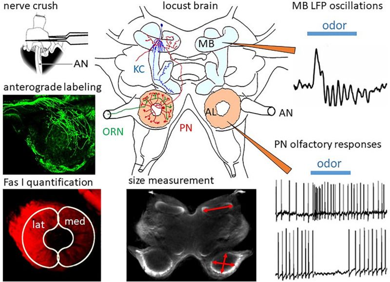
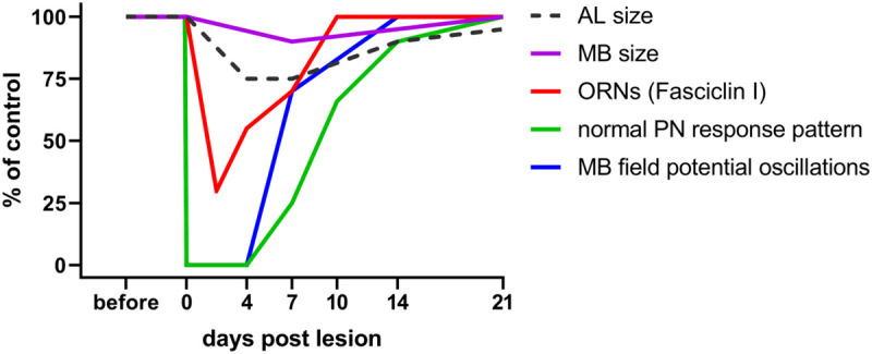
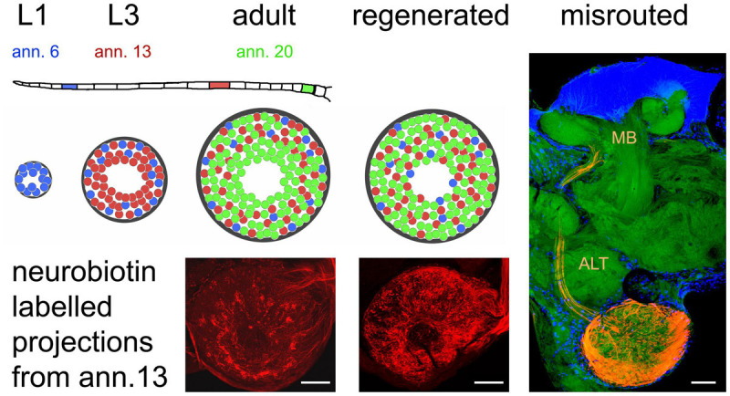
Regeneration after injury is accompanied by transient and lasting changes in the neuroarchitecture of the nervous system and, thus, a form of structural plasticity. In this review, we introduce the olfactory pathway of a particular insect as a convenient model to visualize neural regeneration at an anatomical level and study functional recovery at an electrophysiological level. The olfactory pathway of the locust (Locusta migratoria) is characterized by a multiglomerular innervation of the antennal lobe by olfactory receptor neurons. These olfactory afferents were axotomized by crushing the base of the antenna. The resulting degeneration and regeneration in the antennal lobe could be quantified by size measurements, dye labeling, and immunofluorescence staining of cell surface proteins implicated in axonal guidance during development. Within 3 days post lesion, the antennal lobe volume was reduced by 30% and from then onward regained size back to normal by 2 weeks post injury. The majority of regenerating olfactory receptor axons reinnervated the glomeruli of the antennal lobe. A few regenerating axons project erroneously into the mushroom body on a pathway that is normally chosen by second-order projection neurons. Based on intracellular responses of antennal lobe output neurons to odor stimulation, regenerated fibers establish functional synapses again. Following complete absence after nerve crush, responses to odor stimuli return to control level within 10-14 days. On average, regeneration of afferents, and re-established synaptic connections appear faster in younger fifth instar nymphs than in adults. The initial degeneration of olfactory receptor axons has a trans-synaptic effect on a second order brain center, leading to a transient size reduction of the mushroom body calyx. Odor-evoked oscillating field potentials, absent after nerve crush, were restored in the calyx, indicative of regenerative processes in the network architecture. We conclude that axonal regeneration in the locust olfactory system appears to be possible, precise, and fast, opening an avenue for future mechanistic studies. As a perspective of biomedical importance, the current evidence for nitric oxide/cGMP signaling as positive regulator of axon regeneration in connectives of the ventral nerve cord is considered in light of particular regeneration studies in vertebrate central nervous systems.

Keywords: antennal lobe; fasciclin I; field potential oscillations; mushroom body; semaphorin 1a.


Conflict of interest statement

The authors declare that the research was conducted in the absence of any commercial or financial relationships that could be construed as a potential conflict of interest.

Figures

FIGURE 1
FIGURE 1
Regeneration in the olfactory system of the locust. In the locust brain, olfactory information from the ORNs projecting through the antennal nerves (AN) is processed in the ALs by local interneurons and PNs, which convey information to the mushroom bodies (MB) containing ca. 50,000 Kenyon cells (KC). After ORN axotomy by crushing the AN, degeneration and subsequent regeneration can be studied by measurement of MB and AL size, by anterograde labeling of ORNs, and by quantification of ORN projections into the lateral (lat) and medial (med) part of the AL by measuring immunofluorescence of their cell surface molecule, Fas I. Functional regeneration can be measured by intracellular recording odor-evoked responses from PNs and by recording of extracellular field potentials in the calyx of the MB. Adapted from Wasser et al. (2017) and Wasser and Stern (2017).
FIGURE 2
FIGURE 2
Time course of plastic changes in the adult locust olfactory system after lesion. Values are expressed as percentage of the unlesioned situation (before). On the neuroanatomical level, neuropil volumes and the cell surface protein fasciclin I indicative for ORN terminals in the AL decrease within the first 4 days post lesion, and increase back to normal within 21 days. On the electrophysiological level, responses are completely absent directly after the lesion, begin to return after 4 days, and return back to normal within 21 days as well. For technical reasons in a session of intracellular recordings from AL projection neurons, the chance to hit one of the very few (out of 830) cells with regenerated input is rather low. The mushroom body, however, integrates over all PNs. Thus, the chance to detect a regenerated input is much higher, explaining earlier re-appearance of mushroom body signals. Data from Eickhoff et al. (2012); Wasser et al. (2017), and Wasser and Stern (2017).
FIGURE 3
FIGURE 3
Organization of olfactory receptor neuron projections into the antennal lobe during development and regeneration. The flagellum of the locust antenna consists of 21 rings or annuli (ann.), which increase in number and size during postembryonic development through five larval stages (L1, L3 as examples) till adulthood. Receptor neurons terminate in a distributed fashion in the available glomeruli of the antennal lobe, which also gains size during development. This leads to a weak topographic pattern where older neurons born in younger stages (L1, blue) tend to terminate in the periphery, younger neurons born later (L3, red) terminate peripherally and medially, and terminals of younger neurons born just before the final molt (adult, green) are evenly distributed all over the antennal lobe. After regeneration, this pattern is absent. The photomicrographs of the neurobiotin-labeled sections correspond to the red glomeruli in the schematic. Only a small fraction of receptor neurons (∼15 axons when labeling the complete antennal nerve) fail to terminate in the antennal lobe and are misrouted through the antennal lobe tract (ALT) to the mushroom body (MB). Photomicrographs from Wasser et al. (2017). Scale bars 100 μm.

References

    1. Anton S., Hansson B. S. (1996). Antennal lobe interneurons in the desert locust Schistocerca gregaria (Forskal): processing of aggregation pheromones in adult males and females. J. Comp. Neurol. 370 85–96. - PubMed
    1. Anton S., Ignell R., Hansson B. S. (2002). Developmental changes in the structure and function of the central olfactory system in gregarious and solitary desert locusts. Mic. Res. Tech. 56 281–291. 10.1159/000047222 - DOI - PubMed
    1. Ayaz D., Leyssen M., Koch M., Yan J., Srahna M., Sheeba V., et al. (2008). Axonal injury and regeneration in the adult brain of Drosophila. J. Neurosci. 28 6010–6021. 10.1523/JNEUROSCI.0101-08.2008 - DOI - PMC - PubMed
    1. Bastiani M. J., Harrelson A. L., Snow P. M., Goodman C. S. (1987). Expression of fasciclin I and II glycoproteins on subsets of axon pathways during neuronal development in the grasshopper. Cell 48 745–755. 10.1016/0092-8674(87)90072-9 - DOI - PubMed
    1. Bergmann G. A., Frömbling S., Joseph N., Bode K., Bicker G., Stern M. (2019). An intact insect embryo for developmental neurotoxicity testing of directed axonal elongation. ALTEX 36 643–649. 10.14573/altex.1901292 - DOI - PubMed