. 2020 Dec 10:11:590825.
doi: 10.3389/fneur.2020.590825. eCollection 2020.

Accuracy and Workflow Improvements for Responsive Neurostimulation Hippocampal Depth Electrode Placement Using Robotic Stereotaxy

Affiliations

Accuracy and Workflow Improvements for Responsive Neurostimulation Hippocampal Depth Electrode Placement Using Robotic Stereotaxy

Patrick J Karas et al. Front Neurol. .

Abstract

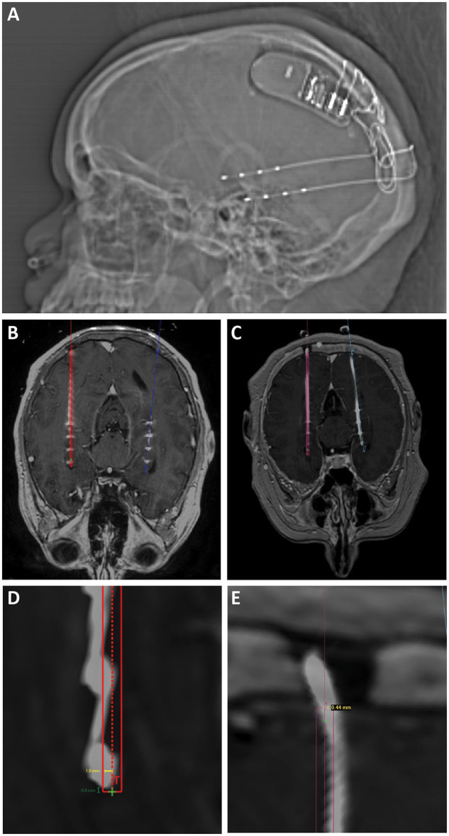
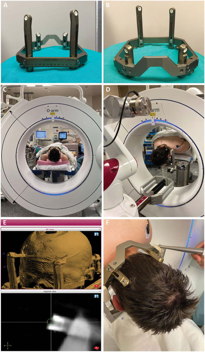
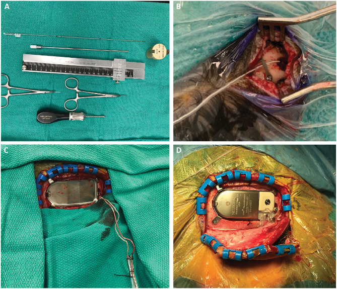
Background: Robotic stereotaxy is increasingly common in epilepsy surgery for the implantation of stereo-electroencephalography (sEEG) electrodes for intracranial seizure monitoring. The use of robots is also gaining popularity for permanent stereotactic lead implantation applications such as in deep brain stimulation and responsive neurostimulation (RNS) procedures. Objective: We describe the evolution of our robotic stereotactic implantation technique for placement of occipital-approach hippocampal RNS depth leads. Methods: We performed a retrospective review of 10 consecutive patients who underwent robotic RNS hippocampal depth electrode implantation. Accuracy of depth lead implantation was measured by registering intraoperative post-implantation fluoroscopic CT images and post-operative CT scans with the stereotactic plan to measure implantation accuracy. Seizure data were also collected from the RNS devices and analyzed to obtain initial seizure control outcome estimates. Results: Ten patients underwent occipital-approach hippocampal RNS depth electrode placement for medically refractory epilepsy. A total of 18 depth electrodes were included in the analysis. Six patients (10 electrodes) were implanted in the supine position, with mean target radial error of 1.9 ± 0.9 mm (mean ± SD). Four patients (8 electrodes) were implanted in the prone position, with mean radial error of 0.8 ± 0.3 mm. The radial error was significantly smaller when electrodes were implanted in the prone position compared to the supine position (p = 0.002). Early results (median follow-up time 7.4 months) demonstrate mean seizure frequency reduction of 26% (n = 8), with 37.5% achieving ≥50% reduction in seizure frequency as measured by RNS long episode counts. Conclusion: Prone positioning for robotic implantation of occipital-approach hippocampal RNS depth electrodes led to lower radial target error compared to supine positioning. The robotic platform offers a number of workflow advantages over traditional frame-based approaches, including parallel rather than serial operation in a bilateral case, decreased concern regarding human error in setting frame coordinates, and surgeon comfort.

Keywords: NeuroPace; RNS workflow; hippocampal depth electrode; responsive neurostimulation (RNS); robotic stereotaxy; robotic stereotaxy accuracy.


Conflict of interest statement

SS is a consultant for Boston Scientific, Abbott, and Zimmer Biomet. The remaining authors declare that the research was conducted in the absence of any commercial or financial relationships that could be construed as a potential conflict of interest.

Figures

Figure 1
Figure 1
(A) Scout x-ray showing lateral view after implantation of bilateral hippocampal RNS electrodes and RNS generator. (B) Intra-operative O-arm fluoroscopic CT projected over preoperative planning MRI is used to confirm target accuracy electrodes compared to operative plans (displayed in red and blue). (C) Post-operative CT projected over preoperative planning MRI was also used to confirm target accuracy in cases where O-arm fluoroscopic CT was not performed. (D) Radial target error (yellow line) was measured as the distance from the planned electrode target to the center of the actual electrode position. Depth target error (green) was measured as the difference in depth between the implanted electrode and the planned electrode tip measured along the trajectory of the implanted electrode. Positive values represent electrodes that were implanted past/deeper to target. Negative values represent electrodes that were implanted more shallow compared to target. (E) Radial entry point error was measured as the distance from the planned electrode entry point at the inner table of the skull to the center of the implanted electrode.
Figure 2
Figure 2
(A,B) The Leksell frame is assembled opposite the traditional manner, with the short fixation posts flanking the curved nasal piece, and long fixation posts flanking the straight piece. Female fixation screws are used and serve as skull fiducials for robot registration after O-arm imaging. (C) After frame placement, the patient is initially positioned supine while still on the stretcher, with the head supported on a radiolucent plastic board, and a pre-operative O-arm image is obtained for registration to the pre-operative CT. (D) The patient is then flipped prone on gel rolls on the OR table, and the Leksell frame is affixed to the robot using the goalpost-shaped Leksell holder. The reverse orientation of the frame allows the three straight edges of the frame to fit within the beveled clamps of the Leksell holder, with the curved nasal piece over the occipital region. (E) Registration points are chosen on the merged fluoroscopic CT image including the frame. Four points are chosen, one for each frame pin, such that the registration marker sits in middle of the divot on the pin with its equator flush with the flat surface of the pin. (F) Registration is then performed using the ball-tip probe robot attachment.
Figure 3
Figure 3
(A) Required stereotactic equipment for the procedure: 2.15 mm PEEK ROSA adapter, 190 mm length slotted canula, RNS depth electrode, stereotactic ruler, bent cranial plate and cut segment of lead cap. The depth electrode is marked at three points: 190 and 200 mm, which flank the wide hub of the cannula, and the point corresponding to the outer table of the skull, measured on the stereotactic plan. This last point is important to mark, as it is the only one visible once the cannula is removed and the robot arm moved away. (B) After the electrode is inserted, a dog-bone cranial fixation miniplate is fastened to the skull to hold the electrode in place. We use the cut segment of the lead cap as a shock absorber around the lead. (C) After creating the craniectomy and securing the ferrule, the RNS leads are retrieved from their subgaleal position. (D) The RNS generator can be placed and connected to the depth electrodes. This can be performed either in the same prone position or after re-positioning the patient supine with the head turned.

References

    1. Fomenko A, Serletis D. Robotic stereotaxy in cranial neurosurgery: a qualitative systematic review. Neurosurgery. (2017) 83:642–50. 10.1093/neuros/nyx576 - DOI - PubMed
    1. Liu L, Mariani SG, De Schlichting E, Grand S, Lefranc M, Seigneuret E, et al. . Frameless ROSA® robot-assisted lead implantation for deep brain stimulation: technique and accuracy. Oper Neurosurg. (2019) 19:57–64. 10.1093/ons/opz320 - DOI - PubMed
    1. Faraji AH, Kokkinos V, Sweat JC, Crammond DJ, Richardson RM. Robotic-assisted stereotaxy for deep brain stimulation lead implantation in awake patients. Oper Neurosurg. (2020) 19:444–52. 10.1093/ons/opaa029 - DOI - PMC - PubMed
    1. Youngerman BE, Oh JY, Anbarasan D, Billakota S, Casadei CH, Corrigan EK, et al. . Laser ablation is effective for temporal lobe epilepsy with and without mesial temporal sclerosis if hippocampal seizure onsets are localized by stereoelectroencephalography. Epilepsia. (2018) 59:595–606. 10.1111/epi.14004 - DOI - PubMed
    1. Karas PJ, Sheth SA, Yoshor D. Epilepsy: mesial temporal. In: Pouratian N, Sheth SA, editors. Stereotactic and Functional Neurosurgery: Principles and Applications. Cham: Springer International Publishing; (2020). p. 339–67.

LinkOut - more resources