. 2020 Dec 23:2020:9358563.
doi: 10.1155/2020/9358563. eCollection 2020.

Ulmus parvifolia Jacq. Exhibits Antiobesity Properties and Potentially Induces Browning of White Adipose Tissue

Affiliations

Ulmus parvifolia Jacq. Exhibits Antiobesity Properties and Potentially Induces Browning of White Adipose Tissue

Yuan Yee Lee et al. Evid Based Complement Alternat Med. .

Abstract

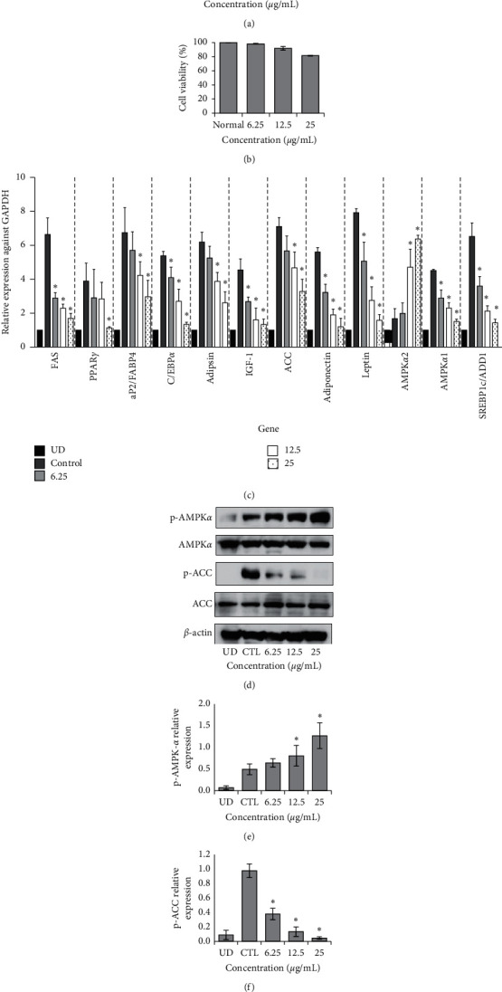
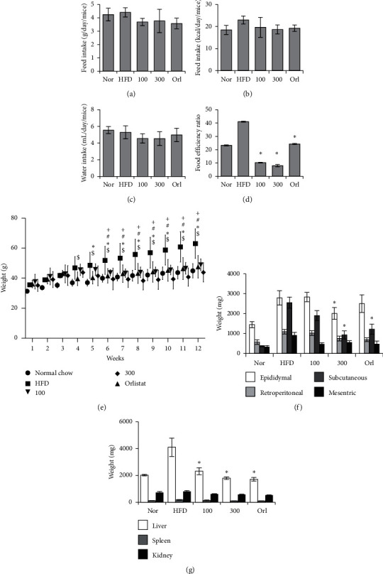
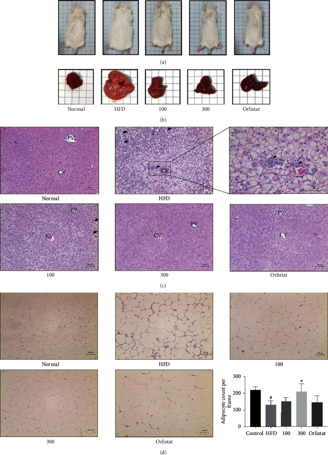
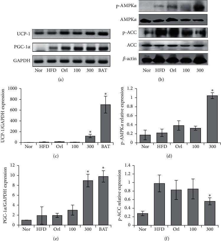
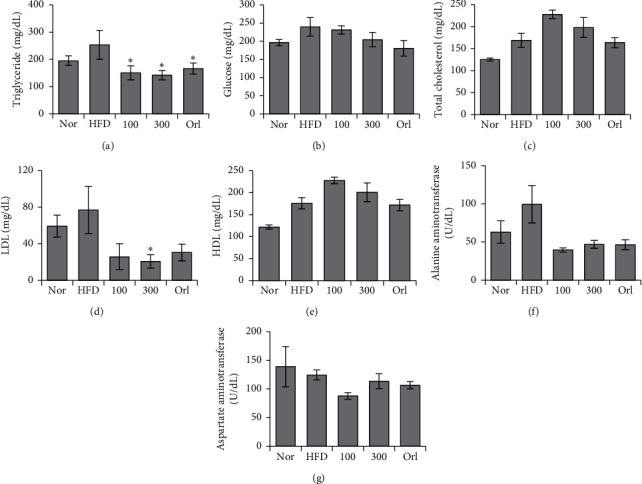
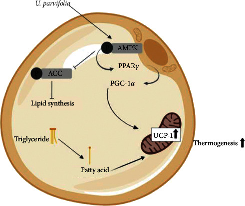
The bark of Ulmus parvifolia Jacq. (UP) was traditionally used as a diuretic and to treat intestinal inflammation. With modern evidence of the correlation of diuretics, gut inflammation, and obesity, our study has shown the antiobesity effects of the bark of UP. UP treatment reduced lipid production and adipogenic genes in vitro. In vivo studies revealed that UP 100 mg/kg and UP 300 mg/kg treatment significantly reduced mouse weight without reducing food intake, indicating increased energy expenditure. UP significantly reduced the weight of epididymal and subcutaneous adipose tissue and decreased liver weight. Histological analysis revealed improvement in the progression of nonalcoholic fatty liver disease and epididymal white adipose tissue hypertrophy induced by a HFD. Real-Time PCR of epididymal adipose tissue revealed significant increases of uncoupling protein-1 (UCP-1) and peroxisome proliferator-activated receptor gamma coactivator 1-alpha (PGC-1α) expression after UP 300 mg/kg treatments. Phosphorylation of AMP-activated protein α (AMPKα) was increased, while phosphorylation of Acetyl-CoA Carboxylase (ACC) was reduced. Our findings reveal the ability of UP to reduce the occurrence of obesity through increased browning of white adipose tissue via increased AMPKα, PPARγ, PGC-1α, and UCP-1 expression.


Conflict of interest statement

All authors declare no conflicts of interest.

Figures

Figure 1
Figure 1
UPLC-QTof MS identification of UP. Peaks detected in ethanol extract of the bark of UP (a). Peaks 1 and 2 were quantified by using a UV detector at 280 nm for target catechin glycosides. Peak 1 was identified as +(-) catechin as compared to its standard shown in (b), and peak 2 was identified as catechin-7-O-β-D-apiofuranoside as compared to its standard as shown in (c).
Figure 2
Figure 2
UP inhibits secretion of lipid droplets in 3T3-L1 preadipocytes. 3T3-L1 preadipocytes were differentiated or undifferentiated (UD) and treated with or without UP for 10 days before they were stained with oil red O (a). Cell viability was confirmed with an MTT assay after 3T3-L1 cells were treated with different concentrations of UP (b). Real-Time PCR was carried out by extracting RNA of differentiated 3T3-L1 cells with or without treatment of UP (c). Western blot analysis of p-AMPKα and p-ACC against the housekeeping gene β-Actin. 3T3-L1 cells were treated with or without UP after differentiation. After 10 days, protein was extracted from the cells, separated using SDS-PAGE, transferred to a PVDF membrane, incubated overnight with the primary antibody, incubated with secondary antibody, and developed (d). Expressions of genes were compared against the differentiated control group. Western blot was repeated in triplicate, and images were quantified using ImageJ (e and f). Statistical analysis was performed using one-way ANOVA with Dunnett's posttest, and P < 0.05 was considered significant.
Figure 3
Figure 3
UP reduced obesity in mice. Food intake and water intake were monitored weekly and averaged as shown in (a–c). Food efficiency ratio was calculated using the weight gain to intake of food ratio (d). Weight of mice for each group was recorded every week for a period of 12 weeks. Mice in the normal group (nor) were given normal chow, whereas all other groups were given a HFD. After 4 weeks, oral administration of orlistat and UP was given daily for 8 weeks (e). After 12 weeks, mice were euthanized, and the fat tissue, liver, spleen, and kidney were harvested and weighed immediately (f and g). Statistics were analyzed using one-way ANOVA with Dunnett's posttest, and P < 0.05 was considered significant. Statistical significance of HFD as compared to the control group in (d) are indicated by $; , for the orlistat group against HFD; #, for UP 100 against HFD; and +, for UP 300 against HFD.
Figure 4
Figure 4
UP reduced the size of mice and mouse livers and improved the histology of the liver and adipose tissue induced by an HFD. Mice were fed with an HFD for a total of 12 weeks, and oral administration of orlistat and UP was carried out after 4 weeks of a daily HFD. Representative images of mice and livers for each group (a-b). Liver tissues and epididymal adipose tissue were dehydrated and fixed in paraffin and then sectioned before staining with H&E (c-d). Liver tissues (c) and epididymal adipose tissue (d) were observed at 100x. Arrows indicate balloon cells, and arrowheads indicate the infiltration of inflammatory cells. CV indicates the location of the central vein, whereas PV indicates the portal veins. Magnified image of the CV area of the liver tissue in HFD-treated group was taken at 200x. Statistical analysis for adipocyte count performed using one-way ANOVA with Dunnett's posttest, and indicates P < 0.05 compared to the HFD group, whereas # indicates a P < 0.05 as compared to the control group.
Figure 5
Figure 5
UP increased expression of browning markers in mice. Extracted RNA was reverse transcribed, and the browning related genes UCP-1 and PGC-1α were investigated using Real-Time PCR conducted using the resultant cDNA (a). Quantification of gel images (c and e). Protein expressions of p-AMPKα and p-ACC in liver tissue were investigated using western blot analysis, as shown in (b), and the gel images were quantified using ImageJ (d and f). All experiments were conducted in triplicate. Statistics were analyzed using one-way ANOVA, and P < 0.05 was considered significant.
Figure 6
Figure 6
UP improved serum biochemical concentrations induced by an HFD in mice. Blood was collected by cardiac puncture, and serum was separated. Levels of (a) triglyceride, (b) glucose, (c) total cholesterol, (d) LDL, (e) HDL, (f) ALT, and (g) AST were investigated using a blood analyzer. Statistical analysis was performed using one-way ANOVA with Dunnett's posttest, and P < 0.05 was considered significant.
Figure 7
Figure 7
Schematic diagram of the proposed mechanism of action of UP. UP increased the browning of WAT by acting through increased AMPKα expression, which, in turn, increases PPARγ expression followed by the increase of PGC-1α expression, which induces upregulation of mitochondrial UCP-1 in white adipocytes and increases the usage of stored triglycerides and thermogenesis. Phosphorylation of ACC was also inhibited, subsequently inhibiting lipid synthesis.

References

    1. Santamour F. S., Jr., Bentz S. E. Updated checklist of elm (Ulmus) cultivars for use in North America. Journal of Arboriculture. 1995;21(3):122–131.
    1. Bate-Smith E. C., Richens R. H. Flavonoid chemistry and taxonomy in Ulmus. Biochemical Systematics and Ecology. 1973;1(3):141–146. doi: 10.1016/0305-1978(73)90004-5. - DOI
    1. Mina S. A. LC/ESI-MS/MS profiling of Ulmus parvifolia extracts and evaluation of its anti-inflammatory, cytotoxic, and antioxidant activities. Zeitschrift für Naturforschung C. 2016;71(11-12):415–421. doi: 10.1515/znc-2016-0057. - DOI - PubMed
    1. Li C.-P. Chinese Herbal Medicine. Washington, DC, USA: US Department of Health, Education, and Welfare, Public Health Service, National Institute of Health; 1974.
    1. Vent W., Duke J. A., Ayensu E. S. Medicinal Plants of China. Vol. 2. Algonac, MI, USA: Reference Publ., Inc.; 1987.