

Visualization of left ventricular Purkinje fiber distribution using widefield optical coherence microscopy
- PMID: 33425102
- PMCID: PMC7791375
Visualization of left ventricular Purkinje fiber distribution using widefield optical coherence microscopy
Abstract
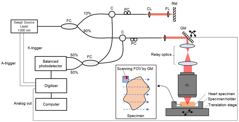
Background: The distribution and connection of ventricular Purkinje fibers are known to be associated with idiopathic left ventricular arrhythmias. Unusual anatomy is one of the important factors associated with catheter ablation success rate. With the widefield high-speed, swept-source optical coherence microscopy (OCM) and light microscope, we visualized the left ventricular Purkinje fiber distribution.
Methods: Left ventricular walls of five adult ovine hearts were incised from the mitral annulus to the apex. Using the widefield OCM technique and light microscopy, we observed the distribution, direction, depth, and dividing patterns of the Purkinje network with multiple tangential angles and without tissue destruction.
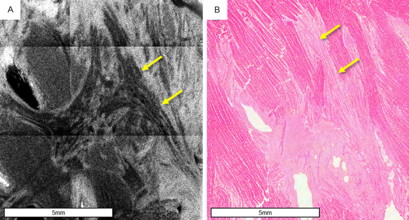
Results: Widefield OCM was used to characterize the ovine heart Purkinje network system in a 4 × 4 mm2 field. Left ventricular Purkinje fibers traveled in the sub-endocardial area near the left-sided peri-membranous septal area and ran like a wide hair bundle. The distal branching fibers penetrated to the endocardium and connected to the contractile muscle. In this distal area, Purkinje fibers were connected to each other, forming multiple layers. Some Purkinje fibers were directly connected within the false tendon between the papillary muscles or between the trabeculations. Some free-running Purkinje fibers were directly connected to the papillary muscle from the left bundle.
Conclusion: Using widefield OCM, we were able to observe the left bundle and its branching patterns in ovine left ventricle without tissue destruction. This might be applied to future cardiac ablation procedures.
Keywords: Purkinje fibers; cardiac arrhythmia; heart conduction system; optical microscopy.
IJCEP Copyright © 2020.
Conflict of interest statement
None.
Figures


Similar articles
-
Morphological study of the atrioventricular conduction system and Purkinje fibers in yak.J Morphol. 2017 Jul;278(7):975-986. doi: 10.1002/jmor.20691. Epub 2017 Apr 26. J Morphol. 2017. PMID: 28444887
-
Role of the conduction system in the endocardial excitation spread in the right ventricle.Am J Cardiol. 1981 Nov;48(5):864-70. doi: 10.1016/0002-9149(81)90351-9. Am J Cardiol. 1981. PMID: 7304434
-
Intracardiac Delineation of Septal Conduction in Left Bundle-Branch Block Patterns.Circulation. 2019 Apr 16;139(16):1876-1888. doi: 10.1161/CIRCULATIONAHA.118.038648. Circulation. 2019. PMID: 30704273
-
Unsolved Questions on the Anatomy of the Ventricular Conduction System.Korean Circ J. 2018 Dec;48(12):1081-1096. doi: 10.4070/kcj.2018.0335. Korean Circ J. 2018. PMID: 30403014 Free PMC article. Review.
-
The normal variants in the left bundle branch system.J Electrocardiol. 2017 Jul-Aug;50(4):389-399. doi: 10.1016/j.jelectrocard.2017.03.004. Epub 2017 Mar 14. J Electrocardiol. 2017. PMID: 28341304 Review.
Cited by
-
Development of Wireless Network Indoor Coverage System Based on Optical Fiber Distribution System.Comput Intell Neurosci. 2022 Jul 5;2022:9728536. doi: 10.1155/2022/9728536. eCollection 2022. Comput Intell Neurosci. 2022. Retraction in: Comput Intell Neurosci. 2023 Jul 12;2023:9891617. doi: 10.1155/2023/9891617. PMID: 35837207 Free PMC article. Retracted.
-
Developing a Mechanistic Approach to Sudden Death Prevention in Mitral Valve Prolapse.J Clin Med. 2022 Feb 26;11(5):1285. doi: 10.3390/jcm11051285. J Clin Med. 2022. PMID: 35268384 Free PMC article. Review.
-
Trigger and Substrate Mapping and Ablation for Ventricular Fibrillation in the Structurally Normal Heart.J Cardiovasc Dev Dis. 2023 May 2;10(5):200. doi: 10.3390/jcdd10050200. J Cardiovasc Dev Dis. 2023. PMID: 37233167 Free PMC article. Review.
References
-
- Boyden PA. Purkinje physiology and pathophysiology. J Interv Card Electrophysiol. 2018;52:255–262. - PubMed
-
- Duan D, Yu S, Cui Y, Li C. Morphological study of the atrioventricular conduction system and Purkinje fibers in yak. J Morphol. 2017;278:975–986. - PubMed
-
- De Almeida MC, Lopes F, Fontes P, Barra F, Guimaraes R, Vilhena V. Ungulates heart model: a study of the Purkinje network using India ink injection, transparent specimens and computer tomography. Anat Sci Int. 2015;90:240–250. - PubMed
-
- Atkinson A, Inada S, Li J, Tellez JO, Yanni J, Sleiman R, Allah EA, Anderson RH, Zhang H, Boyett MR, Dobrzynski H. Anatomical and molecular mapping of the left and right ventricular His-Purkinje conduction networks. J Mol Cell Cardiol. 2011;51:689–701. - PubMed
LinkOut - more resources
Full Text Sources
Miscellaneous