. 2020 Dec 26:2020:8830537.
doi: 10.1155/2020/8830537. eCollection 2020.

Exercise Improves Endothelial Function Associated with Alleviated Inflammation and Oxidative Stress of Perivascular Adipose Tissue in Type 2 Diabetic Mice

Affiliations

Exercise Improves Endothelial Function Associated with Alleviated Inflammation and Oxidative Stress of Perivascular Adipose Tissue in Type 2 Diabetic Mice

Jinju Wang et al. Oxid Med Cell Longev. .

Abstract

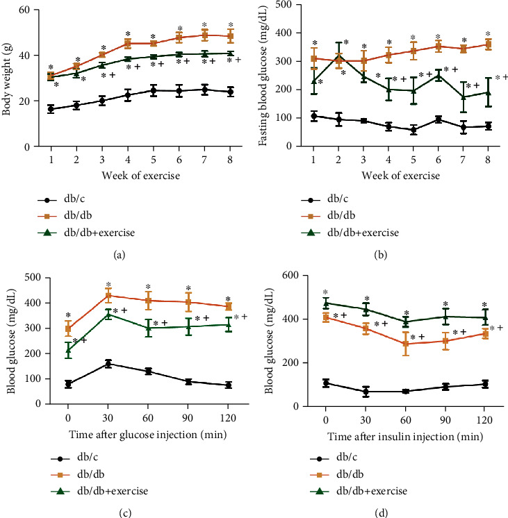
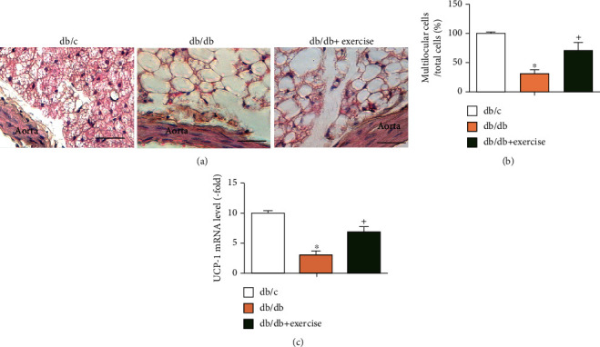
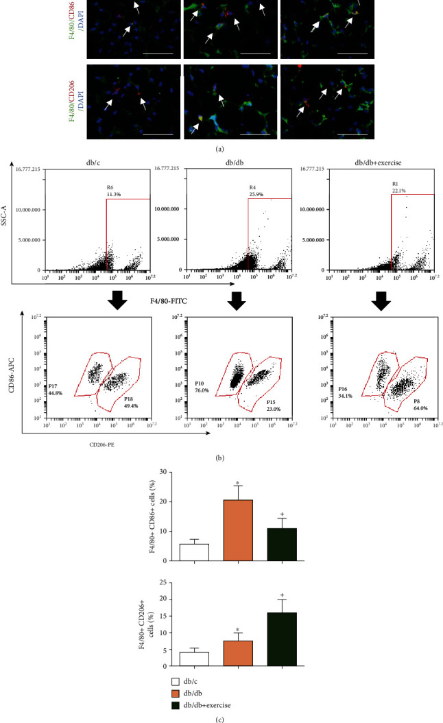
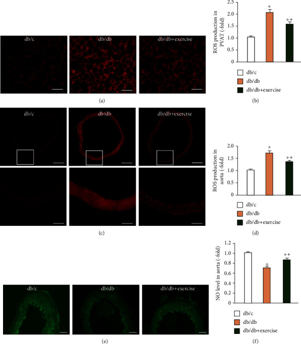
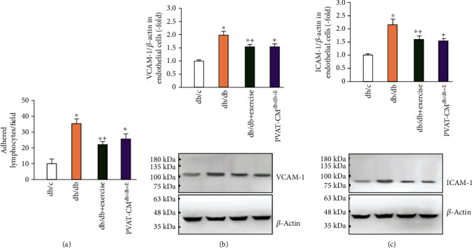
Perivascular adipose tissue (PVAT), a type of adipose tissue that surrounds the blood vessels, has been considered an active component of the blood vessel walls and involved in vascular homeostasis. Recent evidence shows that increased inflammation and oxidative stress in PVAT contribute to endothelial dysfunction in type 2 diabetes (T2D). Exercise is an important nonpharmacological approach for vascular diseases. However, there is limited information regarding whether the beneficial effects of exercise on vascular function is related to the PVAT status. In this study, we investigated whether exercise can decrease oxidative stress and inflammation of PVAT and promote the improvement of endothelial function in a T2D mouse model. Diabetic db/db (5-week old) mice performed treadmill exercise (10 m/min) or keep sedentary for 8 weeks. Body weight, fasting blood glucose levels, glucose, and insulin tolerance were determined. The cytokines (IL-6, IL-10, IFN-γ, and TNF-a) and adiponectin levels, macrophage polarization and adipocyte type in PVAT, oxidative stress, and nitric oxide (NO) expression in the vascular wall were evaluated. The adhesion ability of primary aorta endothelial cells was analyzed. Our data showed that (1) diabetic db/db mice had increased body weight and fasting blood glucose level, compromised glucose tolerance, and insulin sensitivity, which were decreased/improved by exercise intervention. (2) Exercise intervention increased the percentage of multilocular brown adipocytes, promoted M1 to M2 macrophage polarization, associating with an increase of adiponectin and IL-10 levels and decrease of IFN-γ, IL-6, and TNF-a levels in PVAT. (3) Exercise decreased superoxide production in PVAT and the vascular wall of diabetic mice, accompanied with increased NO level. (4) The adhesion ability of aorta endothelial cells to leukocytes was decreased in exercised db/db mice, accompanied by decreased intercellular adhesion molecule 1 (ICAM-1) and vascular cell adhesion molecule 1 (VCAM-1) expressions. Of interesting, coculture with PVAT-culture medium from exercised db/db mice could also reduce ICAM-1 and VCAM-1 expressions in primary endothelial cells. In conclusion, our data suggest that exercise improved endothelial function by attenuating the inflammation and oxidative stress in PVAT.


Conflict of interest statement

The authors declare that they have no conflicts of interest.

Figures

Figure 1
Figure 1
The effects of exercise intervention on body weight and metabolic characters of db/db mice. (a) Body weight of db/c, db/db, and db/db+exercise mice from week 1 to 8 of exercise. (b) Fasting blood glucose of the three groups during the 8-week exercise duration. (c, d) Blood glucose levels at 0, 30, 60, 90, and 120 mins after glucose or insulin injection. Db/db+exercise: db/db mice performed exercise. p < 0.05, vs. db/c; + p < 0.05, vs. db/db. Data are expressed as mean ± SEM. N = 8/group.
Figure 2
Figure 2
Analysis of the characteristics of PVAT. (a) Representative images of H&E staining of aorta PVAT, scale bar: 40 μm. (b) Summarized data showing the percentage of multilocular cells in PVAT. (c) UCP-1 (white adipose tissue gene marker) mRNA levels in PVAT. Data expressed as mean ± SEM, n = 8/group. p < 0.05, vs. db/c, + p < 0.05, vs. db/db.
Figure 3
Figure 3
The effects of exercise intervention on macrophage polarization in PVAT. (a) Representative images showing the double-positive staining of F4/80 and CD86, F4/80 and CD206. Scale bar: 50 μm. White arrows indicate the double-positive cells. Green: F4/80; red: CD86 or CD206; blue: nucleus counterstained by DAPI. (b) Representative flow plot showing the percentage of M1 and M2 macrophages. The gate in the upper panels shows the positive events of F4/80, the down panels showing the double-positive expression of F4/80 and CD86 or CD206. (c, d) Summarized data show the percentage of M1 (F4/80+CD86+) and M2 (F4/80+CD206+) cells in PVAT in the three groups. Db/db+exercise: db/db mice performed exercise. p < 0.05, vs. db/c; + p < 0.05, vs. db/db. Data are expressed as mean ± SEM. N = 8/group.
Figure 4
Figure 4
Analysis of ROS and/or NO level in PVAT and aorta. (a, b) Representative images and summarized data showing ROS level in PVAT in db/c, db/db, and db/db+exercise mice after 8-week exercise. Scale bar: 50 μm. (c) Representative images showing ROS expression in the aorta; bottom panels are enlarged from the relative box areas of upper panels. Scale bar in the aorta: 200 μm (upper panels) and 50 μm (bottom panels). (d) Summarized data of the ROS level in the aorta in the three groups. (e) Representative images showing NO expression in the aorta. Scale bar in aorta: 40 μm. (f) NO level in aorta from db/c, db/db, and db/db+exercise mice after 8-week exercise; Db/db+exercise: db/db mice performed exercise. p < 0.05, vs. db/c; + p < 0.05, vs. db/db. Data are expressed as mean ± SEM. N = 8/group.
Figure 5
Figure 5
The effects of exercise intervention on aorta endothelial cell inflammation. (a) The adhesion ability of primary aorta endothelial cells to lymphocytes. (b, c) Protein levels of ICAM-1 and VCAM-1 in the primary aorta endothelial cells from the three mouse groups and pre-treated by PVAT-CM. Db/db+exercise: endothelial cells cultured from exercised db/db mice. PVAT-CMdb/db+E: endothelial cells cultured from db/db mice were pretreated with the culture medium of PVAT isolated from exercised db/db mice. p < 0.05, vs. db/c; + p < 0.05, vs. db/db. Data are expressed as mean ± SEM. N = 8/group.

Similar articles

Cited by

References

    1. Franzoni F., Ghiadoni L., Galetta F., et al. Physical activity, plasma antioxidant capacity, and endothelium-dependent vasodilation in young and older men. American Journal of Hypertension. 2005;18(4):510–516. doi: 10.1016/j.amjhyper.2004.11.006. - DOI - PubMed
    1. Knowler W. C., Barrett-Connor E., Fowler S. E., et al. Reduction in the incidence of type 2 diabetes with lifestyle intervention or metformin. The New England Journal of Medicine. 2002;346(6):393–403. doi: 10.1056/NEJMoa012512. - DOI - PMC - PubMed
    1. Wang J., Liu H., Chen S., Zhang W., Chen Y., Yang Y. Moderate exercise has beneficial effects on mouse ischemic stroke by enhancing the functions of circulating endothelial progenitor cell-derived exosomes. Experimental Neurology. 2020;330, article 113325 doi: 10.1016/j.expneurol.2020.113325. - DOI - PMC - PubMed
    1. Pedralli M. L., Marschner R. A., Kollet D. P., et al. Different exercise training modalities produce similar endothelial function improvements in individuals with prehypertension or hypertension: a randomized clinical trial. Scientific Reports. 2020;10(1):p. 7628. doi: 10.1038/s41598-020-64365-x. - DOI - PMC - PubMed
    1. Siegel-Axel D. I., Haring H. U. Perivascular adipose tissue: an unique fat compartment relevant for the cardiometabolic syndrome. Reviews in Endocrine & Metabolic Disorders. 2016;17(1):51–60. doi: 10.1007/s11154-016-9346-3. - DOI - PubMed

MeSH terms