

A rare case of acute idiopathic colocolic intussusception in an adult patient
- PMID: 33425321
- PMCID: PMC7778517
- DOI: 10.1093/jscr/rjaa547
A rare case of acute idiopathic colocolic intussusception in an adult patient
Abstract
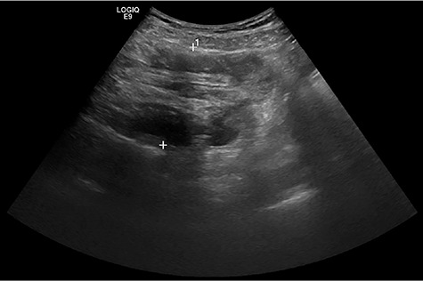
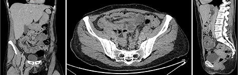
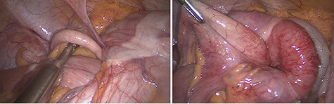
Adult intussusception is a rare condition that is frequently associated with malignancy and requires surgical approach. Symptoms are often non-specific and of subacute or chronic character. Therefore, computerized tomography (CT) scan is the most commonly used modality for identifying adult intussusception. A 51-year-old female presented with a 1-day history of increasing abdominal pain. Abdominal ultrasound and CT scan revealed intussusception. Intra-operatively, colocolic intussusception was present and laparoscopically reduced. A lead point was found neither intra-operatively nor in post-operative ileocolonoscopy and resection of involved bowel segments was not necessary.
Published by Oxford University Press and JSCR Publishing Ltd. All rights reserved. © The Author(s) 2020.
Figures
References
-
- Blanch AJM, Perel SB, Acworth JP. Paediatric intussusception: epidemiology and outcome. Emerg Med Australas 2007;19:45–50. - PubMed
-
- Moulin D, Paul Barbette MD. A seventeenth-century Amsterdam author of best-selling textbooks. Bull Hist Med 1985;59:506–14. - PubMed
-
- Hutchinson J. A successful case of abdominal section for intussusception. Proc R Med Chirur Soc 1873;7:195–8.
-
- Dean DL. Intussusception in adults. Arch Surg 1956;73:6. - PubMed
-
- Holcomb GW, Murphy JP, St. Peter SD, Gatti JM. Holcomb and Ashcraft's Pediatric Surgery, 7th edn. Philadelphia, PA: Elsevier, 2020,
Publication types
LinkOut - more resources
Full Text Sources
