. 2020 Dec 17;9(12):e1229.
doi: 10.1002/cti2.1229. eCollection 2020.

Pro-inflammatory dopamine-2 receptor-specific T cells in paediatric movement and psychiatric disorders

Affiliations

Pro-inflammatory dopamine-2 receptor-specific T cells in paediatric movement and psychiatric disorders

Deepti Pilli et al. Clin Transl Immunology. .

Abstract

Objectives: A dysregulated inflammatory response against the dopamine-2 receptor (D2R) has been implicated in movement and psychiatric disorders. D2R antibodies were previously reported in a subset of these patients; however, the role of T cells in these disorders remains unknown. Our objective was to identify and characterise pro-inflammatory D2R-specific T cells in movement and psychiatric disorders.

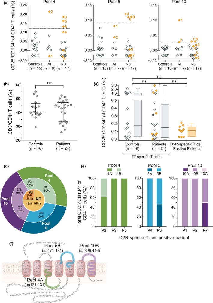
Methods: Blood from paediatric patients with movement and psychiatric disorders of suspected autoimmune and neurodevelopmental aetiology (n = 24) and controls (n = 16) was cultured in vitro with a human D2R peptide library, and D2R-specific T cells were identified by flow cytometric quantification of CD4+CD25+CD134+ T cells. Cytokine secretion was analysed using a cytometric bead array and ELISA. HLA genotypes were examined in D2R-specific T-cell-positive patients. D2R antibody seropositivity was determined using a flow cytometry live cell-based assay.

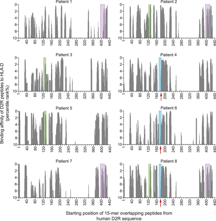
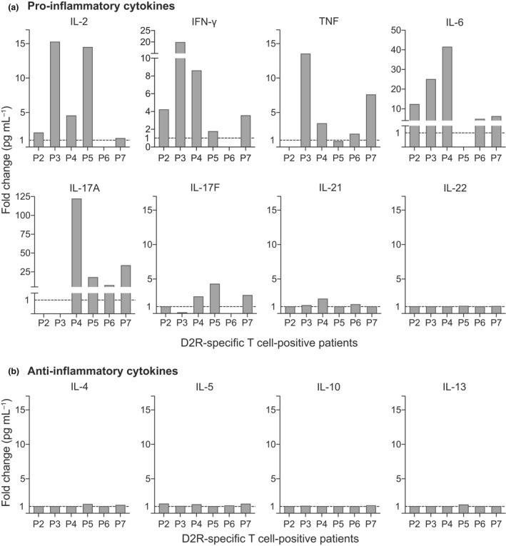
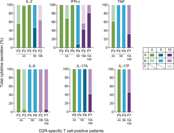
Results: Three immunodominant regions of D2R, amino acid (aa)121-131, aa171-181 and aa396-416, specifically activated CD4+ T cells in 8/24 patients. Peptides corresponding to these regions were predicted to bind with high affinity to the HLA of the eight positive patients and had also elicited the secretion of pro-inflammatory cytokines IL-2, IFN- γ, TNF, IL-6, IL-17A and IL-17F. All eight patients were seronegative for D2R antibodies.

Conclusion: Autoreactive D2R-specific T cells and a pro-inflammatory Th1 and Th17 cytokine profile characterise a subset of paediatric patients with movement and psychiatric disorders, further underpinning the theory of immune dysregulation in these disorders. These findings offer new perspectives into the neuroinflammatory mechanisms of movement and psychiatric disorders and can influence patient diagnosis and treatment.

Keywords: autoimmune encephalitis; autoimmunity; dopamine‐2 receptor antibodies; neurodevelopmental disorders; pro‐inflammatory T cells.


Conflict of interest statement

DP reports funding from the Neville Brown Scholarship (Australia). SR reports fellowship research funding from the National Health and Medical Research Council (Australia). RCD and FB have received research funding from the Trish Multiple Sclerosis Research Foundation, Multiple Sclerosis Research Australia, the Petre Foundation and the National Health Medical Research Council (Australia). They have received honoraria from Biogen Idec and Merck Serono as invited speakers. AZ, RD, JAL, FT, GL, FXZL, VM, SDH, AP, HFJ, SM, ADK and SIA declare no competing interests.

Figures

Figure 1
Figure 1
T cells in patients with movement and psychiatric disorders recognised dopamine‐2 receptor (D2R) at three distinct regions. D2R peptide pools that elicited T‐cell activation in patients with movement and psychiatric disorders of an autoimmune (AI) and neurodevelopmental (ND) aetiology were determined by assessing the co‐expression of CD25 and CD134 on CD4+ T cells using the CD25/CD134 assay. (a) Patient whole blood (n = 24) was stimulated with the 10 master pools of D2R peptides and eight patients (numbered orange diamonds) exhibited activation against at least one of three pools: pool 4, pool 5 and pool 10. Positive patients were defined as a frequency of CD25+CD134+CD4+ T cells above the threshold (dashed line; mean + 3SD of controls (n = 16)) for each peptide pool. Samples were tested once soon after their collection to preserve sample integrity. (b) The percentage of total CD3+CD4+ T cells in controls and patients was not significantly (ns) different (Mann–Whitney U‐tests, P = 0.86; error bars = mean ± SD). (c) Activation of antigen‐specific T cells in response to the recall antigen tetanus toxoid (TT) was tested concurrently to the activation of D2R‐specific T cells. The frequency of D2R‐specific T cells in patients was comparable to the frequency of TT‐specific T cells in controls, in patients with D2R‐specific T cells (orange diamonds) and in patients without D2R‐specific T cells (Kruskal–Wallis tests, P = 0.46; whiskers = minimum to maximum). (d) 6/8 D2R‐specific T‐cell‐positive patients had an ND aetiology, and 2/8 D2R‐specific T‐cell‐positive patients had an AI aetiology. (e) The peripheral blood mononuclear cells of 7/8 positive patients were re‐assessed against sub‐pool peptide of pools 4, 5 and 10. Due to sample limitation, samples were tested once, while ensuring all relevant sub‐pools were tested. Of the total activated D2R‐specific T cells, there was preferential activation by pool 4A (3/3), pool 5B (2/2) and pool 10B (2/3). (f) These three immunogenic sub‐pools corresponded to three discrete regions of thehuman D2R (green = pool 4A; blue = pool 5B; purple = pool 10B).
Figure 2
Figure 2
Immunodominant dopamine‐2 receptor (D2R) peptides identified in vitro were high‐affinity binders of HLA‐D molecules. In D2R‐specific T‐cell‐positive patients (n = 8), the likelihood of HLA‐DRB1, HLA‐DQA1, HLA‐DQB1, HLA‐DPA1 and HLA‐DPB1 alleles to present D2R 15‐mer peptides encompassing the full‐length protein was predicted using the Immune Epitope Database (IEDB). The top 10 percentile ranks are shown on the y‐axis and represent the binding affinity of D2R peptides to HLA‐D. A lower percentile rank is indicative of a higher binding affinity to a HLA‐D molecule. Shaded areas denote the peptide pool which elicited CD4+T‐cell activation in that patient using the CD25/CD134 assay (green = peptide pool 4A; blue = peptide pool 5B; purple = peptide pool 10B; red arrow indicates peptides within blue shaded area).
Figure 3
Figure 3
Dopamine‐2 receptor (D2R)‐specific T‐cell‐positive patients exhibited a pro‐inflammatory cytokine profile. Peripheral blood mononuclear cells of D2R‐specific T‐cell‐positive patients (n = 6) were stimulated with immunodominant sub‐pools 4, 5 and 10. The supernatant of each sub‐pool stimulation was tested once in duplicates on a multiplex cytometric bead array and IFN‐γ ELISA to characterise the cytokine secretions. Compared to the controls (dashed line = normalised median of controls), D2R‐specific T‐cell‐positive patients had (a) elevated levels of pro‐inflammatory cytokines IL‐2 (4/6), IFN‐γ (1/3), TNF (4/6), IL‐6 (5/6), IL‐17A (4/6) and IL‐17F (3/6), and slightly higher IL‐21 (1/6). (b) Contrastingly, D2R‐specific T‐cell‐positive patients had similar concentration of Th2 cytokines (6/6) as the controls.
Figure 4
Figure 4
An elevated pro‐inflammatory cytokine secretion was associated with the dopamine‐2 receptor (D2R) peptide sub‐pools which also activated D2R‐specific T cells. Peripheral blood mononuclear cells (PBMCs) from D2R‐specific T‐cell‐positive patients (n = 6) were stimulated with the sub‐pools of immunodominant master pools 4, 5 and 10. The supernatant of each sub‐pool stimulation was tested in duplicates on a multiplex cytometric bead array and IFN‐γ ELISA to characterise the cytokine secretions. Elevated levels of pro‐inflammatory cytokines in each patient were found to be predominantly secreted when the PBMCs were stimulated by D2R sub‐pools 4A, 5B and 10B as compared to the other sub‐pools 4B, 5A, 10A and 10C. These sub‐pools that induced these increased cytokine secretions largely corresponded to the sub‐pools that also elicited activation of D2R‐specific T cells, as specified beneath each patient.

References

    1. Balint B, Vincent A, Meinck HM, Irani SR, Bhatia KP. Movement disorders with neuronal antibodies: syndromic approach, genetic parallels and pathophysiology. Brain 2018; 141: 13–36. - PMC - PubMed
    1. Cellucci T, Van Mater H, Graus F et al Clinical approach to the diagnosis of autoimmune encephalitis in the pediatric patient. Neurol Neuroimmunol Neuroinflamm 2020; 7: e663. - PMC - PubMed
    1. Dalmau J, Graus F. Antibody‐mediated encephalitis. N Engl J Med 2018; 378: 840–851. - PubMed
    1. Graus F, Titulaer MJ, Balu R et al A clinical approach to diagnosis of autoimmune encephalitis. Lancet Neurol 2016; 15: 391–404. - PMC - PubMed
    1. Jeppesen R, Benros ME. Autoimmune diseases and psychotic disorders. Front Psychiatry 2019; 10: e131. - PMC - PubMed