

Middle Ear Neuroendocrine Adenoma: A Case Report and Literature Review
- PMID: 33425416
- PMCID: PMC7772049
- DOI: 10.1155/2020/8863188
Middle Ear Neuroendocrine Adenoma: A Case Report and Literature Review
Abstract
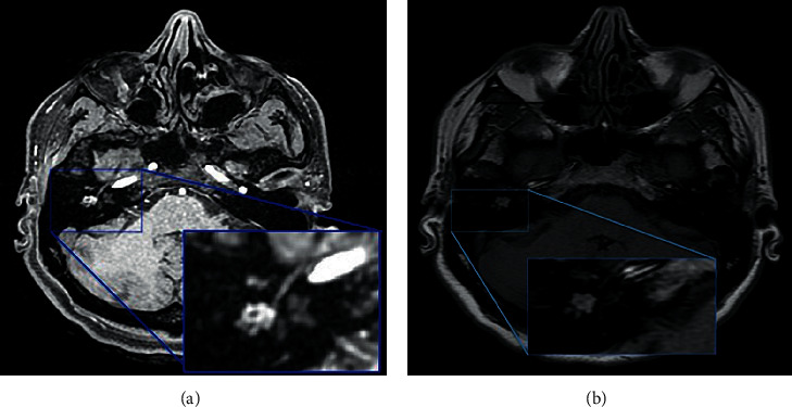
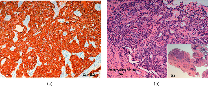
Neuroendocrine adenomas of the middle ear are rare tumors that represent less than 2% of primary tumors of the ear. In this paper, we describe a case of a 40-year-old woman who developed neuroendocrine adenoma of the middle ear. The specific management strategy for this rare tumor is unclear; information in the available literature on the management of this tumor is varied. However, an extensive demolition seems to be the gold standard treatment for this tumor to avoid recurrence and regional metastases in the lymph node or distant metastases. For the present case, we performed an incisional biopsy to confirm the diagnosis, and thereafter, we performed a canal-wall-down tympanoplasty. For cases like the present one, careful long-term clinical and instrumental follow-up is required to monitor progress and facilitate patient recovery.
Copyright © 2020 Luca Bruschini et al.
Conflict of interest statement
The authors declare that they have no conflicts of interest.
Figures


Similar articles
-
A rare case of middle ear adenoma.Nagoya J Med Sci. 2014 Aug;76(3-4):355-60. Nagoya J Med Sci. 2014. PMID: 25741045 Free PMC article.
-
Middle ear adenomatous neuroendocrine tumors: suggestion for surgical strategy.Braz J Otorhinolaryngol. 2022 Jan-Feb;88(1):83-88. doi: 10.1016/j.bjorl.2020.05.011. Epub 2020 Jun 15. Braz J Otorhinolaryngol. 2022. PMID: 32605830 Free PMC article.
-
Neuro-endocrine adenoma of the middle ear: a case study.Eur Arch Otorhinolaryngol. 2007 Dec;264(12):1525-8. doi: 10.1007/s00405-007-0390-1. Epub 2007 Jul 17. Eur Arch Otorhinolaryngol. 2007. PMID: 17639440
-
Middle Ear Neuroendocrine Tumor: A Case Report and Review of the Literature in Pediatric Population.J Int Adv Otol. 2021 Mar;17(2):150-155. doi: 10.5152/JIAO.2021.8491. J Int Adv Otol. 2021. PMID: 33893785 Free PMC article. Review.
-
Primary pleomorphic adenoma of the external ear canal. Report of a case and literature review.Am J Otolaryngol. 2008 Mar-Apr;29(2):142-6. doi: 10.1016/j.amjoto.2007.04.005. Am J Otolaryngol. 2008. PMID: 18314029 Review.
Cited by
-
Middle Ear Cavity and Mastoid Neuroendocrine Tumor Presenting as Otomastoiditis with Cholesteatoma: A Clinicoradiological and Histopathological Correlation.World J Nucl Med. 2023 Dec 26;22(4):310-315. doi: 10.1055/s-0043-1777695. eCollection 2023 Dec. World J Nucl Med. 2023. PMID: 38152101 Free PMC article.
-
Middle Ear Neuroendocrine Tumor Mimicking As Chronic Otitis Media.Cureus. 2023 Jul 22;15(7):e42296. doi: 10.7759/cureus.42296. eCollection 2023 Jul. Cureus. 2023. PMID: 37609079 Free PMC article.
-
A rare finding of pulmonary nodules in a middle ear neuroendocrine tumor: a case report and review of the literature.J Surg Case Rep. 2024 Oct 21;2024(10):rjae662. doi: 10.1093/jscr/rjae662. eCollection 2024 Oct. J Surg Case Rep. 2024. PMID: 39435307 Free PMC article.
References
-
- Ketabchi S., Massi D., Franchi A. Middle ear adenoma is an amphicrine tumor: why call it adenoma? Ultrastructural Pathology. 2001;25:73–78. - PubMed
Publication types
LinkOut - more resources
Full Text Sources
