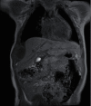
Chest Wall Mass in Infancy: The Presentation of Bone-Tumor-Like BCG Osteitis
- PMID: 33425419
- PMCID: PMC7780224
- DOI: 10.1155/2020/8884770
Chest Wall Mass in Infancy: The Presentation of Bone-Tumor-Like BCG Osteitis
Abstract
Chest wall mass in infancy is rare. Malignant lesions are more common than infection or benign tumors. This is a case of a 12-month-old girl who presented with a 2 cm mass at the right costal margin and poor weight gain. Chest radiograph demonstrated a moth-eaten osteolytic lesion at the 8th rib. The resection was performed, and a mass with pus content was found. The positive acid fast stain (AFB) organism was noted. Pathology confirmed caseous granulomatous inflammation compatible with mycobacterial infection. However, QuantiFERON-TB Gold was negative, so Mycobacterium bovis (M. bovis) osteitis is highly suspected. She was treated with antimycobacterium drugs and showed good results. Osteomyelitis can manifest by mimicking bone tumors. Without a biopsy, the pathogen may go undetected. So, interventions such as biopsy are warranted and avoid mass resection without indication. High C-reactive protein (CRP), alkaline phosphatase (ALP), periosteal reaction of radiating spicules, and penumbra sign in magnetic resonance imaging (MRI) are helpful for discriminating osteomyelitis from bone tumor.
Copyright © 2020 Phumin Chaweephisal et al.
Conflict of interest statement
The authors declare that they have no conflicts of interest.
Figures


Similar articles
-
Unusual cause of chest pain: empyema necessitans and tubercular osteomyelitis of the rib in an immunocompetent man.BMJ Case Rep. 2016 Jan 4;2016:bcr2015212311. doi: 10.1136/bcr-2015-212311. BMJ Case Rep. 2016. PMID: 26729824 Free PMC article.
-
Clinical Manifestations, Management, and Outcomes of Osteitis/Osteomyelitis Caused by Mycobacterium bovis Bacillus Calmette-Guérin in Children: Comparison by Site(s) of Affected Bones.J Pediatr. 2019 Apr;207:97-102. doi: 10.1016/j.jpeds.2018.11.042. Epub 2018 Dec 18. J Pediatr. 2019. PMID: 30577978
-
Chest wall pseudotumor: a case of non-tuberculous mycobacterial infection.BMC Infect Dis. 2021 Feb 19;21(1):196. doi: 10.1186/s12879-021-05843-z. BMC Infect Dis. 2021. PMID: 33607951 Free PMC article.
-
Imaging findings of vertebral osteomyelitis caused by nontuberculous mycobacterial organisms: Three case reports and literature review.Medicine (Baltimore). 2022 Jun 17;101(24):e29395. doi: 10.1097/MD.0000000000029395. Medicine (Baltimore). 2022. PMID: 35713445 Free PMC article. Review.
-
Current and Emerging Therapies for HER2-Positive Women With Metastatic Breast Cancer.J Adv Pract Oncol. 2017 Mar;8(2):164-168. Epub 2017 Mar 1. J Adv Pract Oncol. 2017. PMID: 29900024 Free PMC article. Review.
References
Publication types
LinkOut - more resources
Full Text Sources
Research Materials
Miscellaneous

