Review
. 2020 Nov 13;20(6):e44.
doi: 10.4110/in.2020.20.e44. eCollection 2020 Dec.

Recent Insights into Cellular Crosstalk in Respiratory and Gastrointestinal Mucosal Immune Systems

Affiliations
Review

Recent Insights into Cellular Crosstalk in Respiratory and Gastrointestinal Mucosal Immune Systems

Sae-Hae Kim et al. Immune Netw. .

Abstract

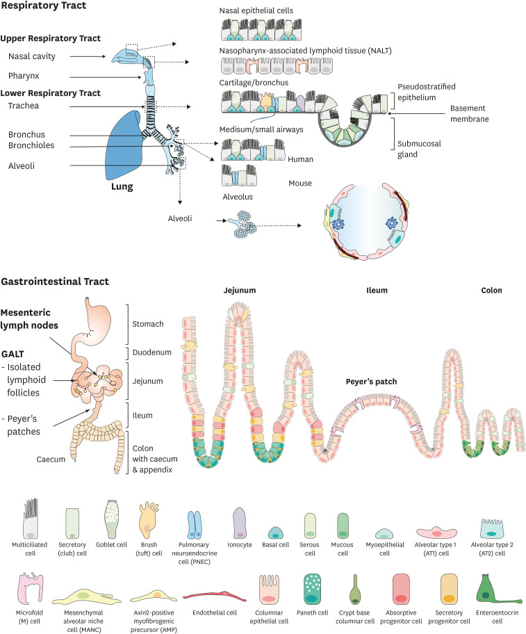
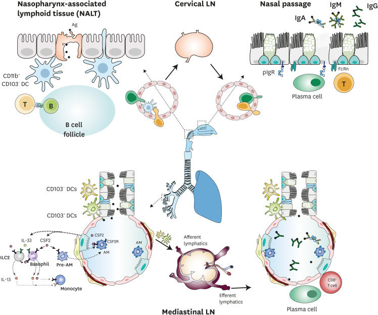
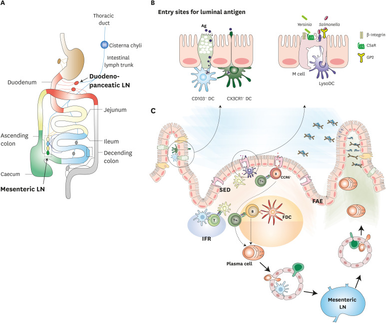
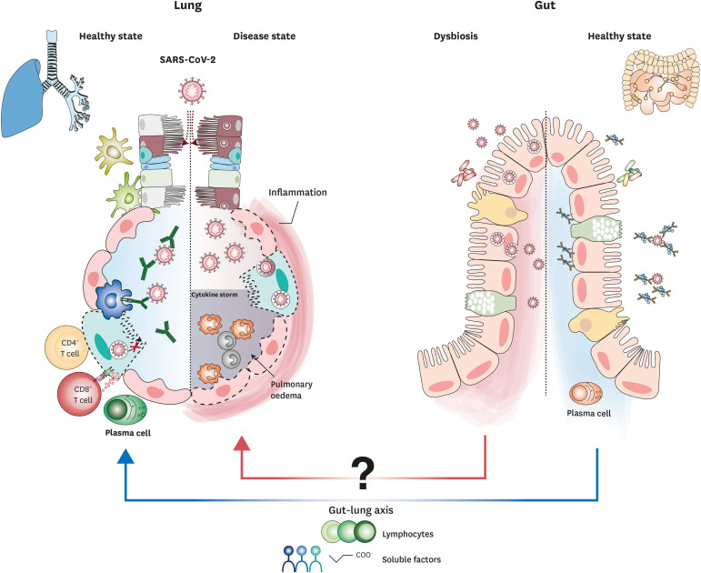
The human body is continuously threatened by pathogens, and the immune system must maintain a balance between fighting infection and becoming over-activated. Mucosal surfaces cover several anatomically diverse organs throughout the body, such as the respiratory and gastrointestinal tracts, and are directly exposed to the external environment. Various pathogens invade the body through mucosal surfaces, making the mucosa the frontline of immune defense. The immune systems of various mucosal tissues display distinctive features that reflect the tissues' anatomical and functional characteristics. This review discusses the cellular components that constitute the respiratory and gastrointestinal tracts; in particular, it highlights the complex interactions between epithelial and immune cells to induce Ag-specific immune responses in the lung and gut. This information on mucosal immunity may facilitate understanding of the defense mechanisms against infectious agents that invade mucosal surfaces, such as severe acute respiratory syndrome coronavirus 2, and provide insight into effective vaccine development.

Keywords: Gastrointestinal tract; Infection; Mucosal immunity; Respiratory tract.


Conflict of interest statement

Conflict of Interest: The authors declare no potential conflicts of interest.

Figures

Figure 1
Figure 1
Cellular composition of the respiratory and gastrointestinal tracts. (A) The respiratory tract consists of the upper (nasal cavity, pharynx) and lower (trachea, bronchus, bronchioles, alveoli) tracts. They are covered by a single layer of epithelial cells, such as multiciliated cells, club cells, goblet cells, basal cells, PNECs, tuft cells, ionocytes, serous cells, myoepithelial cells, AT1 and AT2 cells, mesenchymal alveolar niche cells, and AMPs. (B) The small and large intestines of the gastrointestinal tract are distinguished by the crypt-villus structure and are composed of columnar epithelial, microfold, Paneth, goblet, enteroendocrine, and tuft cells. AMP, axin2-positive myofibrogenic precursor.
Figure 2
Figure 2
Induction of mucosal immune responses in the respiratory tract. In the NALT, nasal DCs take up Ags via M cells and their dendrites, and then migrate to B cell follicles and CLNs. Ag-specific effector cells are recruited to the nasal passage, where they secrete Ag-specific IgG, IgA, and IgM. In the lower respiratory tract, Ag-stimulated DCs migrate to mediastinal LNs and initiate the Ag-specific immune response via activation of naïve T cells. Effector cells then home to alveolar sacs.
Figure 3
Figure 3
Induction of mucosal immune responses in the gastrointestinal tract. (A) Lymph from mucosal lymphatics collects for transport to the draining LNs. (B) Luminal Ags and pathogens, such as Yersinia and Salmonella, can be delivered to PP by M cells. This is facilitated by GP2, C5aR, and integrin β1. Close to the M cells, Ags encounter lysozyme-expressing DCs. Luminal soluble Ags can be also transported into the LP via both the goblet cell-associated Ag passages of goblet cells and extending dendrites from CX3CR1+ cells. (C) In PPs, the Ag-specific immune response was initiated in the GC, SED of PPs, and MLNs. Ag-specific effector cells then home to LP.
Figure 4
Figure 4
Hypothetical immunity in the lung and gut during SARS-CoV-2 infection. SARS-CoV-2 infects both ACE2- and TMPRSS2-expressing epithelial cells, including ciliated cells, AT2 cells, and enterocytes. In mucosal compartments, induction of the adaptive immune response can promote viral clearance.

References

    1. Brandtzaeg P, Kiyono H, Pabst R, Russell MW. Terminology: nomenclature of mucosa-associated lymphoid tissue. Mucosal Immunol. 2008;1:31–37. - PubMed
    1. Macpherson AJ, McCoy KD, Johansen FE, Brandtzaeg P. The immune geography of IgA induction and function. Mucosal Immunol. 2008;1:11–22. - PubMed
    1. Phalipon A, Corthésy B. Novel functions of the polymeric Ig receptor: well beyond transport of immunoglobulins. Trends Immunol. 2003;24:55–58. - PubMed
    1. Michaud E, Mastrandrea C, Rochereau N, Paul S. Human secretory IgM: an elusive player in mucosal immunity. Trends Immunol. 2020;41:141–156. - PubMed
    1. Heron M, Grutters JC, ten Dam-Molenkamp KM, Hijdra D, van Heugten-Roeling A, Claessen AM, Ruven HJ, van den Bosch JM, van Velzen-Blad H. Bronchoalveolar lavage cell pattern from healthy human lung. Clin Exp Immunol. 2012;167:523–531. - PMC - PubMed