

Onyx Embolization of a Distal Middle Cerebral Artery Pseudoaneurysm in a Five-Week-Old: A Case Report and Review of Current Treatment Options
- PMID: 33425546
- PMCID: PMC7790325
- DOI: 10.7759/cureus.11974
Onyx Embolization of a Distal Middle Cerebral Artery Pseudoaneurysm in a Five-Week-Old: A Case Report and Review of Current Treatment Options
Abstract
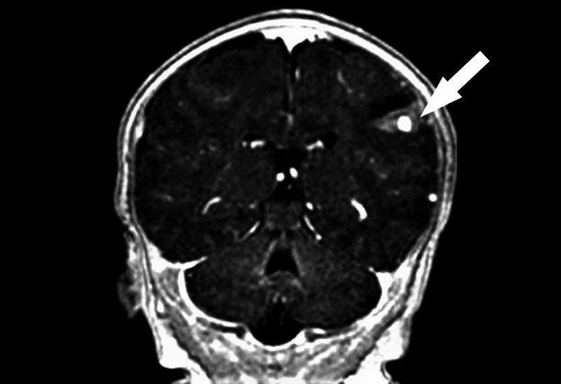
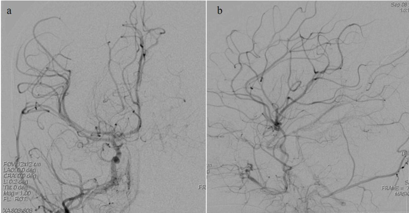
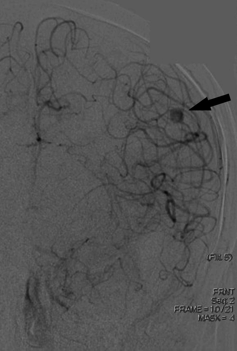
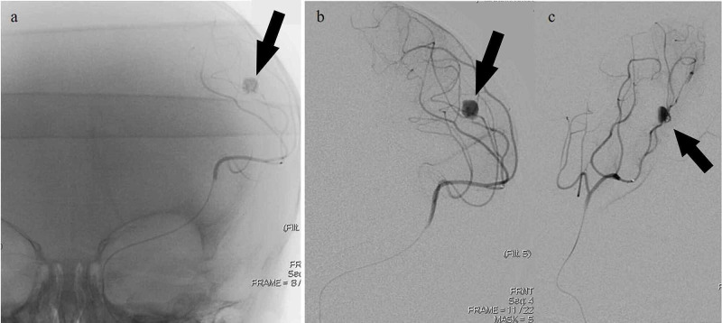
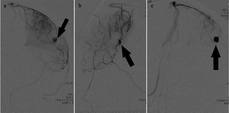
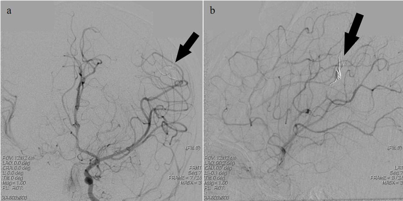
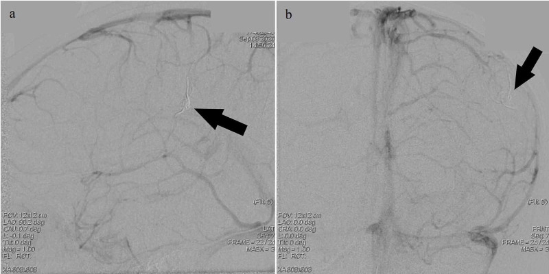
Intracranial pseudoaneurysms secondary to traumatic birth are a rare finding in infants. Definitive diagnosis of such findings is challenging, and no standard management is delineated for management of pseudoaneurysms in the pediatric population. Commonly attempted treatments include endovascular embolization or surgical clipping. A 5-week-old female presented with a two day history of right hand focal seizures. The patient was found to have a dysplastic superficial intra-axial aneurysm arising from the distal left middle cerebral artery (MCA) branch in the setting of a left posterior frontal lobe hemorrhage noted on brain magnetic resonance imaging/magnetic resonance angiography (MRI/MRA). The patient underwent diagnostic cerebral angiogram demonstrating a left distal MCA pseudoaneurysm, which was treated with Onyx embolization. Post-embolization period was complicated by recurrent left central localized seizures and a left hemispheric temporoparietal hemorrhagic infarction. The patient was managed on levetiracetam, phenytoin, phenobarbital with stable seizure control. Herein, we highlight the youngest case to date of a 5-week-old infant with a left distal MCA pseudoaneurysm treated with Onyx embolization. Pseudoaneurysmal incidence, diagnosis and accepted management is discussed.
Keywords: middle cerebral artery infarct; pediatric vascular malformation; pseudoaneurysm.
Copyright © 2020, Elsawaf et al.
Conflict of interest statement
The authors have declared that no competing interests exist.
Figures
References
-
- Pediatric intracranial pseudoaneurysms: a report of 15 cases and review of the literature. Chen R, Zhang S, Guo R, You C, Ma L. World Neurosurg. 2018;116:951–959. - PubMed
-
- Dorsal internal carotid artery aneurysms with special reference to angiographic presentation and surgical management. Shigeta H, Kyoshima K, Nakagawa F, Kobayashi S. Acta Neurochir. 1992;119:42–48. - PubMed
-
- Surgical management of traumatic intracranial pseudoaneurysms: a report of 12 cases. Wang X, Chen JX, You C, He M. https://pubmed.ncbi.nlm.nih.gov/18310837/ Neurol India. 2008;56:47–51. - PubMed
-
- Flow diverter treatment of intracranial vertebral artery dissecting pseudoaneurysms. Cerejo R, Bain M, Moore N, et al. J Neurointerv Surg. 2017;9:1064–1068. - PubMed
Publication types
LinkOut - more resources
Full Text Sources