

Dolichoectasia and Its Diagnostic Criteria: A Case Report and Literature Review
- PMID: 33425563
- PMCID: PMC7788005
- DOI: 10.7759/cureus.12516
Dolichoectasia and Its Diagnostic Criteria: A Case Report and Literature Review
Abstract
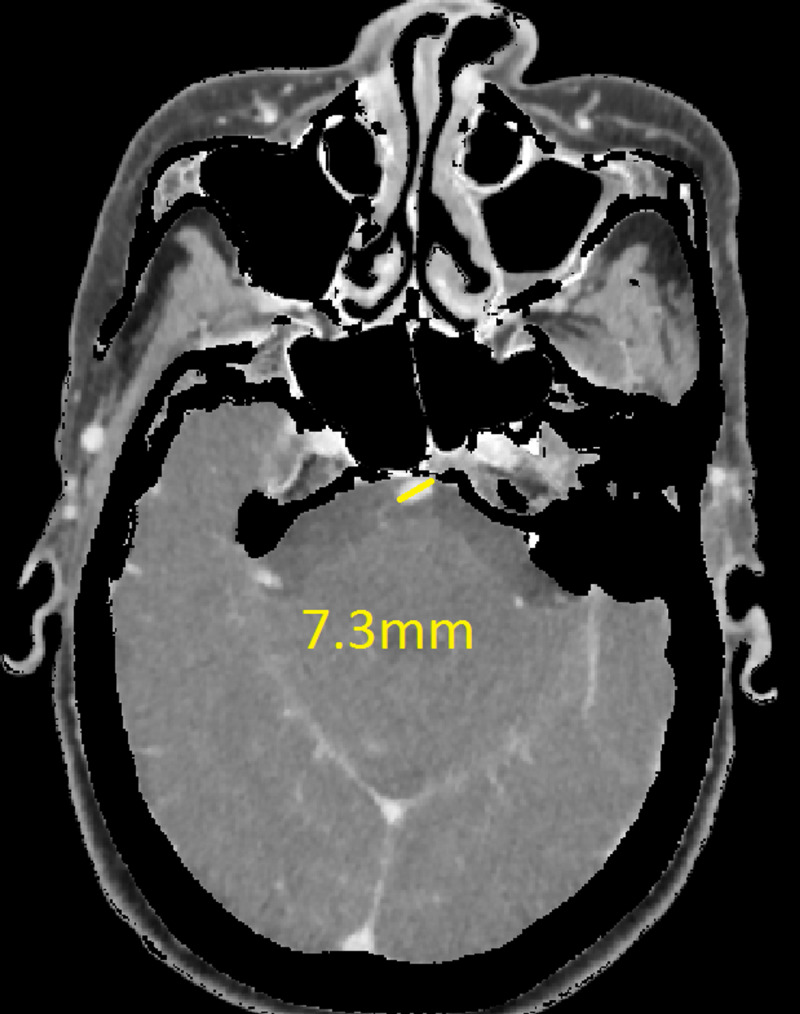
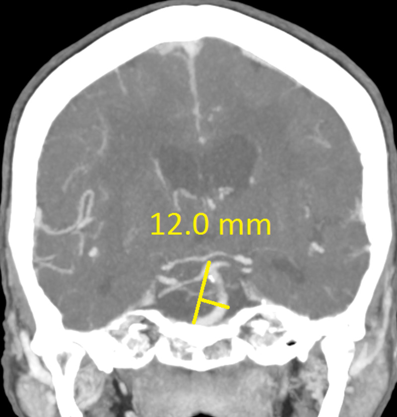
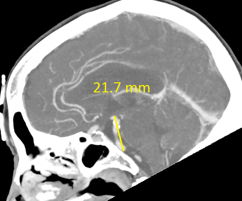
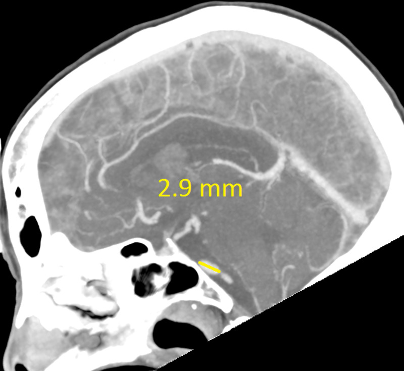
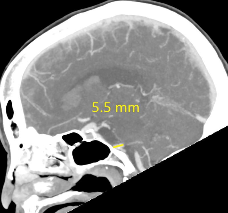
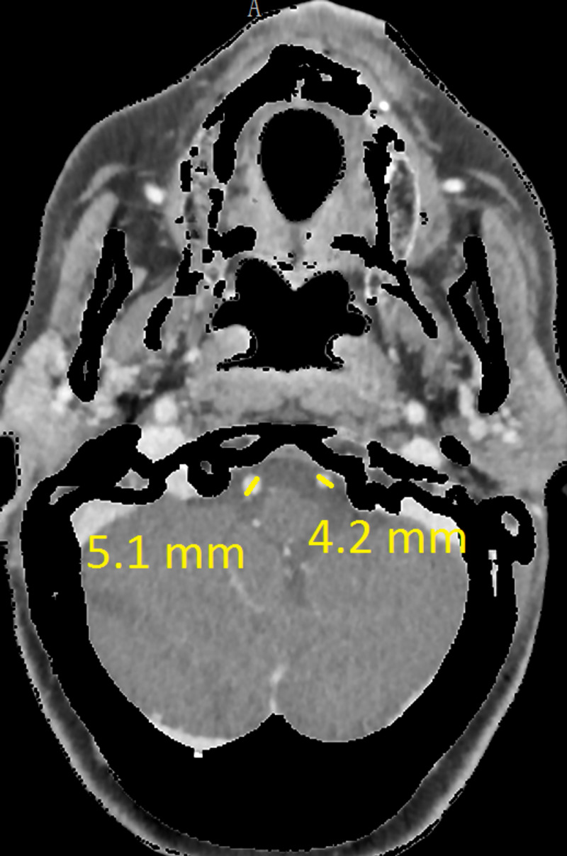
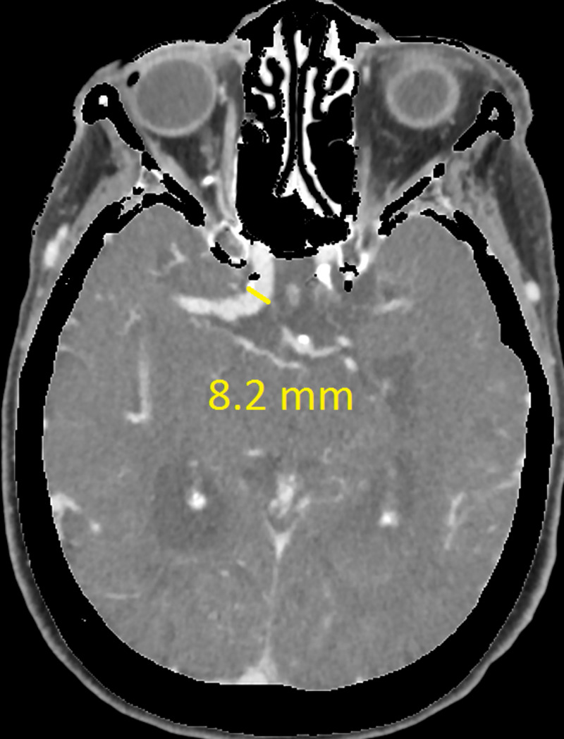
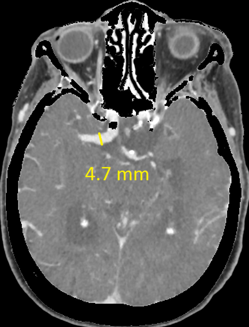
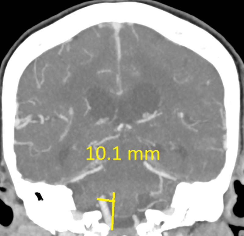
Dolichoectasia (DE) is a rare disorder of cerebral vasculature and involves dilation and elongation of the blood vessels. It is mostly reported in the vertebrobasilar circulation, but it can occur in the anterior circulation. This report describes a case involving both anterior and posterior vessel dilation with the suspicion of DE. Here the vessels were enlarged - but not grossly - as in some cases where the diagnosis is obvious. Thus a closer look had to be taken. We refer to multiple studies that attempt to provide some guideline for diagnosis assisting us with our assessment. This illustrates the importance of objective evaluation to prevent missing important pathologies that can change treatment and prognosis if identified.
Keywords: aneurysm; dolichoectasia; vasculopathy.
Copyright © 2021, Conradie et al.
Conflict of interest statement
The authors have declared that no competing interests exist.
Figures
References
-
- Dilatative arteriopathy (dolichoectasia): what is known and not known. Caplan LR. Ann Neurol. 2005;57:469–471. - PubMed
-
- Vertebrobasilar dolichoectasia diagnosed by magnetic resonance angiography and risk of stroke and death: a cohort study. Ubogu E, Zaidat O. https://jnnp.bmj.com/content/75/1/22.short Neurol Neurosurg Psychiatry. 2004:22–26. - PMC - PubMed
-
- Basilar and bilateral carotid dolichoectasia with spontaneous dissection of C2 segment of the internal carotid artery. Borota L, Jonasson P. http://www.ajnr.org/content/27/6/1241.short. AJNR Am J Neuroradiol. 2006;27:1241–1244. - PMC - PubMed
Publication types
LinkOut - more resources
Full Text Sources
Other Literature Sources