

Basal Cell Carcinoma with Sebaceous Differentiation: A Case Report and Review of Literature
- PMID: 33425577
- PMCID: PMC7787308
- DOI: 10.1097/GOX.0000000000003234
Basal Cell Carcinoma with Sebaceous Differentiation: A Case Report and Review of Literature
Abstract
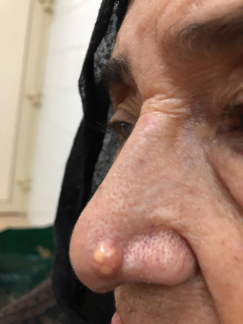
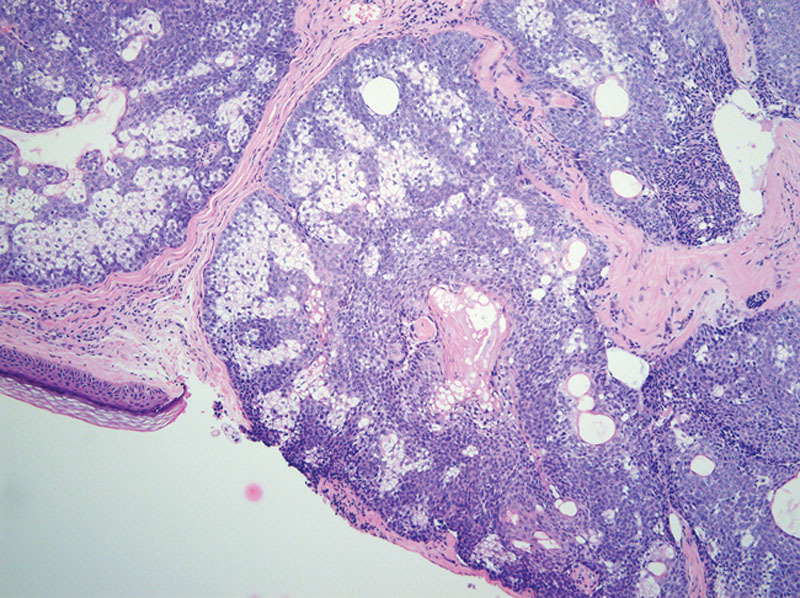
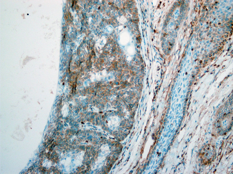
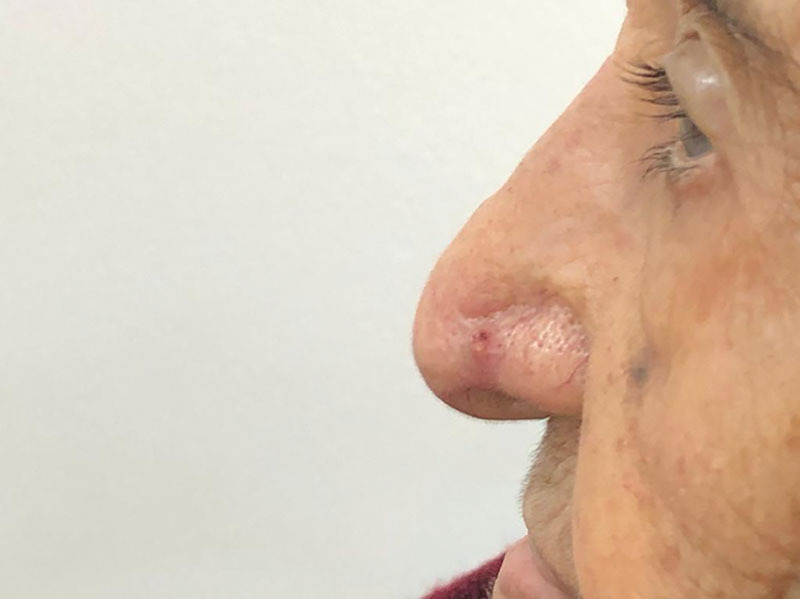
We report a very rare type of tumor in the left nasal ala in an elderly patient. An 81-year-old Saudi woman known to have hypertension, osteoporosis, and rheumatoid disease (who had been compliant to her medications) presented with a 0.5-cm fixed, firm, round well-defined nodule on the left ala of the nose (with crusting, erosion, and telangiectasia of the overlying skin), whose size had been gradually increasing for 2 years. The patient underwent excisional biopsy, and the specimen was sent for a histopathologic analysis. Macroscopic examination showed a round tan-white homogenous nodule, measuring 0.6 × 0.5 × 0.5 cm3. Microscopic examination revealed a fairly circumscribed unencapsulated dermal lesion, featuring basaloid cells with peripheral palisading, and focal stromal clefting. The final diagnosis of basal cell carcinoma with sebaceous differentiation was made. The patient was managed with Mohs surgery with clear margins, and full-thickness skin graft was done. Four months after surgery, the patient had a recurrence, which was managed with a surgical excision (with 4-mm margin) and covered by a full-thickness skin graft.
Copyright © 2020 The Authors. Published by Wolters Kluwer Health, Inc. on behalf of The American Society of Plastic Surgeons.
Conflict of interest statement
Figures
Similar articles
-
Cutaneous leiomyosarcoma, trichoblastoma, and syringocystadenoma papilliferum arising from nevus sebaceus.Int J Dermatol. 2007 Mar;46(3):306-8. doi: 10.1111/j.1365-4632.2007.03151.x. Int J Dermatol. 2007. PMID: 17343592
-
Muir-Torre syndrome: a case of this uncommon entity.Int J Dermatol. 2006 Mar;45(3):311-3. doi: 10.1111/j.1365-4632.2006.01797.x. Int J Dermatol. 2006. PMID: 16533237
-
A case of sebaceous adenoma of the eyelid showing excessively rapid growth.Clin Ophthalmol. 2013;7:667-70. doi: 10.2147/OPTH.S42135. Epub 2013 Apr 3. Clin Ophthalmol. 2013. PMID: 23579539 Free PMC article.
-
Sebaceous carcinoma in children.J Am Acad Dermatol. 2002 Dec;47(6):950-3. doi: 10.1067/mjd.2002.114615. J Am Acad Dermatol. 2002. PMID: 12451386 Review.
-
Role of In Vivo Reflectance Confocal Microscopy in the Analysis of Melanocytic Lesions.Acta Dermatovenerol Croat. 2018 Apr;26(1):64-67. Acta Dermatovenerol Croat. 2018. PMID: 29782304 Review.
Cited by
-
Sebaceous Hyperplasia of the Face: A Case Report.Cureus. 2024 Oct 10;16(10):e71196. doi: 10.7759/cureus.71196. eCollection 2024 Oct. Cureus. 2024. PMID: 39525119 Free PMC article.
-
Basal cell carcinoma: Comprehensive clinical and histopathological aspects, novel imaging tools and therapeutic approaches (Review).Exp Ther Med. 2022 Jan;23(1):60. doi: 10.3892/etm.2021.10982. Epub 2021 Nov 18. Exp Ther Med. 2022. PMID: 34917186 Free PMC article. Review.
References
-
- Bickers DR, Lim HW, Margolis D, et al. American Academy of Dermatology Association; Society for Investigative Dermatology. The burden of skin diseases: 2004 a joint project of the American Academy of Dermatology Association and the Society for Investigative Dermatology. J Am Acad Dermatol. 2006;55:490–500. - PubMed
-
- Rubin AI, Chen EH, Ratner D. Basal-cell carcinoma. N Engl J Med. 2005;353:2262–2269. - PubMed
-
- Misago N, Mihara I, Ansai S, et al. Sebaceoma and related neoplasms with sebaceous differentiation: a clinicopathologic study of 30 cases. Am J Dermatopathol. 2002;24:294–304. - PubMed
Publication types
LinkOut - more resources
Full Text Sources