Review
. 2020 Dec 9:8:529244.
doi: 10.3389/fbioe.2020.529244. eCollection 2020.

Biomaterials in Valvular Heart Diseases

Affiliations
Review

Biomaterials in Valvular Heart Diseases

Bita Taghizadeh et al. Front Bioeng Biotechnol. .

Abstract

Valvular heart disease (VHD) occurs as the result of valvular malfunction, which can greatly reduce patient's quality of life and if left untreated may lead to death. Different treatment regiments are available for management of this defect, which can be helpful in reducing the symptoms. The global commitment to reduce VHD-related mortality rates has enhanced the need for new therapeutic approaches. During the past decade, development of innovative pharmacological and surgical approaches have dramatically improved the quality of life for VHD patients, yet the search for low cost, more effective, and less invasive approaches is ongoing. The gold standard approach for VHD management is to replace or repair the injured valvular tissue with natural or synthetic biomaterials. Application of these biomaterials for cardiac valve regeneration and repair holds a great promise for treatment of this type of heart disease. The focus of the present review is the current use of different types of biomaterials in treatment of valvular heart diseases.

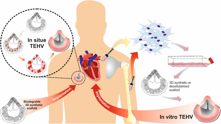
Keywords: biomaterials; cardiac valve regeneration; heart valve replacement; tissue-engineered heart valves; valvular heart diseases.


Conflict of interest statement

The authors declare that the research was conducted in the absence of any commercial or financial relationships that could be construed as a potential conflict of interest.

Figures

FIGURE 1
FIGURE 1
Design and development of biomaterials used in heart valves.
FIGURE 2
FIGURE 2
(A) Schematic micro-structured Nickle-Titanium (NiTi) thin film as a matrix scaffold for tissue engineered hybrid heart valves. (a) Formation of 3D valve leaflets by a steel mold. (b) Fabricated NiTi film valve leaflets. Reprinted with permission from Loger et al. (2016). Copyright (2016), Springer Nature. (B) Leaflets and stents assembly for fabricating heart valves (left), Top view of each six HVs with different arch profiles. Reprinted with permission from Yousefi et al. (2017). Copyright (2017), Springer Nature.
FIGURE 3
FIGURE 3
Expandable polytetrafluoroethylene (ePTFE) was used to fabricate the leaflets of the prototype, which were sewed to a stent made from stainless steel. (A) An arterial valve based on ePTFE located in the heart. (B) Photograph of implanted valve’s outflow surface. (C) The method of device sectioning after performing plastic embedding. (D–F) Valve expansion geometry for the primary valve geometry, X-ray images of laser-cut stainless steel functional valve prototypes being expanded via serial balloon dilation and representative right ventricular angiograms in a lamb. (G–I) Valve expansion geometry for the primary valve geometry and functional prototype, in vitro flow loop testing of functional prototype at two polar expansion states, and representative right ventricular and pulmonary artery pressures recorded at two states of valve expansion. Reprinted with permission from Hofferberth et al. (2020). Copyright (2020), American Association for the advancement of Science.
FIGURE 4
FIGURE 4
Three types of bioprosthetic valve replacements: stented, stentless, and percutaneous.
FIGURE 5
FIGURE 5
TEHV approaches comparison: in situ vs. in vitro.
FIGURE 6
FIGURE 6
An engineered heterogeneous valve scaffold with poly-ethylene glycol-diacrylate (PEG-DA) hydrogels/alginate, supplemented with porcine aortic valve interstitial cells (PAVIC) by 3D-printing/photocrosslinking technique (a) 3D Printer setup of heterogeneous valve scaffolds. (b–f) Axisymmetric valve STL file of valve, micro-CT scan of aortic valve, the leaflet and root regions, the printable STL geometries of the threshold regions and the printing software sliced the geometries into layers and generated extrusion paths for each layer. Reprinted with permission from Hockaday et al. (2012). Copyright (2012), IOP science Publishing Ltd.

References

    1. Aagaard J. (2004). The carbomedics aortic heart valve prosthesis: a review. J. Cardiovasc. Surg. 45 531–534. - PubMed
    1. Akins R. J. (1979). Nickel-Titanium Alloy, Heart Valve. Prosthetic device couplings, 4233690A.
    1. Alferiev I., Vyavahare N., Song C., Connolly J., Hinson J. T., Lu Z., et al. (2001). Bisphosphonate derivatized polyurethanes resist calcification. Biomaterials 22 2683–2693. 10.1016/S0142-9612(01)00010-2 - DOI - PubMed
    1. Alves P., Cardoso R., Correia T. R., Antunes B. P., Correia I. J., Ferreira P. (2014). Surface modification of polyurethane films by plasma and ultraviolet light to improve haemocompatibility for artificial heart valves. Colloids Surf. B Biointerfaces 113 25–32. 10.1016/j.colsurfb.2013.08.039 - DOI - PubMed
    1. Andersen H. R. (2009). History of percutaneous aortic valve prosthesis. Herz 34 343–346. 10.1007/s00059-009-3251-4 - DOI - PubMed