

Biogenic Silver Nanoparticles Synthesized by Lysinibacillus xylanilyticus MAHUQ-40 to Control Antibiotic-Resistant Human Pathogens Vibrio parahaemolyticus and Salmonella Typhimurium
- PMID: 33425864
- PMCID: PMC7793659
- DOI: 10.3389/fbioe.2020.597502
Biogenic Silver Nanoparticles Synthesized by Lysinibacillus xylanilyticus MAHUQ-40 to Control Antibiotic-Resistant Human Pathogens Vibrio parahaemolyticus and Salmonella Typhimurium
Abstract
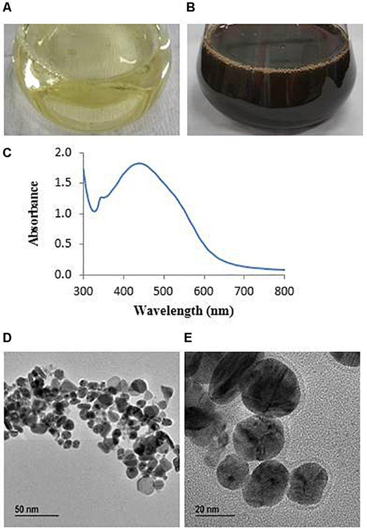
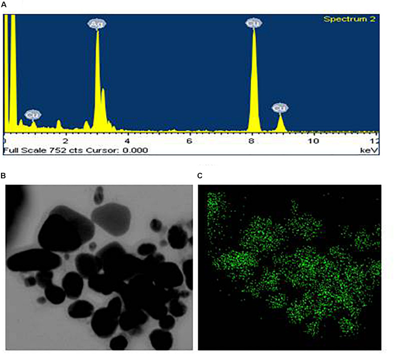
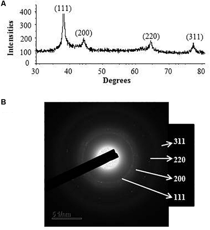
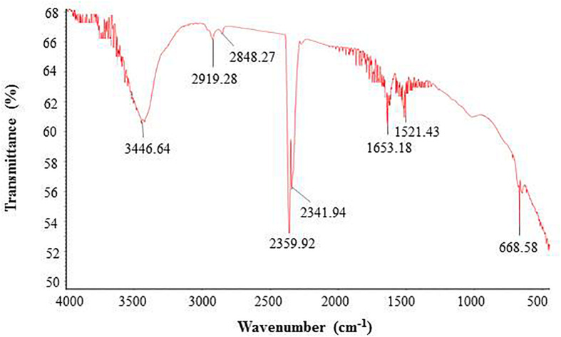
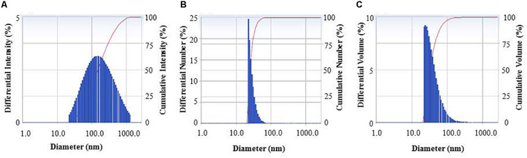
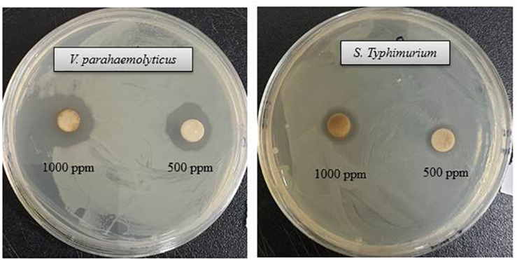
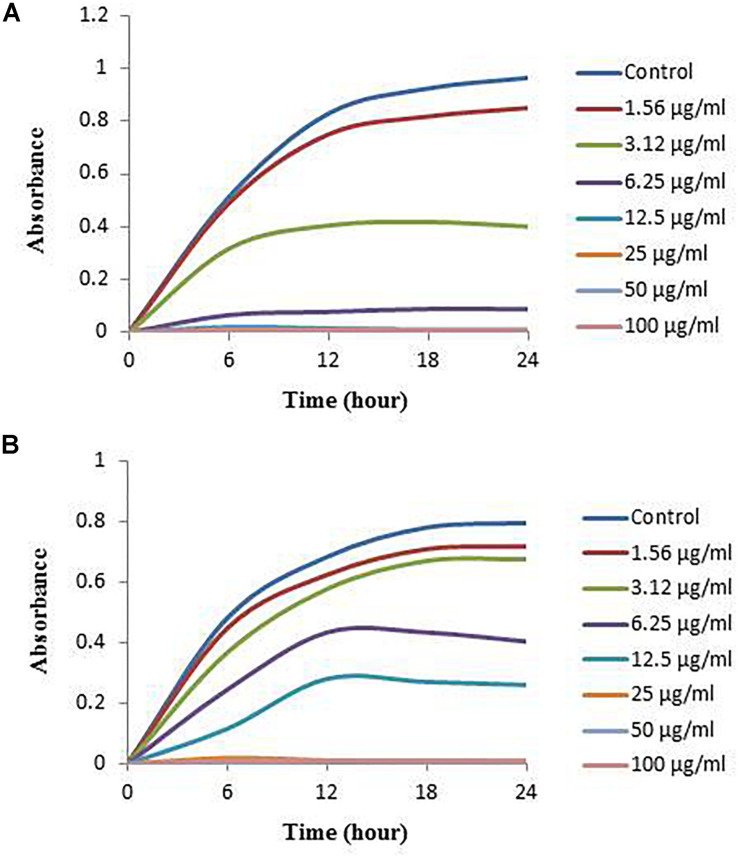
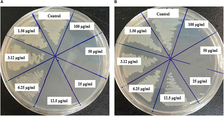
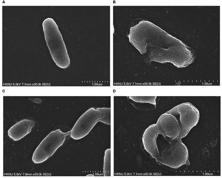
The present study highlights a simple and eco-friendly method for the biosynthesis of silver nanoparticles (AgNPs) using Lysinibacillus xylanilyticus strain MAHUQ-40. Also, the synthesized AgNPs were used to investigate their antibacterial activity and mechanisms against antibiotic-resistant pathogens. Biosynthesis of AgNPs was confirmed by ultraviolet-visible spectroscopy, and then, they were characterized by field emission-transmission electron microscopy (FE-TEM), X-ray diffraction (XRD), dynamic light scattering (DLS), and fourier transform-infrared (FTIR). The toxicity of AgNPs against two pathogenic bacteria was evaluated. The UV-vis spectral scanning showed the peak for synthesized AgNPs at 438 nm. Under FE-TEM, the synthesized AgNPs were spherical with diameter ranges from 8 to 30 nm. The XRD analysis revealed the crystallinity of synthesized AgNPs. FTIR data showed various biomolecules including proteins and polysaccharides that may be involved in the synthesis and stabilization of AgNPs. The resultant AgNPs showed significant antibacterial activity against tested pathogens. The MICs (minimum inhibitory concentrations) and MBCs (minimum bactericidal concentrations) of the AgNPs synthesized by strain MAHUQ-40 were 3.12 and 12.5 μg/ml, respectively, against Vibrio parahaemolyticus and 6.25 and 25 μg/ml, respectively, against Salmonella Typhimurium. FE-TEM analysis showed that the biogenic AgNPs generated structural and morphological changes and damaged the membrane integrity of pathogenic bacteria. Our findings showed the potentiality of L. xylanilyticus MAHUQ-40 to synthesis AgNPs that acted as potent antibacterial material against pathogenic bacterial strains.
Keywords: AgNPs; Lysinibacillus xylanilyticus MAHUQ-40; antimicrobial activity; eco-friendly synthesis; human pathogens.
Copyright © 2020 Huq.
Conflict of interest statement
The author declares that the research was conducted in the absence of any commercial or financial relationships that could be construed as a potential conflict of interest.
Figures

References
-
- Abdel-Raouf N., Al-Enazi N. M., Ibraheem I. B. (2017). Green biosynthesis of gold nanoparticles using Galaxaura elongata and characterization of their antibacterial activity. Arab. J. Chem. 10(Suppl. 2) S3029–S3039. 10.1016/j.arabjc.2013.11.044 - DOI
-
- Akter S., Huq M. A. (2020). Biologically rapid synthesis of silver nanoparticles by Sphingobium sp. MAH-11T and their antibacterial activity and mechanisms investigation against drug-resistant pathogenic microbes. Artif. Cells Nanomed. Biotechnol. 48 672–682. 10.1080/21691401.2020.1730390 - DOI - PubMed
-
- Ali D. M., Sasikala M., Gunasekaran M., Thajuddin N. (2011). Biosynthesis and characterization of silver nanoparticles using marine cyanobacterium, Oscillatoria willei NTDM01. Dig. J. Nanomater. Biostruct. 6 385–390.
-
- Ansari M. A., Baykal A., Asiri S. (2018). Synthesis and characterization of antibacterial activity of spinel chromium-substituted copper ferrite nanoparticles for biomedical application. J. Inorg. Organomet. Polym. Mater. 28 2316–2327. 10.1007/s10904-018-0889-5 - DOI
LinkOut - more resources
Full Text Sources
Molecular Biology Databases