. 2020 Dec 14:8:603598.
doi: 10.3389/fbioe.2020.603598. eCollection 2020.

hBMSC-Derived Extracellular Vesicles Attenuate IL-1β-Induced Catabolic Effects on OA-Chondrocytes by Regulating Pro-inflammatory Signaling Pathways

Affiliations

hBMSC-Derived Extracellular Vesicles Attenuate IL-1β-Induced Catabolic Effects on OA-Chondrocytes by Regulating Pro-inflammatory Signaling Pathways

Shushan Li et al. Front Bioeng Biotechnol. .

Abstract

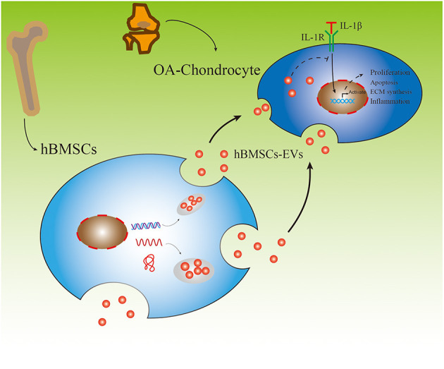
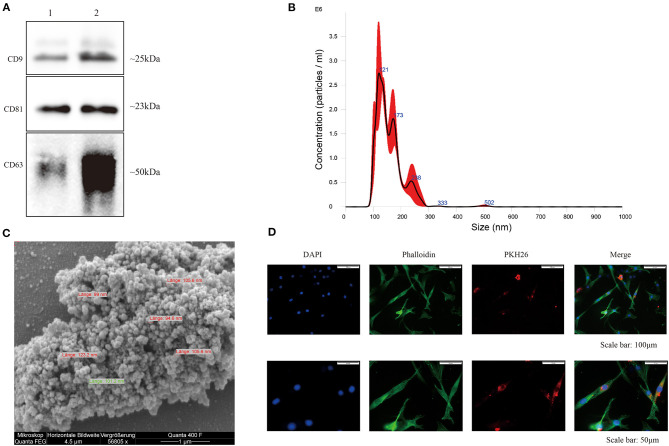
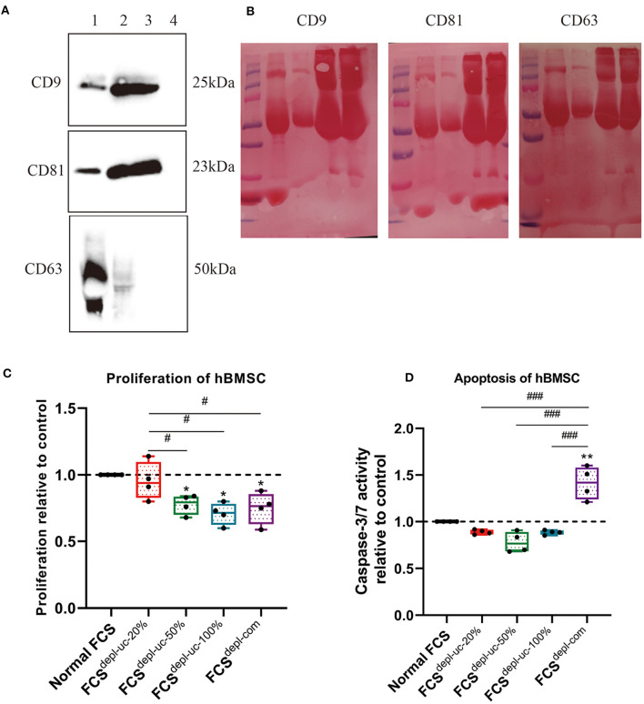
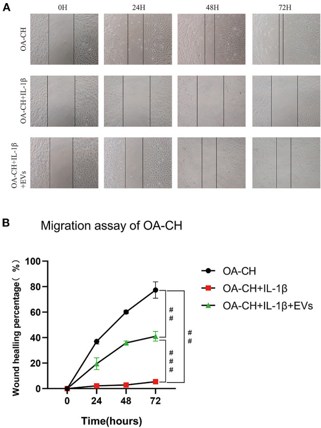
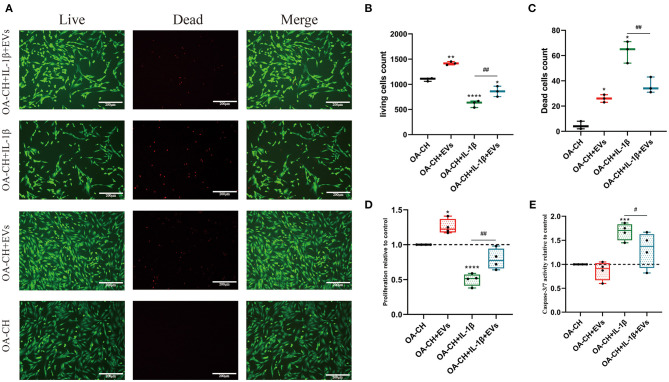
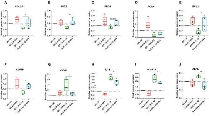
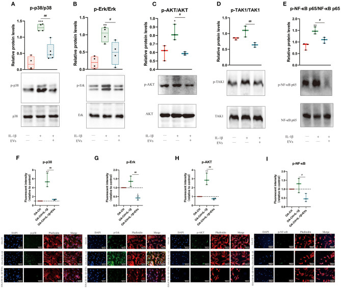
Background: Human bone marrow-derived mesenchymal stromal cells (hBMSCs) provide a promising therapeutic approach in the cell-based therapy of osteoarthritis (OA). However, several disadvantages evolved recently, including immune responses of the host and regulatory hurdles, making it necessary to search for alternative treatment options. Extracellular vesicles (EVs) are released by multiple cell types and tissues into the extracellular microenvironment, acting as message carriers during intercellular communication. Here, we investigate putative protective effects of hBMSC-derived EVs as a cell-free approach, on IL-1β-stimulated chondrocytes obtained from OA-patients. Methods: EVs were harvested from the cell culture supernatant of hBMSCs by a sequential ultracentrifugation process. Western blot, scanning electron microscopy (SEM), and nanoparticle tracking analysis (NTA) were performed to characterize the purified particles as EVs. Intracellular incorporation of EVs, derived from PHK26-labeled hBMSCs, was tested by adding the labeled EVs to human OA chondrocytes (OA-CH), followed by fluorescence microscopy. Chondrocytes were pre-stimulated with IL-1β for 24 h, followed by EVs treatment for 24 h. Subsequently, proliferation, apoptosis, and migration (wound healing) were analyzed via BrdU assay, caspase 3/7 assay, and scratch assay, respectively. With qRT-PCR, the relative expression level of anabolic and catabolic genes was determined. Furthermore, immunofluorescence microscopy and western blot were performed to evaluate the protein expression and phosphorylation levels of Erk1/2, PI3K/Akt, p38, TAK1, and NF-κB as components of pro-inflammatory signaling pathways in OA-CH. Results: EVs from hBMSCs (hBMSC-EVs) promote proliferation and reduce apoptosis of OA-CH and IL-1β-stimulated OA-CH. Moreover, hBMSC-EVs attenuate IL-1β-induced reduction of chondrocyte migration. Furthermore, hBMSC-EVs increase gene expression of PRG4, BCL2, and ACAN (aggrecan) and decrease gene expression of MMP13, ALPL, and IL1ß in OA-CH. Notably, COL2A1, SOX9, BCL2, ACAN, and COMP gene expression levels were significantly increased in IL-1β+ EV groups compared with those IL-1β groups without EVs, whereas the gene expression levels of COLX, IL1B, MMP13, and ALPL were significantly decreased in IL-1β+ EV groups compared to IL-1β groups without EVs. In addition, the phosphorylation status of Erk1/2, PI3K/Akt, p38, TAK1, and NF-κB signaling molecules, induced by IL-1β, is prevented by hBMSC- EVs. Conclusion: EVs derived from hBMSCs alleviated IL-1β-induced catabolic effects on OA-CH via promoting proliferation and migration and reducing apoptosis, probably via downregulation of IL-1ß-activated pro-inflammatory Erk1/2, PI3K/Akt, p38, TAK1, and NF-κB signaling pathways. EVs released from BMSCs may be considered as promising cell-free intervention strategy in cartilage regenerative medicine, avoiding several adverse effects of cell-based regenerative approaches.

Keywords: IL-1ß; chondrocytes; extracellular vesicles; hBMSC; osteoarthritis; signaling pathways.


Conflict of interest statement

The authors declare that the research was conducted in the absence of any commercial or financial relationships that could be construed as a potential conflict of interest.

Figures

Figure 1
Figure 1
Experimental outline of responses of IL-1β-induced osteoarthritic chondrocytes (OA-CH) treated with hBMSC-derived EVs (hBMSC-EVs).
Figure 2
Figure 2
Characterization of hBMSC-derived EVs. (A) Representative western blot image (n = 4) demonstrates standard surface markers (CD9, CD63, and CD81) of hBMSC-derived EVs (lane 1: hBMSC lysate; lane 2: hBMSC-EVs). (B) Particle size distribution of hBMSC-EVs was measured by NTA. (C) Morphology of hBMSC-EVs was monitored by SEM, scale bar: 1 μm. (D) Cell nuclei were stained with DAPI (blue) and chondrocytes were stained with phalloidin (green) to visualize the cytoskeleton. PKH26-labeled hBMSC-derived EVs (red) internalized by chondrocytes were visualized with fluorescent microscopy.
Figure 3
Figure 3
Characterization of EV-depleted FCS (FCSdepl-uc). Prior to ultracentrifugation, normal FCS was diluted in culture medium to different concentrations (20, 50%, and undiluted = 100%); different FCSdepl-uc groups were identified after ultracentrifugation. (A,B) FCSdepl-uc-20% was controlled for EV surface makers (CD9, CD63, and CD81) detected by western blotting (lane 1: hBMSC lysate; lane 2: hBMSC-EVs; lane 3: undepleted FCS; lane 4: FCSdepl-uc-20%). Representative western blot image (A) and Ponceau Red-stained images for each surface marker (B) are shown; n = 3. (C,D) Proliferation and apoptosis of hBMSC were determined by BrdU assay and caspase-3/7 activity assay separately after being incubated in culture medium supplemented with the different FCS groups for 24 h. All values represent mean ± standard deviation. *Significant difference to control: *p < 0.05; **p < 0.01; #Significant difference between groups: #p < 0.05; ###p < 0.001 one-way ANOVA with Newman–Keuls Multiple Comparison Test; n = 4.
Figure 4
Figure 4
Effect of hBMSC-EVs on migration of OA-CH and IL-1β-induced OA-CH. (A) A scratch (wound healing) assay was used to evaluate the effect of hBMSC-EVs on migration of IL-1β-induced OA-CH. Pictures of gaps were taken 0, 24, 48, and 72 h after treatment of cells with EVs and IL-1β. (B) Gap closure percentage was used to calculate the migration ability of each group. The OA-CH group without treatment is used as migration control (black bars); #p < 0.05; ##p < 0.01; ###p < 0.001. One-way ANOVA with Newman–Keuls Multiple Comparison Test; n = 3.
Figure 5
Figure 5
Effect of hBMSC-EVs on vitality, proliferation, and apoptosis of OA-CH and IL-1β-induced OA-CH. (A–C) Live and dead cells were visualized by fluorescence microscopy after labeling cells with calcein and ethidium homodimer. Living cells were labeled with calcein (green fluorescence) and dead cells were stained with ethidium homodimer (red fluorescence). Scale bar = 200 μm. (D,E) BrdU incorporation assay and Caspase-3/7 activity assay were used to determine the proliferation and apoptosis rate of OA-CH after treatment with hBMSC-EVs or IL-1β for 24 h. The OA-CH group without treatment is used as control and set to 1.0 (black bars). *Significant difference to control (OA-CH): *p < 0.05; **p < 0.01; ***p < 0.001; ****p < 0.001; #significant difference between groups: #p < 0.05; ##p < 0.01; one-way ANOVA with Newman–Keuls Multiple Comparison Test; n = 4.
Figure 6
Figure 6
hBMSC-EV effect on gene expression of anabolic and catabolic genes. (A–F) After 24 h treatment with IL-1β and/or hBMSC-EVs, gene expression levels of COL2A1, SOX9, PRG4, ACAN, BCL2, and COMP (anabolic) and COLX, IL1B, MMP13, and ALPL (catabolic) were determined using real-time PCR analysis (G–J). All values represent mean ± standard deviation. *Significant difference to control (OA-CH): *p < 0.05; **p < 0.01; ***p < 0.001; #significant difference between groups: #p < 0.05; ##p < 0.01; ###p < 0.001. One-way ANOVA with Newman–Keuls Multiple Comparison Test (n = 4–6); EVs: hBMSC-EVs.
Figure 7
Figure 7
hBMSC-EV effects on IL-1β-induced activation of Erk1/2, PI3K/Akt p38, TAK1, and NF-κB signaling pathways in OA-CH. The phosphorylation levels of Erk1/2, PI3K/Akt, p38, TAK1, and NF-κB in OA-CH were detected by western blotting and immunofluorescence staining. (A–E) Quantification of protein phosphorylation level (upper panel) and representative western blot images (lower panel) of Erk1/2, P13K/Akt, p38, TAK1, and NF-κB after 30 min of stimulation with Il-1β and EVs. (F–I) Immunofluorescence staining (lower panel) and quantification of protein expression of the phosphorylated forms of Erk1/2, P13K/Akt, p38, and NF-κB (upper panel) in OA-CH after treatment with hBMSC-EVs and/or IL-1β. Scale bar = 100 μm. All values represent mean ± standard deviation. *Significant differences to OA-CH group (control group): *p < 0.05; **p < 0.01; ***p < 0.001; #significant differences between groups: #p < 0.05; ##p < 0.01; one-way ANOVA with Newman–Keuls Multiple Comparison Test; n = 3.

Similar articles

Cited by

References

    1. Bereza P. L., Ekiel A., Auguściak-Duma A., Aptekorz M., Wilk I., Kusz D. J., et al. (2013). Identification of silent prosthetic joint infection: preliminary report of a prospective controlled study. Int. Orthop. 37, 2037–2043. 10.1007/s00264-013-1955-9 - DOI - PMC - PubMed
    1. Cai C., Min S., Yan B., Liu W., Yang X., Li L., et al. . (2019). MiR-27a promotes the autophagy and apoptosis of IL-1β treated-articular chondrocytes in osteoarthritis through PI3K/AKT/mTOR signaling. Aging 11, 6371–6384. 10.18632/aging.102194 - DOI - PMC - PubMed
    1. Daheshia M., Yao J. Q. (2008). The interleukin 1beta pathway in the pathogenesis of osteoarthritis. J. Rheumatol. 35, 2306–2312. 10.3899/jrheum.080346 - DOI - PubMed
    1. de Araujo Farias V., O'Valle F., Serrano-Saenz S., Anderson P., Andres E., Lopez-Penalver J., et al. . (2018). Exosomes derived from mesenchymal stem cells enhance radiotherapy-induced cell death in tumor and metastatic tumor foci. Mol. Cancer 17:122. 10.1186/s12943-018-0867-0 - DOI - PMC - PubMed
    1. Glyn-Jones S., Palmer A. J. R., Agricola R., Price A. J., Vincent T. L., Weinans H., et al. . (2015). Osteoarthritis. Lancet 386, 376–387. 10.1016/S0140-6736(14)60802-3 - DOI - PubMed