Review
. 2020 Dec 23:8:595896.
doi: 10.3389/fcell.2020.595896. eCollection 2020.

The Ocular Neural Crest: Specification, Migration, and Then What?

Affiliations
Review

The Ocular Neural Crest: Specification, Migration, and Then What?

Antionette L Williams et al. Front Cell Dev Biol. .

Abstract

During vertebrate embryonic development, a population of dorsal neural tube-derived stem cells, termed the neural crest (NC), undergo a series of morphogenetic changes and extensive migration to become a diverse array of cell types. Around the developing eye, this multipotent ocular NC cell population, called the periocular mesenchyme (POM), comprises migratory mesenchymal cells that eventually give rise to many of the elements in the anterior of the eye, such as the cornea, sclera, trabecular meshwork, and iris. Molecular cell biology and genetic analyses of congenital eye diseases have provided important information on the regulation of NC contributions to this area of the eye. Nevertheless, a complete understanding of the NC as a contributor to ocular development remains elusive. In addition, positional information during ocular NC migration and the molecular pathways that regulate end tissue differentiation have yet to be fully elucidated. Further, the clinical challenges of ocular diseases, such as Axenfeld-Rieger syndrome (ARS), Peters anomaly (PA) and primary congenital glaucoma (PCG), strongly suggest the need for better treatments. While several aspects of NC evolution have recently been reviewed, this discussion will consolidate the most recent current knowledge on the specification, migration, and contributions of the NC to ocular development, highlighting the anterior segment and the knowledge obtained from the clinical manifestations of its associated diseases. Ultimately, this knowledge can inform translational discoveries with potential for sorely needed regenerative therapies.

Keywords: anterior segment; neural crest; ocular development; ocular diseases; periocular mesenchyme.


Conflict of interest statement

The authors declare that the research was conducted in the absence of any commercial or financial relationships that could be construed as a potential conflict of interest.

Figures
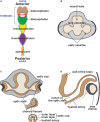
FIGURE 1
FIGURE 1
Vertebrate eye development (human eye). (A) During early vertebrate development, the eye field is established at the boundary between the telencephalon (brown) and the diencephalon (green). (B) Optic vesicles bilaterally protrude from either side of the forebrain approaching the thickened surface ectoderm (lens placodes). (C) The interaction between the optic vesicle and the lens placode of the surface ectoderm results in optic vesicle invagination, optic cup formation and lens placode evagination (lens pit). Simultaneously, the optic fissure is formed along the inferonasal aspect of the optic cup, which surrounds the hyaloid artery. (D) Continued evagination of surface ectoderm leads to the formation of an independent lens vesicle.
FIGURE 2
FIGURE 2
Cranial neural crest migration into the head. Time-lapse imaging of NC cell migration in zebrafish has shown that cranial NC cells from the diencephalon (green dashed lines and arrows) and anterior mesencephalon (anterior yellow dashed lines and arrows) travel either dorsal or ventral to the optic cup to establish the POM. A subpopulation of these cells migrates anteriorly into the frontonasal process (brown dashed lines and arrows). NC cells from the posterior mesencephalon (posterior yellow dashed lines and arrows) and rhombencephalon (purple dashed lines and arrows) migrate toward the pharyngeal arches on the ventral side of the embryo. Some of the factors implicated in the regulation of NC migration (Pax6, Pitx2, Foxd3, Sox10, Foxc1, etc.) are shown (inset, upper right). The signaling gradient that maintains the integrity of cranial NC cell migration [i.e., thyroid hormone (TH; blue) and retinoic acid (RA; red)] into the posterior pharyngeal arches is also shown.
FIGURE 3
FIGURE 3
Ocular neural crest migration and establishment of the ocular anterior segment. POM NC cells (purple), along with mesoderm cells (red), migrate adjacent to the hyaloid vasculature within the optic fissure and between the surface ectoderm and the optic cup contributing to the corneal stroma and endothelium, the iris stroma and the trabecular meshwork. In this figure, these structures are all indicated in purple to illustrate their ocular NC derivation. Notably, as previously discussed in the text above, the patterns of ocular NC cell migration vary between species (three distinct waves of NC cells in humans, two NC cell waves in mice and a single continuous migratory wave in chick). However, for simplicity, these patterns are not depicted here.
FIGURE 4
FIGURE 4
Congenital eye diseases associated with neural crest defects in the anterior segment. (A) Axenfeld-Rieger syndrome is characterized by anteriorization of Schwalbe’s line (posterior embryotoxon, black arrowheads) and iris hypoplasia, causing corectopia and pseudopolycoria (white arrows). Over half of affected individuals develop glaucoma, and many require placement of a glaucoma drainage device (asterisk) to control intraocular pressure. (B) Primary congenital glaucoma is due to developmental abnormalities in the trabecular meshwork and aqueous outflow tracts. As a result, elevated intraocular pressures in infants cause corneal edema and buphthalmos (increased eye size). (C) Peters anomaly shows central corneal opacification (black arrows) reflecting the abnormal separation of the lens vesicle from the surface ectoderm.

Similar articles

Cited by

References

    1. Akula M., Park J. W., West-Mays J. A. (2019). Relationship between neural crest cell specification and rare ocular diseases. J. Neurosci. Res. 97 7–15. 10.1002/jnr.24245 - DOI - PMC - PubMed
    1. Alallah J., Mohtisham F., Khan Z. (2020). Spontaneous bilateral corneal perforation in a neonate: a case report. Int. J. Pediatr. Adolesc. Med. 7 53–55. - PMC - PubMed
    1. Arikawa A., Yoshida S., Yoshikawa H., Ishikawa K., Yamaji Y., Arita R. I., et al. (2010). Case of novel PITX2 gene mutation associated with Peters’ anomaly and persistent hyperplastic primary vitreous. Eye 24 391–393. 10.1038/eye.2009.114 - DOI - PubMed
    1. Baulmann D. C., Ohlmann A., Flugel-Koch C., Goswami S., Cvekl A., Tamm E. R. (2002). Pax6 heterozygous eyes show defects in chamber angle differentiation that are associated with a wide spectrum of other anterior eye segment abnormalities. Mech. Dev. 118 3–17. 10.1016/s0925-4773(02)00260-5 - DOI - PubMed
    1. Berker N., Alanay Y., Elgin U., Volkan-Salanci B., Simsek T., Akarsu N., et al. (2009). A new autosomal dominant Peters’ anomaly phenotype expanding the anterior segment dysgenesis spectrum. Acta Ophthalmol. 87 52–57. 10.1111/j.1600-0420.2007.01082.x - DOI - PubMed

LinkOut - more resources