. 2020 Dec 8:8:605084.
doi: 10.3389/fcell.2020.605084. eCollection 2020.

Lack of FAM20A, Ectopic Gingival Mineralization and Chondro/Osteogenic Modifications in Enamel Renal Syndrome

Affiliations

Lack of FAM20A, Ectopic Gingival Mineralization and Chondro/Osteogenic Modifications in Enamel Renal Syndrome

Victor Simancas Escorcia et al. Front Cell Dev Biol. .

Abstract

Enamel renal syndrome (ERS) is a rare recessive disorder caused by loss-of-function mutations in FAM20A (family with sequence similarity 20 member A, OMIM #611062). Enamel renal syndrome is characterized by amelogenesis imperfecta, delayed or failed tooth eruption, intrapulpal calcifications, gingival overgrowth and nephrocalcinosis. Although gingival overgrowth has consistently been associated with heterotopic calcifications the pathogenesis, structure and interactions of the mineral deposits with the surrounding connective tissue are largely unknown. We here report a novel FAM20A mutation in exon 1 (c.358C > T) introducing a premature stop codon (p.Gln120*) and resulting in a complete loss of FAM20A. In addition to the typical oral findings and nephrocalcinosis, ectopic calcified nodules were also seen in the cervical and thoracic vertebrae regions. Histopathologic analysis of the gingiva showed an enlarged papillary layer associated with aberrant angiogenesis and a lamina propria displaying significant changes in its extracellular matrix composition, including disruption of the collagen I fiber network. Ectopic calcifications were found throughout the connective gingival tissue. Immunomorphological and ultrastructural analyses indicated that the calcification process was associated with epithelial degeneration and transformation of the gingival fibroblasts to chondro/osteoblastic-like cells. Mutant gingival fibroblasts cultures were prone to calcify and abnormally expressed osteoblastic markers such as RUNX2 or PERIOSTIN. Our findings expand the previously reported phenotypes and highlight some aspects of ERS pathogenesis.

Keywords: Fam20A; calcification; cell death; enamel renal syndrome; fibroblast; gingiva; osteoblast.


Conflict of interest statement

The authors declare that the research was conducted in the absence of any commercial or financial relationships that could be construed as a potential conflict of interest.

Figures

FIGURE 1
FIGURE 1
Clinical features of the proband. Front (A) and side (B) photos showing facial asymmetry, ocular hypertelorism and dense, bushy eyebrows. (C,D) Generalized gingival enlargement and a yellow–brown discoloration of the unique erupted tooth. (E) CBCT reconstruction; agenesis of 11, 14, 18, 28, 31, 36, 41, 42, 45, and 46; multiple unerupted teeth; large radiolucent pericoronal areas associated with impacted molars. (F) Mandibular and maxillary CBCT reconstruction. Note the displacement of the premolars into the maxillary sinus and the molars in the mandibular canal. (G) Renal axial CT-scan and (H,I) ultrasound showing bilateral medullary nephrocalcinosis (arrowheads). (J) Sagittal and (K) para-sagittal CBCT scans reveal round bilateral calcifications within the foramens of the cranial vertebrae (arrows). (L,M) Sequencing chromatograms of FAM20A exon 1 of a control (L) and proband (M). The homozygous nonsense mutation, c.358C > T causes a premature termination at p.Gln120*. (N) Vesicular distribution of FAM20A in control gingival fibroblasts. (O) Loss of FAM20A staining in the proband’s gingival fibroblasts. Scale bars: (O), P = 20 μm.
FIGURE 2
FIGURE 2
FAM20A and FAM20C expression in normal gingiva. (A) FAM20A expression is detected in epithelial cells (epi) and vascular cells in the papillary layer (pl). (B) FAM20A (green) is expressed in S100A9 (red) cells (arrowhead). Thin arrows bordered a blood vessel. (C) In fibroblasts, FAM20A is found in vesicles within elongated processes. (D) In the lamina propria, FAM20A is also expressed in endothelial and perivascular cells of blood vessels. (E) FAM20C distribution in epithelial, vascular (arrowheads) and fibroblastic cells of the papillary layer and of the lamina propria (lp). (F) FAM20C is found in vesicles within elongated processes of fibroblasts. (G) FAM20A (green) and FAM20C (red) are co-expressed in epithelial cells. (H) FAM20C expression in S100A9 positive populations (arrowheads). (I,J) Absence of FAM20A expression (green) in the proband’s gingiva; FAM20C (red) is detected in enlarged blood vessels (I) and gingival epithelium (J). Arrows indicated FAM20C expression in fibroblasts. (K) Western-blot of FAM20A and FAM20C in control and proband gingiva. V: vessel. Scale bars: (AD,GJ) = 100 μm; (E) = 200 μm; (F) = 20 μm.
FIGURE 3
FIGURE 3
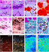
Histologic features of proband gingiva. (A) Hematoxylin-eosin staining shows numerous, tortuous capillary vessels with an abnormally large diameter in the papillary layer (arrowheads). Numerous ovoid calcified particles (ec) of various size are found in the reticular layer (rl) of the lamina propria. Epithelial islands (asterisks) are adjacent to the calcified particles. (B) Inset of A showing ovoid calcified particles in gingiva. (C) Alizarin red staining identifies calcium deposits in the reticular layer of the lamina propria. (D) High power photomicrograph showing that cells between calcified particles are heavily loaded in calcium. (E,F) Von Kossa staining indicates the presence of calcium phosphate. (E) Abnormal calcium deposits were located in the reticular layer of the lamina propria. Arrowheads indicate abnormally large blood vessels in the papillary layer (pl). (F) High power photomicrograph indicates the presence of cells intermixed with calcium deposits of various sizes. (G,H) Goldner Trichrome allows the detection of collagen in green. (G) In the papillary layer the light green coloration indicates a low amount of collagen. In the reticular layer collagen fibers do not assemble in large bundles and surround ectopic calcifications (ec). Arrowheads indicated abnormally large blood vessels and asterisks epithelial islands. (H) High power photomicrograph indicated that mineralized nodules (green) are surrounded by an osteoid-like layer (red/orange) (I,J) Picrosirius red polarizing staining. (I) Arrows indicate thick reddish bundles of collagen fibers in the normal gingiva lamina propria (rl and pl). (J) In the proband’s lamina propria, areas of loosely packed green–yellow disoriented collagen fibers and of yellow–red large collagen bundles were found. Asterisks indicate areas of no birefringence superimposing with calcified particles. Note the great diminution of collagen fibers in proband’s papillary layer. (K,L) F-TIR analysis at 1246 cm–1 indicates the presence of SO42- residues in the extracellular matrix. In control gingiva (K) the IR spectra indicate a moderate to low concentration of SO42- in the matrix. In proband gingiva (L) an increase of SO42- residues is observed within the calcified areas (arrows). bv = blood vessel; epi = epithelium. Scale bars: (A,C,E,G) = 200 μm; (B) = 50 μm; (D,H) = 20 μm; (F) = 15 μm; (I,J) = 300 μm; (K,L) = 500 μm.
FIGURE 4
FIGURE 4
(A) COLLAGEN IV immunoreactivty in the basement membrane of the papillae (PL). (B) Punctuate patterns of COLLAGEN IV in proband gingiva. (C) Sirius red staining of the collagen network in the normal papillae. (D) Sirius Red staining in proband shows a disheveled network (arrowhead). Asterisks showed the tortuosity and abnormal diameters of vessels. Ep: Epithelium layer. Scale bars: (A,B) = 50 μm; (C) = 40 μm; (D) = 80 μm.
FIGURE 5
FIGURE 5
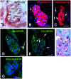
(A) Sirius red staining identifies clusters of cells with a yellowish cytoplasm typical of epithelial cell surrounded by loosely packed collagen fibers. Asterisks indicate large vacuoles associated with a crescent-shaped nucleus. Arrows indicate pyknotic nuclei. (B,C) KERATIN-14 (K14) immunostaining. (B) Little vacuolized islands strongly express K14. Arrows indicate pyknotic nuclei. (C) Increased vacuolization, loss of K14 and presence of numerous pyknotic nuclei (arrows). (D,E) CALNEXIN immunostaining suggests absence of endoplasmic reticulum in the vacuoles. (D) Vacuoles associated with crescent-shaped nuclei. (E) In highly vacuolized clusters, vacuoles fuse and numerous pyknotic nuclei are observed. Arrows indicate pyknotic nuclei. (F) Alcian Blue pH 2.5 staining indicates that epithelial and calcified particles are embedded in sulfated glycoproteins. Arrow indicated calcified nodules. (G) Absence of COLLAGEN IV immunoreactivity surrounding the epithelial islands. C, calcified nodules; e, epithelial island. Scale bars: (A) = 40 μm; (B,D,G) = 50 μm; (C,E) = 80 μm; (F) = 120 μm.
FIGURE 6
FIGURE 6
(A,B) SEM shows heavily mineralized entities in proband gingiva. (C) SEM of a mineralized sphericule displaying clear concentric layers in an inner coat around an amorphous nucleus. The outer coat is thick and displays a granular texture. TEM (D) and SAED pattern (E) of a small calcified sphericule. SAED indicates a hydroxyapathite-like material.
FIGURE 7
FIGURE 7
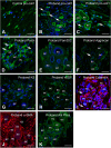
Immunocytochemical features of gingival calcifications. (A) Pro-COLLAGEN I expression in fibroblasts in normal gingiva. (B,C) In proband gingiva, pro-COLLAGEN I is expressed in cells with a round morphology, in the first external ring of calcified particles and small structures surrounding pyknotic bodies (thin arrows). (D) POSTN is localized in the extracellular matrix surrounding mineralized nodules and cells (thin arrows). (E) FAM20C is localized in the cytoplasm of cells associated with mineralized nodules. (F) ACAN, (G), and Keratan sulfate expressions are associated with the extracellular matrix of gingival calcifications. Thin arrows in F indicated pyknotic body. (H) An abnormal expression of VEGF is seen within mineralized structures. (I) CALBINDIN and (J) α-SMA is strongly expressed in cells between the ectopic calcifications. Thin arrows in panel (I) indicated pyknotic bodies. (K) ALPL staining is also found as dots in cells between the ectopic calcifications. ec, ectopic calcification. Scale bars: (AD,F,H–K) = 100 μm; (E,G) = 20 μm.
FIGURE 8
FIGURE 8
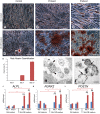
An osteogenic-like process is activated in FAM20A deficient gingival fibroblasts (GFs) grown in mineralization-inducing conditions. (A,B) Representative bright field images of normal (A) and FAM20A deficient (B,C) GFs grown for 21 days in standard medium colored with Red Alizarin. (D–F) Representative bright field images of normal (D) and FAM20A deficient (E,F) GFs grown 21 days in a mineralization-inducing medium colored with Red Alizarin. (D) Arrowhead indicates rare and small calcium deposits in control GFs. (E) Calcium deposits of various sizes are visualized in FAM20A deficient cultures. (F) High power photomicrograph of calcium deposits intermixed with cells. (G) Quantitative analysis of Red Alizarin expression by morphometric analysis on the pooled results of three separate experiments; the value represents the means ± SD. *p < 0.05. (H,I) Transmission electron microscopy images showing extracellular matrix vesicles containing electron dense bodies. Initial calcium-phosphate deposition takes place at the interior membrane (arrows). Depositions are also observed around cell debris. (J–L) qRT/PCR of ALPL (J), RUNX2 (K), and POSTN (L) at 7 and 21 days of culture. Data are presented as the mean ± SD of three separate experiments. ***p < 0.001. Scale bars: (AF) = 150 μm; (H,I) = 2 μm.

Similar articles

Cited by

References

    1. Acevedo A. C., Poulter J. A., Alves P. G., de Lima C. L., Castro L. C., Yamaguti P. M., et al. (2015). Variability of systemic and oro-dental phenotype in two families with non-lethal Raine syndrome with FAM20C mutations. BMC Med. Genet. 16:8. 10.1186/s12881-015-0154-5 - DOI - PMC - PubMed
    1. Azuma K., Casey S. C., Urano T., Horie-Inoue K., Ouchi Y., Blumberg B., et al. (2015). Pregnane X receptor knockout mice display aging-dependent wearing of articular cartilage. PLoS One 10:e0119177. 10.1371/journal.pone.0119177 - DOI - PMC - PubMed
    1. Chen J.-H., Simmons C. A. (2011). Cell-matrix interactions in the pathobiology of calcific aortic valve disease: critical roles for matricellular, matricrine, and matrix mechanics cues. Circ. Res. 108 1510–1524. 10.1161/CIRCRESAHA.110.234237 - DOI - PubMed
    1. Cho S. H., Seymen F., Lee K.-E., Lee S.-K., Kweon Y.-S., Kim K. J., et al. (2012). Novel FAM20A mutations in hypoplastic amelogenesis imperfecta. Hum. Mutat. 33 91–94. 10.1002/humu.21621 - DOI - PubMed
    1. Couchourel D., Aubry I., Delalandre A., Lavigne M., Martel-Pelletier J., Pelletier J.-P., et al. (2009). Altered mineralization of human osteoarthritic osteoblasts is attributable to abnormal type I collagen production. Arthritis Rheum. 60 1438–1450. 10.1002/art.24489 - DOI - PMC - PubMed

LinkOut - more resources