

PPAR-δ agonist affects adipo-chondrogenic differentiation of human mesenchymal stem cells through the expression of PPAR-γ
- PMID: 33426208
- PMCID: PMC7770446
- DOI: 10.1016/j.reth.2020.07.003
PPAR-δ agonist affects adipo-chondrogenic differentiation of human mesenchymal stem cells through the expression of PPAR-γ
Abstract
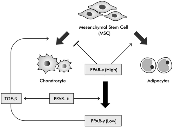
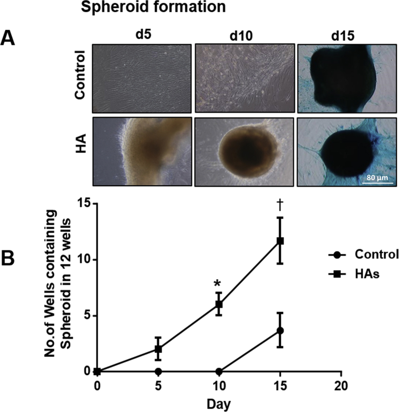
Introduction: Peroxisome proliferator-activated receptor (PPAR) subfamily play an important role in chondrogenesis. Previous study has reported that mixture of GW0742 (PPAR-δ agonist), hyaluronic acid (HA) and mesenchymal stem cells (MSCs) enhance chondrogenesis. The purpose of this study is to compare with efficacies of commercially available HA and demonstrate correlation of PPAR-γ and PPAR-δ.
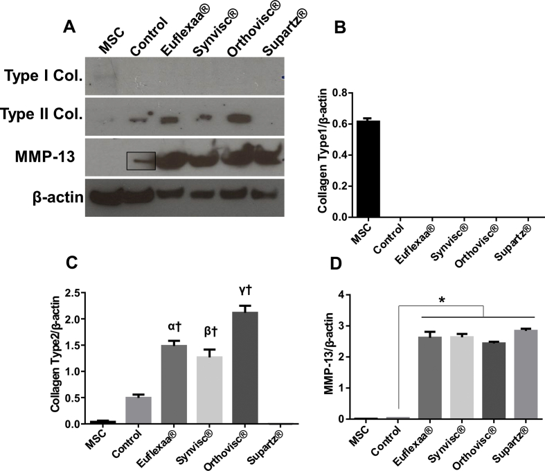
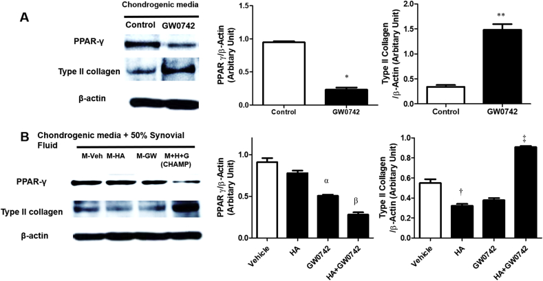
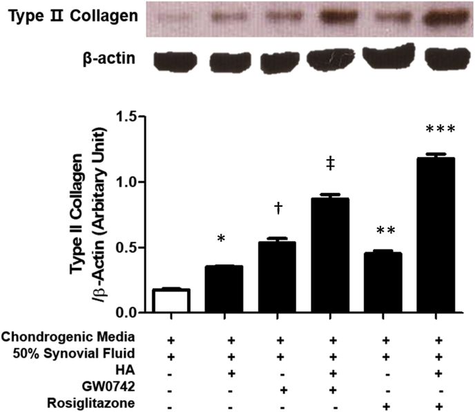
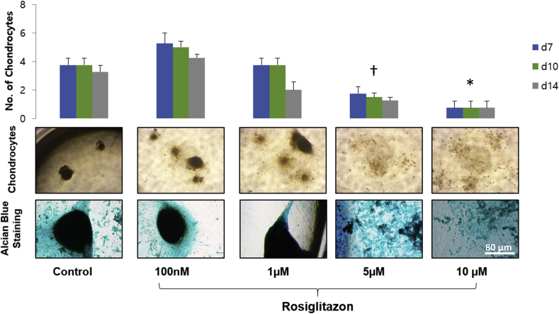
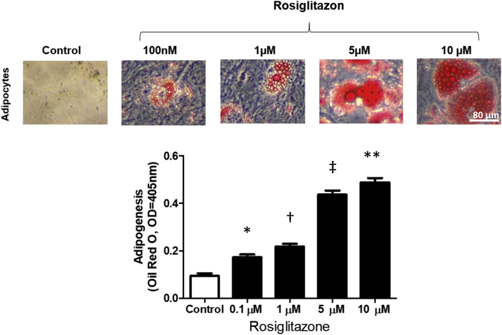
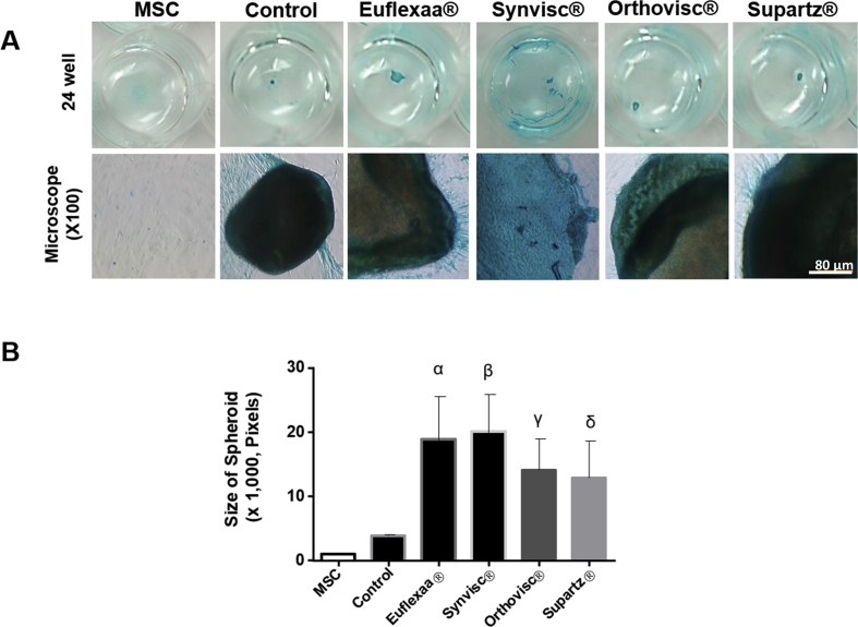
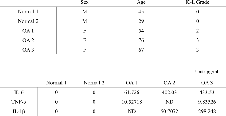
Methods: In this experimental study, MSCs were cultured with chondrogenic media and clinical HA gels (Euflexxa®, Synvisc®, Orthovisc® and Supartz®) using micormass culture method. Expression of type Ⅰ, Ⅱ collagen and matrix metalloprotease-13 (MMP-13) was measured by immunoblotting. MSCs were cultured with chondrogenic media and/or HA and/or GW0742 and/or rosiglitazone (PPAR-γ agonist) and/or human osteoarthritis synovial fluid. Immunoblotting was used to measure expression of type Ⅱ collagen and PPAR-γ. To identify the effective dose for chondrogenesis and adipogenesis, either 0.1, 1, 5 or 10 μM of rosiglitazone was added to MSCs in chondrogenic media or adipogenic media.
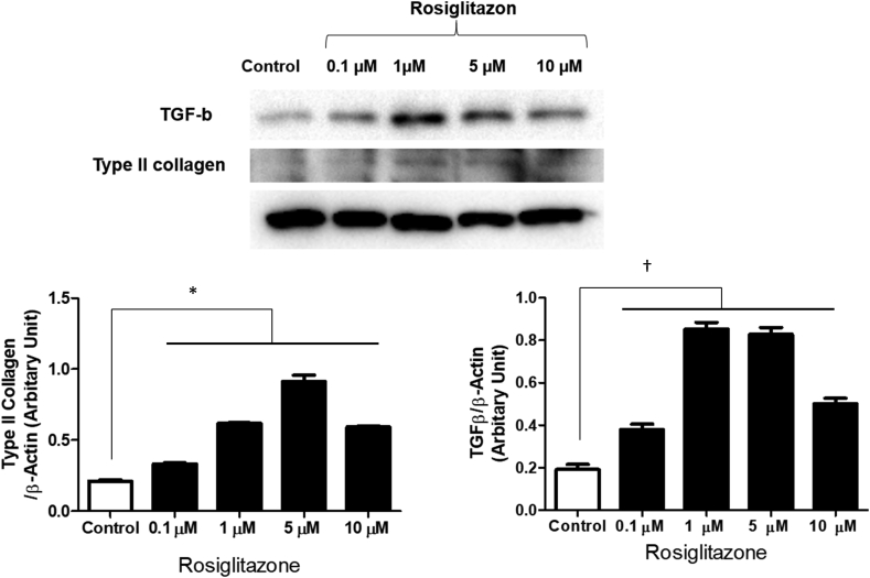
Results: Clinical HA gels inhibited expression of type Ⅰ collagen and enhanced the expression of MMP-13. Type Ⅱ collagen expression was significantly elevated in all treatment groups except Supartz®. GW0742 decreased the expression of PPAR-γ with/without inflammation condition. Rosiglitazone enhanced adipogenesis in a dose-dependent manner and enhanced the expression of type Ⅱ collagen under inflammation condition. Otherwise, the expression of type Ⅱ collagen and formation of chondrocyte spheroids showed a dose-dependent manner with a peak at 1 μM of rosiglitazone.
Conclusions: PPAR-γ has a considerable anti-inflammatory effect and a strong pro-adipogenic effect, which inhibits the chondrogenic effect. PPAR-γ is related with PPAR-δ and shows a chondrogenic effect at lower concentrations. And clinical HA gels shows various efficacy of chondrogenesis. This study suggested that PPAR-γ and PPAR-δ are key regulatory factors of chondrogenesis.
Keywords: Adipogenesis; Chondrogenesis; DJD, degenerative joint disease; ECM, extracellular matrix; FBS, fetal bovine serum; GAG, glycosaminoglycans; HA, hyaluronic acid; MMP, matrix metalloprotease; MSC, mesenchymal stem cells; Mesenchymal stem cells; OA, osteoarthritis; PBS, phosphate-buffered saline; PPAR, Peroxisome proliferator–activated receptor; PPAR-γ; PPAR-δ; TGF, Transforming growth factor; Type Ⅱ collagen; α-MEM, α-minimum essential medium.
© 2020 The Japanese Society for Regenerative Medicine. Production and hosting by Elsevier B.V.
Figures
Similar articles
-
PPAR-δ Agonist With Mesenchymal Stem Cells Induces Type II Collagen-Producing Chondrocytes in Human Arthritic Synovial Fluid.Cell Transplant. 2017 Aug;26(8):1405-1417. doi: 10.1177/0963689717720278. Cell Transplant. 2017. PMID: 28901183 Free PMC article.
-
Mechanical stimulation by ultrasound enhances chondrogenic differentiation of mesenchymal stem cells in a fibrin-hyaluronic acid hydrogel.Artif Organs. 2013 Jul;37(7):648-55. doi: 10.1111/aor.12041. Epub 2013 Mar 15. Artif Organs. 2013. PMID: 23495957
-
Chondrogenic differentiation of bovine bone marrow mesenchymal stem cells (MSCs) in different hydrogels: influence of collagen type II extracellular matrix on MSC chondrogenesis.Biotechnol Bioeng. 2006 Apr 20;93(6):1152-63. doi: 10.1002/bit.20828. Biotechnol Bioeng. 2006. PMID: 16470881
-
Hyaluronic acid facilitates chondrogenesis and matrix deposition of human adipose derived mesenchymal stem cells and human chondrocytes co-cultures.Acta Biomater. 2017 Apr 1;52:130-144. doi: 10.1016/j.actbio.2017.01.064. Epub 2017 Jan 25. Acta Biomater. 2017. PMID: 28131943
-
Chondrogenic differentiation of ATDC5 and hMSCs could be induced by a novel scaffold-tricalcium phosphate-collagen-hyaluronan without any exogenous growth factors in vitro.J Biomed Mater Res A. 2014 Aug;102(8):2725-35. doi: 10.1002/jbm.a.34948. Epub 2013 Sep 24. J Biomed Mater Res A. 2014. PMID: 24026971
Cited by
-
Fighting age-related orthopedic diseases: focusing on ferroptosis.Bone Res. 2023 Mar 1;11(1):12. doi: 10.1038/s41413-023-00247-y. Bone Res. 2023. PMID: 36854703 Free PMC article. Review.
-
PPARδ Agonist Promotes Type II Cartilage Formation in a Rabbit Osteochondral Defect Model.Cells. 2022 Sep 20;11(19):2934. doi: 10.3390/cells11192934. Cells. 2022. PMID: 36230897 Free PMC article.
-
5-aminosalicylic acid suppresses osteoarthritis through the OSCAR-PPARγ axis.Nat Commun. 2024 Feb 3;15(1):1024. doi: 10.1038/s41467-024-45174-6. Nat Commun. 2024. PMID: 38310093 Free PMC article.
-
Role of Peroxisome Proliferator-Activated Receptor α-Dependent Mitochondrial Metabolism in Ovarian Cancer Stem Cells.Int J Mol Sci. 2024 Nov 1;25(21):11760. doi: 10.3390/ijms252111760. Int J Mol Sci. 2024. PMID: 39519311 Free PMC article.
-
Therapeutic Single Compounds for Osteoarthritis Treatment.Pharmaceuticals (Basel). 2021 Feb 6;14(2):131. doi: 10.3390/ph14020131. Pharmaceuticals (Basel). 2021. PMID: 33562161 Free PMC article. Review.
References
-
- Brooks P.M. Impact of osteoarthritis on individuals and society: how much disability? Social consequences and health economic implications. Curr Opin Rheumatol. 2002;14(5):573–577. - PubMed
-
- Freyria A.M., Mallein-Gerin F. Chondrocytes or adult stem cells for cartilage repair: the indisputable role of growth factors. Injury. 2012;43(3):259–265. - PubMed
-
- Frean S.P., Abraham L.A., Lees P. In vitro stimulation of equine articular cartilage proteoglycan synthesis by hyaluronan and carprofen. Res Vet Sci. 1999;67(2):183–190. - PubMed
LinkOut - more resources
Full Text Sources
