. 2020 Oct 16:15:202-209.
doi: 10.1016/j.reth.2020.09.006. eCollection 2020 Dec.

LncRNA MALAT1 promotes wound healing via regulating miR-141-3p/ZNF217 axis

Affiliations

LncRNA MALAT1 promotes wound healing via regulating miR-141-3p/ZNF217 axis

Zun-Hong Liang et al. Regen Ther. .

Abstract

Background: The process of wound healing is complex. Increasing evidences have shown that lncRNA MALAT1 is abundant in fibroblasts and may be engaged in wound healing process. Therefore, we explored the mechanism of MALAT1 affecting wound healing.

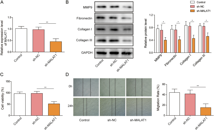
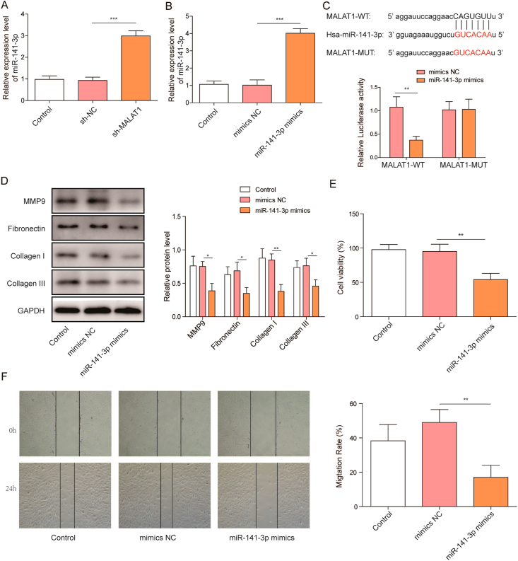
Methods: The expression levels of MALAT1, miR-141-3p as well as ZNF217 in human fibroblast cells (HFF-1) were quantified by qRT-PCR. HFF-1 proliferation was measured by MTT, while migration was detected by wound healing assay. SMAD2 activation and matrix proteins expression were detected by western blotting. The interaction between miR-141-3p and MALAT1 or ZNF217 was further confirmed using the luciferase reporter gene assay. In vivo wound healing was assessed by full-thickness wound healing model on C57BL/6 mice.

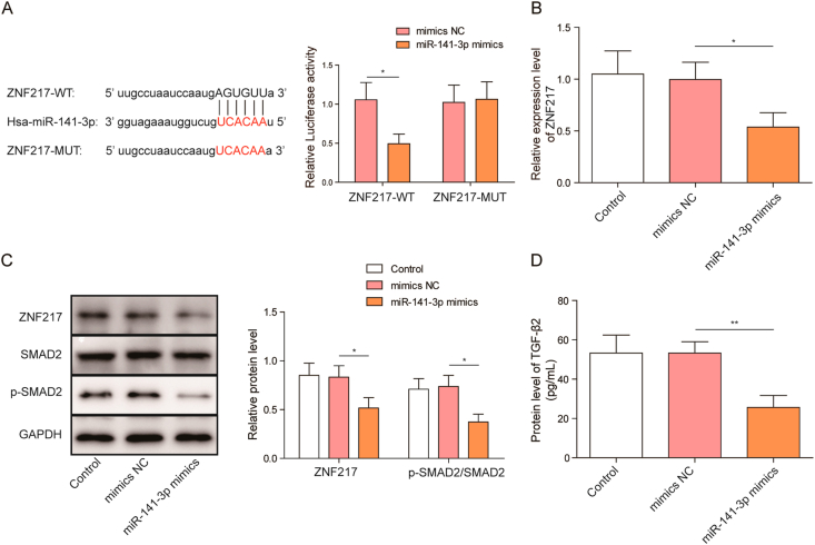
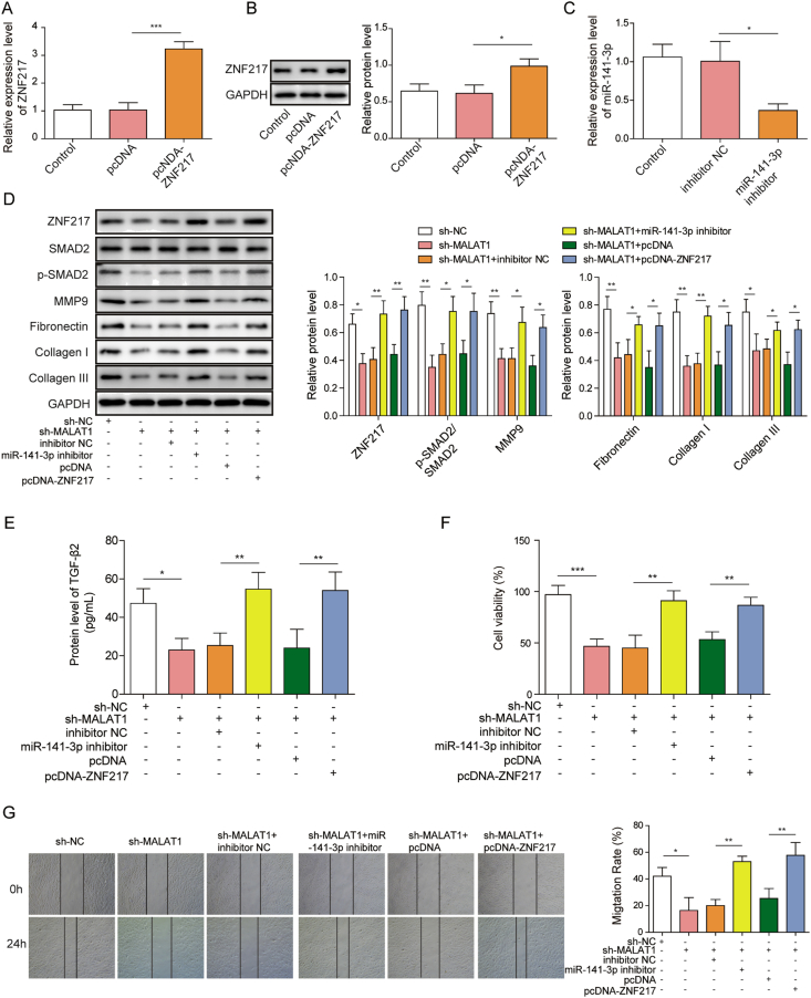
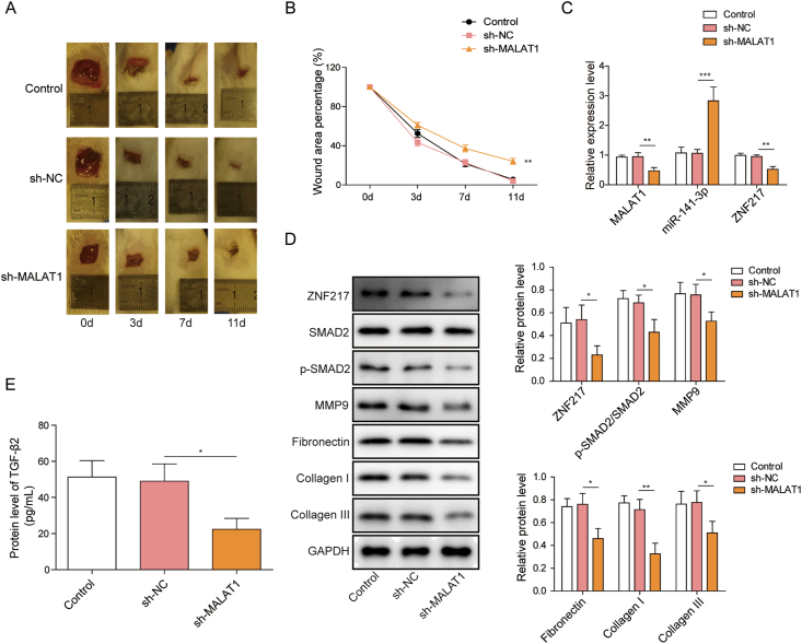
Result: Knockdown of MALAT1 as well as overexpression miR-141-3p remarkably inhibited the proliferation, migration and matrix protein expression in HFF-1 cells. MALAT1 directly targeted and inhibited the expression of miR-141-3p. MiR-141-3p suppressed the activation of TGF-β2/SMAD2 signaling pathway by targeting ZNF217. Knockdown of MALAT1 inhibited wound healing process in mice.

Conclusions: MALAT1 up-regulates ZNF217 expression by targeting miR-141-3p, thus enhances the activity of TGF-β2/SMAD2 signaling pathway and promotes wound healing process. This investigation shed new light on the understanding of the role of MALAT1 in wound healing, and may provide potential target for the diagnosis or therapy of chronic wounds.

Keywords: ECM, extra cellular matrix; ELISA, enzyme linked immunosorbent assay; EMT, epithelial mesenchymal transition; HFF-1, human fibroblast cells; MALAT1; MALAT1, metastasis-associated lung adenocarcinoma transcript 1; MTT, 3-(4,5-dimethyl-2-thiazolyl)-2,5-diphenyl-2-H-tetrazolium bromide; PVDF, polyvinylidene fluoride; SDS-PAGE, sodium dodecyl sulfate-polyacrylamide gel electrophoresis; TGF-β2, Transforming Growth Factor-β2; Wound healing; ZEB1, E-box binding homeobox 1; ZNF217; ZNF217, zinc-finger protein 217; lncRNA, long non-coding RNA; miR-141-3p; qRT-PCR, quantitative real-time PCR.


Conflict of interest statement

The authors have no conflict of interest to disclose.

Figures

Fig. 1
Fig. 1
MALAT1 knockdown decreases proliferation, migration and matrix protein expression of fibroblasts (A) MALAT1 level was measured by qRT-PCR in HFF-1 cells transfected with MALAT1 knockdown vector (sh-MALAT1) or the vector contains a non-targeting shRNA sequence (sh-NC) (B) Matrix protein expressions were determined by western blotting in HFF-1 cells (C) The viability of HFF-1 cells was detected by MTT assay after sh-MALAT1 or sh-NC treatment (D) The effect of MALAT1 on cell migration in HFF-1 cells. The data were presented as mean ± SD in triplicate. One-way ANOVA test was performed for statistical analysis. ∗P < 0.05, ∗∗P < 0.01, ∗∗∗P < 0.001.
Fig. 2
Fig. 2
MiR-141-3p binds to MALAT1 and inhibits proliferation, migration, and matrix proteins expression of fibroblast (A) QRT-PCR analyzed endogenous miR-141-3p levels in HFF-1 cells treated with sh-MALAT1 or sh-NC (B) Effect of miR-141-3p mimics on the expression of miR-141-3p (C) Interaction of MALAT1 and miR-141-3p was determined by StarBase virtual screening and dual-luciferase reporter gene assay (D) Western blotting was performed to determine the expression of matrix proteins in HFF-1 cells after miR-141-3p mimics treatment (E) Effect of miR-141-3p on HFF-1 cell proliferation (F) Effect of miR-141-3p on HFF-1 cell migration. The data were presented as mean ± SD in triplicate. One-way ANOVA test and student t-test analysis were performed for statistical analysis. ∗P < 0.05, ∗∗P < 0.01, ∗∗∗P<0.001.
Fig. 3
Fig. 3
MiR-141-3p regulates TGF-β2/SMAD2 signaling pathway by targeting ZNF217 (A) Interaction of ZNF217 and miR-141-3p was determined by StarBase virtual screening and dual-luciferase reporter gene assay (B) Effect of miR-141-3p mimics on the expression of ZNF217 (C) Effect of miR-141-3p mimics on the expression of ZNF217, SMAD2 and p-SMAD2 (D) Effect of miR-141-3p mimics on the expression of TGF-β2 in HFF-1 cells. The data were presented as mean ± SD in triplicate. One-way ANOVA test and student t-test analysis were performed for statistical analysis. ∗P < 0.05, ∗∗P < 0.01, ∗∗∗P < 0.001.
Fig. 4
Fig. 4
MALAT1 promotes fibroblast proliferation, migration, and matrix protein expression by activating ZNF217/TGF-β2/SMAD2 signal pathway (A) ZNF217 expression after transfection of pcDNA-ZNF217 and the empty plasmids in HFF-1 cells (B) ZNF217 protein levels were tested by western blotting (C) Effect of miR-141-3p inhibitor on the expression of miR-141-3p in HFF-1 (D) Fibroblast ZNF217, matrix protein and SMAD2 phosphorylation levels were detected by western blotting after indicated treatments (E) Expression levels of TGF-β2 were determined by ELISA in HFF-1 cells after indicated treatments (F) Proliferation of HFF-1 cells was subjected to MTT assays after indicated treatments (G) Cell migration of HFF-1 cells after indicated treatments. The data were presented as mean ± SD in triplicate. One-way ANOVA test was performed for statistical analysis. ∗P < 0.05, ∗∗P < 0.01, ∗∗∗P < 0.001.
Fig. 5
Fig. 5
Knockdown of MALAT1 inhibits the wound healing of mice by inhibiting ZNF217/TGF-β2/SMAD2 signal pathway (A) Images of the mouse skin wounds in sh-NC and sh-MALAT1 treated groups 3, 7, and 11 days after surgery (B) The comparison of the percentage of wound area remaining in wounds transfection with sh-NC and sh-MALAT1 (C) MALAT1, ZNF217 and miR-141-3p levels in granulation tissues after indicated treatments (D) The protein levels of ZNF217, matrix proteins and SMAD2 phosphorylation were determined by western blotting after indicated treatments (E) Expression levels of TGF-β2 in granulation tissues were determined by ELISA in HFF-1 cells after indicated treatments. The data were presented as mean ± SD in triplicate. One-way ANOVA test was performed for statistical analysis. ∗P < 0.05, ∗∗P < 0.01, ∗∗∗P < 0.001.

References

    1. Gurtner G.C., Werner S., Barrandon Y., Longaker M.T. Wound repair and regeneration. Nature. 2008;453(7193):314–321. - PubMed
    1. Jones R.E., Foster D.S., Longaker M.T. Management of chronic wounds-2018. J Am Med Assoc. 2018;320(14):1481–1482. - PubMed
    1. Frykberg R.G., Banks J. Challenges in the treatment of chronic wounds. Adv Wound Care. 2015;4(9):560–582. - PMC - PubMed
    1. Bainbridge P. Wound healing and the role of fibroblasts. J Wound Care. 2013;22(8):407–408. 410-12. - PubMed
    1. Tao S.C., Rui B.Y., Wang Q.Y., Zhou D., Zhang Y., Guo S.C. Extracellular vesicle-mimetic nanovesicles transport LncRNA-H19 as competing endogenous RNA for the treatment of diabetic wounds. Drug Deliv. 2018;25(1):241–255. - PMC - PubMed