

Saving the lower limb with GlassBONE™ - Successful surgical revision of pseudarthrosis after infected open proximal tibia fracture type IIIC with bioactive glass grafting - A case report
- PMID: 33426259
- PMCID: PMC7779823
- DOI: 10.1016/j.tcr.2020.100382
Saving the lower limb with GlassBONE™ - Successful surgical revision of pseudarthrosis after infected open proximal tibia fracture type IIIC with bioactive glass grafting - A case report
Erratum in
-
Erratum regarding missing Declaration of Competing Interest statements in previously published articles.Trauma Case Rep. 2023 Feb 17;45:100798. doi: 10.1016/j.tcr.2023.100798. eCollection 2023 Jun. Trauma Case Rep. 2023. PMID: 37234584 Free PMC article.
Abstract
Background: The management of bone defect due to trauma or surgical debridement is a current problem in orthopedic trauma surgery, often complicated by infection and bone nonunion. The graft is one of the most challenging variables in surgical treatment. Bioactive Glass (BAG) as a biocompatible and osteogenic product is a promising bone substitute showing good results in maxillo-facial-, spine surgery and treatment of osteomyelitis. Surprisingly, there is very little data on BAG use in trauma surgery.
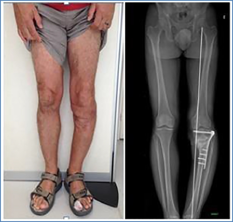
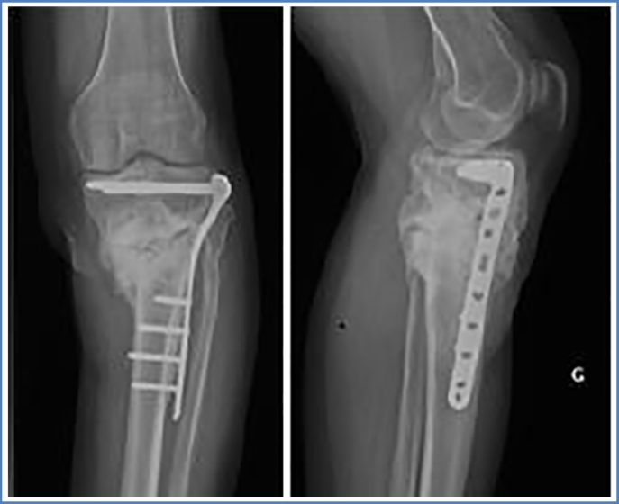
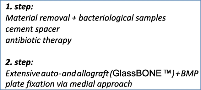
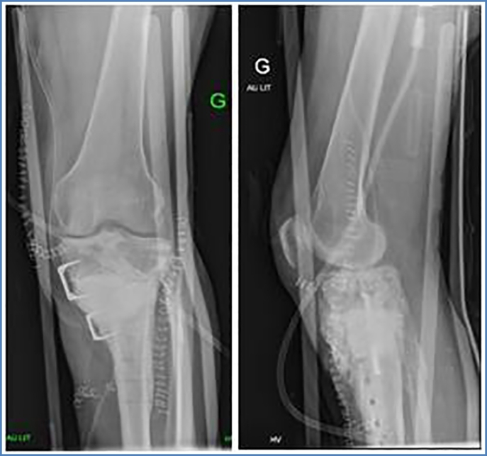
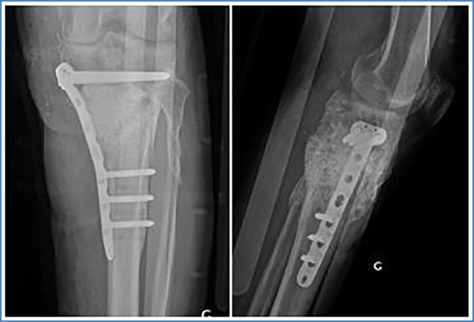
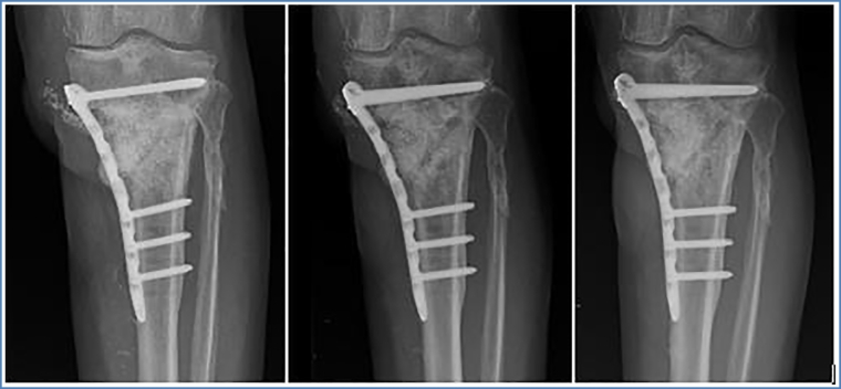
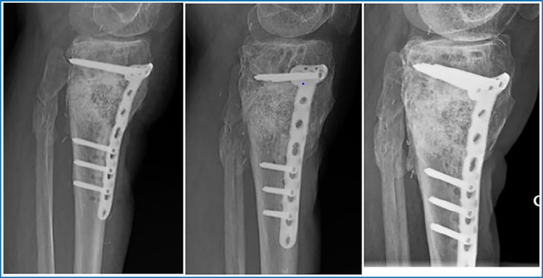
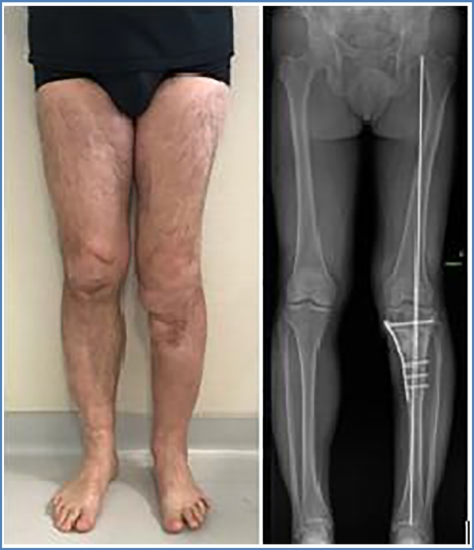
Case presentation: A 51-year-old male patient, involved in a motorcycle accident, suffered an open proximal tibia fracture, type IIIC, of the left leg. Patient was admitted in January of 2013 to a general orthopedic department for surgical treatment. After several surgical revisions due to infection, vascular damage, and bone nonunion, the patient was successfully treated with Masquelet therapy followed by GlassBONE™ grafting (GlassBONE™ 45S5; Norarker). The patient demonstrated excellent results over the course of a two-year follow-up.
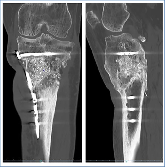
Conclusions: In our experience, GlassBONE™ 45S5 has proven to be an effective bone substitute even in difficult grafting conditions, including multiple surgical revisions for bone nonunion and infection. In our case, at the end of 2 years and 3 months of follow-up, the patient reported no pain, and had no signs of infection. Bone union and full weight bearing was achieved.This case report is oriented by the CARE guidelines for clinical case reports; the patient gave consent for publication.
Keywords: BAG; Bone nonunion; Tibia fracture.
© 2020 The Authors.
Figures
References
-
- Einhorn T.A., Gerstenfeld L.C., Fracture healing: mechaniEinhorn, T. A, Gerstenfeld L.C. Fracture healing: mechanisms and interventions. Nat. Rev. Rheumatol. 2015;11(1):45–54. doi: 10.1038/nrrheum.2014.164. (http://doi.org/10.1038/nrrheum.2014.164sms and interventions. Nat Rev Rheumatol (2015)) - DOI - PMC - PubMed
-
- Audigé L., Griffin D., Bhandari M., Kellam J., Rüedi T.P. Path analysis of factors for delayed healing and nonunion in 416 operatively treated tibial shaft fractures. Clin. Orthop. Relat. Res. 2005;(438):221–232. - PubMed
-
- Schlundt C. Clinical and research approaches to treat non-union fracture. Curr. Osteoporos. Rep. 2018;16:155–168. - PubMed
-
- P, G., E, W., RA, E., MM, M. & CM, C.-B Fractures of the tibia. Can their outcome be predicted. J. Bone Joint Surg. Br. 1999:81. - PubMed
Publication types
LinkOut - more resources
Full Text Sources
Other Literature Sources
