. 2020 Dec 29;6(12):e05810.
doi: 10.1016/j.heliyon.2020.e05810. eCollection 2020 Dec.

Methods for isolation and transcriptional profiling of individual cells from the human heart

Affiliations

Methods for isolation and transcriptional profiling of individual cells from the human heart

Neha Pimpalwar et al. Heliyon. .

Abstract

Background: Global transcriptional profiling of individual cells represents a powerful approach to systematically survey contributions from cell-specific molecular phenotypes to human disease states but requires tissue-specific protocols. Here we sought to comprehensively evaluate protocols for single cell isolation and transcriptional profiling from heart tissue, focusing particularly on frozen tissue which is necessary for study of human hearts at scale.

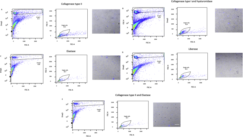
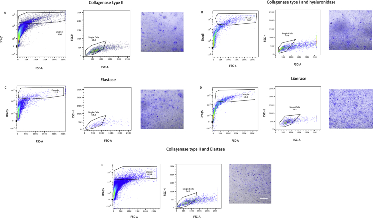
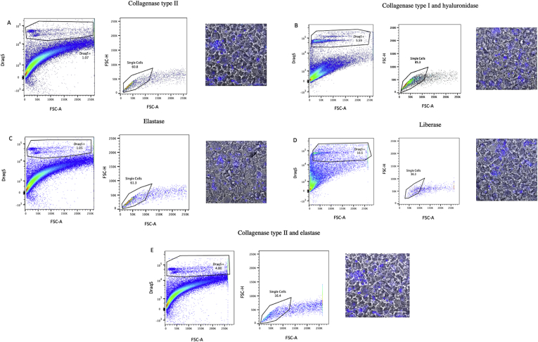
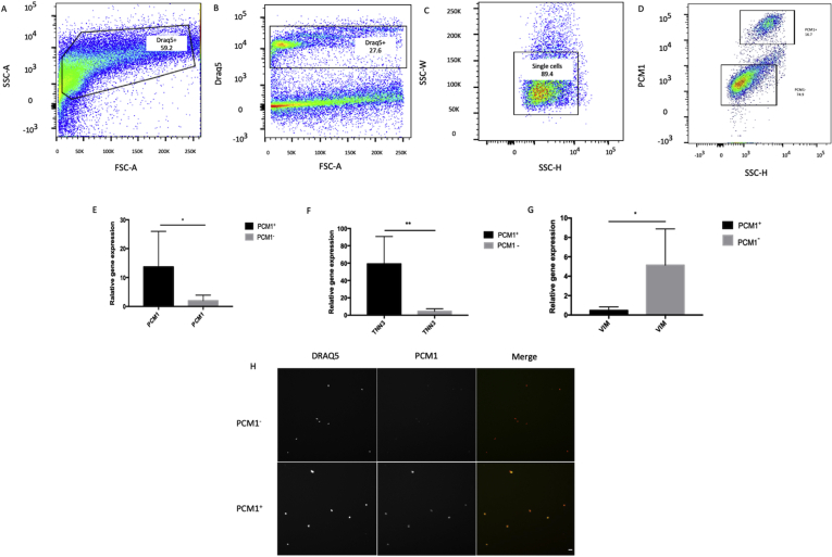
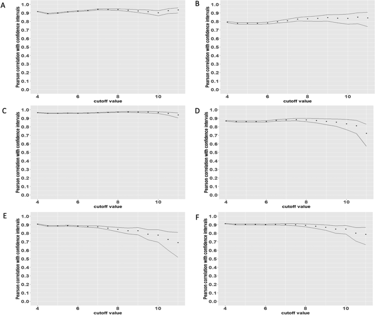
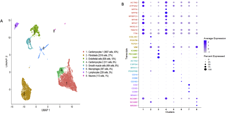
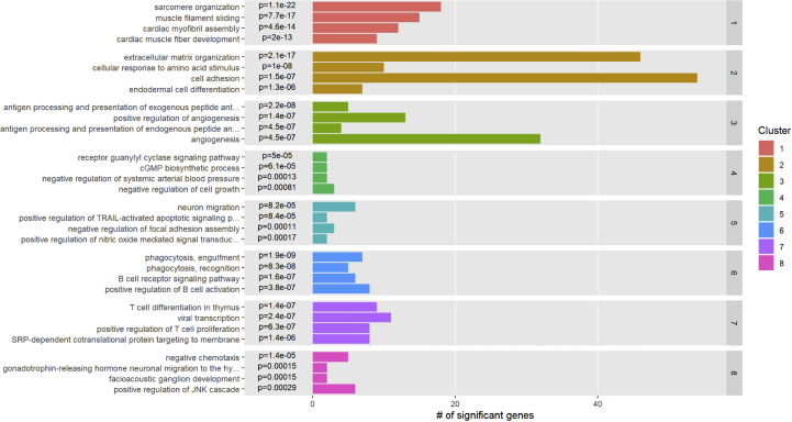
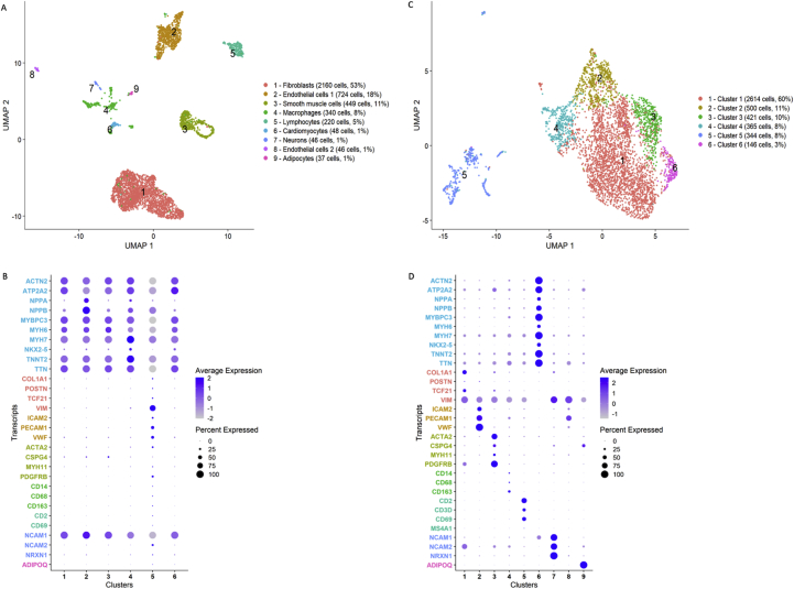
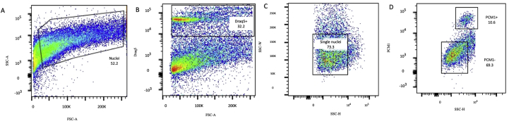
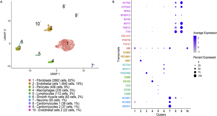
Methods and results: Using flow cytometry and high-content screening, we found that enzymatic dissociation of fresh murine heart tissue resulted in a sufficient yield of intact cells while for frozen murine or human heart resulted in low-quality cell suspensions across a range of protocols. These findings were consistent across enzymatic digestion protocols and whether samples were snap-frozen or treated with RNA-stabilizing agents before freezing. In contrast, we show that isolation of cardiac nuclei from frozen hearts results in a high yield of intact nuclei, and leverage expression arrays to show that nuclear transcriptomes reliably represent the cytoplasmic and whole-cell transcriptomes of the major cardiac cell types. Furthermore, coupling of nuclear isolation to PCM1-gated flow cytometry facilitated specific cardiomyocyte depletion, expanding resolution of the cardiac transcriptome beyond bulk tissue transcriptomes which were most strongly correlated with PCM1+ transcriptomes (r = 0.8). We applied these methods to generate a transcriptional catalogue of human cardiac cells by droplet-based RNA-sequencing of 8,460 nuclei from which cellular identities were inferred. Reproducibility of identified clusters was confirmed in an independent biopsy (4,760 additional PCM1- nuclei) from the same human heart.

Conclusion: Our results confirm the validity of single-nucleus but not single-cell isolation for transcriptional profiling of individual cells from frozen heart tissue, and establishes PCM1-gating as an efficient tool for cardiomyocyte depletion. In addition, our results provide a perspective of cell types inferred from single-nucleus transcriptomes that are present in an adult human heart.

Keywords: Cardiology; Cardiovascular System; Health sciences; Heart; Human; Methods; Protocol; Single cell; Transcriptome; Transcriptomics.


Conflict of interest statement

The authors declare no conflict of interest.

Figures

Figure 1
Figure 1
Flow cytometry analysis of mouse cells isolated from frozen heart tissue samples pretreated with an RNA-stabilizing agent. Murine heart tissue was digested with (A) collagenase type II, (B) collagenase type I and hyaluronidase, (C) elastase, (D) Liberase, or (E) collagenase type II and elastase, and analysed by flow cytometry and high content screening (HCS). Isolated cells were identified and gated first by forward scatter area (FSC-A) vs DRAQ5, second on FSC-A vs FSC-height (FSC-H) to distinguish single cells. The morphology and integrity of DRAQ5-stained cell suspensions were analysed using a HCS fluorescent microscope (scale bar = 200 μm). Prior to digestion, hearts had been treated with an RNA-stabilizing agent and frozen to -80 °C.
Figure 2
Figure 2
Flow cytometry analysis of mouse cells isolated from snap frozen heart tissue samples. Murine heart tissue was digested with (A) collagenase type II, (B) collagenase type I and hyaluronidase, (C) elastase, (D) Liberase, or (E) collagenase type II and elastase, and analysed by flow cytometry and high content screening (HCS). Isolated cells were identified and gated first by forward scatter area (FSC-A) vs DRAQ5, second on FSC-A vs FSC-height (FSC-H) to distinguish single cells. The morphology and integrity of DRAQ5-stained cell suspensions were analysed using a HCS fluorescent microscope (scale bar = 200 μm).
Figure 3
Figure 3
Flow cytometry analysis of human cells isolated from frozen heart tissue samples pretreated with an RNA-stabilizing agent. Human heart tissue was digested with (A) collagenase type II, (B) collagenase type I and hyaluronidase, (C) elastase, (D) Liberase, or (E) collagenase type II and elastase, and analysed by flow cytometry and high content screening (HCS). Isolated cells were identified and gated first by forward scatter area (FSC-A) vs DRAQ5, second on FSC-A vs FSC-height (FSC-H) to distinguish single cells. The morphology and integrity of DRAQ5-stained cell suspensions were analysed using a HCS fluorescent microscope (scale bar = 200 μm). Prior to digestion, hearts had been treated with an RNA-stabilizing agent and frozen to -80 °C.
Figure 4
Figure 4
Flow cytometry analysis of mouse cells isolated from fresh heart tissue. Murine heart tissue was digested with (A) collagenase type II, (B) collagenase type II and hyaluronidase, (C) elastase, (D) Liberase, or (E) collagenase type II and elastase, and analysed by flow cytometry and high content screening (HCS). Isolated cells were identified and gated first by forward scatter area (FSC-A) vs DRAQ5, second on FSC-A vs FSC-height (FSC-H) to distinguish single cells. The morphology and integrity of DRAQ5-stained cell suspensions were analysed using a HCS fluorescent microscope (scale bar = 200 μm).
Figure 5
Figure 5
Evaluation of nuclei isolated from human cardiac left ventricle tissue. (A) Nuclei were first identified by forward scatter area (FSC-A) vs side scatter area (SSC-A). (B) In the second gate DRAQ5 positive nuclei were separated from debris. (C) Single nuclei were gated by SSC-height (SSC-H) versus SSC-width (SSC-W). (D) Cardiomyocyte nuclei were separated from non-cardiomyocyte nuclei based on PCM1-staining. (E,F,G) Gene expression of sorted nuclei revealed that the PCM1+ fraction was enriched for the cardiomyocyte marker genes PCM1 and TNNT3, whereas the cardiac fibroblast marker gene VIM was enriched in the PCM1- fraction. Data is presented as mean ± SD from three independent experiments. (H) FACS-sorted nuclei, stained with DRAQ5 and PCM1 were scanned with high content screening microscopy. Scale bar = 10 μm.
Figure 6
Figure 6
Correlation of nuclear with cytoplasmic and whole-cell transcriptomes. Pearson's correlation coefficients with 95% confidence intervals for nuclear transcriptomes with cytoplasmic and whole-cell transcriptomes in induced pluripotent stem cell derived cardiomyocytes (cytoplasmic, A; whole-cell, B), primary human cardiac fibroblasts (cytoplasmic, C; whole-cell, D), and cardiac microvascular endothelial cells (cytoplasmic, E; whole-cell, F). X-axis indicates different thresholds for non-expressed transcripts representing technical noise.
Figure 7
Figure 7
RNA-sequencing of human heart cells. Panel A shows a Uniform Manifold Approximation and Projection (UMAP) plot based on RNA-sequencing of 8,460 individual nuclei from a human heart. Of the nuclei, the proportion of cardiomyocyte nuclei has been enriched such that 4,390 had been selected from PCM1+ and 4,070 from PCM1- nuclear fractions by fluorescence-activated cell sorting. Panel B shows a dot plot of expression across clusters of preselected, established marker genes representing the major expected cell types in human heart: cardiomyocytes (red), fibroblasts (yellow), endothelial cells (green), vascular smooth muscle cells and pericytes (cyan), macrophages (blue), lymphocytes (purple), and neurons (pink).
Figure 8
Figure 8
Pathway enrichment analysis of single-nucleus clusters. Pathway enrichment analysis across clusters, incorporating all expressed transcripts and pathway annotations from the Gene Ontology project.
Figure 9
Figure 9
Clustering of nuclei by PCM1-staining. Uniform Manifold Approximation and Projection (UMAP) projections based on RNA-sequencing of 4,070 PCM1- (panels A and B) and 4,390 PCM1+ (C and D) nuclear fractions from fluorescence-activated cell sorting. Below UMAP plots are corresponding dot plots (panels B and D) illustrating expression across clusters of preselected, established marker genes representing the major expected cell types in human heart, with color codes representing the PCM1- clusters: cardiomyocytes (cyan), fibroblasts (red), endothelial cells (yellow), vascular smooth muscle cells and pericytes (light green), leukocyte populations (dark green), neurons (blue) and adipocytes (pink).
Figure 10
Figure 10
Flow cytometry analysis of a second sample from the same human heart. (A) Nuclei were first identified by forward scatter area (FSC-A) vs side scatter area (SSC-A). (B) In the second gate DRAQ5 positive nuclei were separated from debris. (C) Single nuclei were gated by SSC-height (SSC-H) versus SSC-width (SSC-W). (D) Cardiomyocyte nuclei were separated from non-cardiomyocyte nuclei based on PCM1-staining.
Figure 11
Figure 11
Clustering of nuclei from the second sample. Uniform Manifold Approximation and Projection (UMAP) projection based on RNA-sequencing of 4,760 PCM1- nuclei from fluorescence-activated cell sorting of a second, separate biopsy from the same heart as in Figure 4. The corresponding dot plot illustrates expression across clusters of preselected, established marker genes representing the major expected cell types in human heart: cardiomyocytes (pink), fibroblasts (red), endothelial cells (yellow), vascular smooth muscle cells and pericytes (cyan), leukocyte populations (green), and neurons (blue).

Similar articles

Cited by

References

    1. Ackers-Johnson M., Tan W.L.W., Foo R.S. Following hearts, one cell at a time: recent applications of single-cell RNA sequencing to the understanding of heart disease. Nat. Commun. 2018;9:4434. - PMC - PubMed
    1. Ashburner M., Ball C.A., Blake J.A., Botstein D., Butler H., Cherry J.M., Davis A.P., Dolinski K., Dwight S.S., Eppig J.T., Harris M.A., Hill D.P., Issel-Tarver L., Kasarskis A., Lewis S., Matese J.C., Richardson J.E., Ringwald M., Rubin G.M., Sherlock G. Gene ontology: tool for the unification of biology. The Gene Ontology Consortium. Nat. Genet. 2000;25:25–29. - PMC - PubMed
    1. Asp M., Giacomello S., Larsson L., Wu C., Furth D., Qian X., Wardell E., Custodio J., Reimegard J., Salmen F., Osterholm C., Stahl P.L., Sundstrom E., Akesson E., Bergmann O., Bienko M., Mansson-Broberg A., Nilsson M., Sylven C., Lundeberg J. A spatiotemporal organ-wide gene expression and cell Atlas of the developing human heart. Cell. 2019;179:1647–1660. e19. - PubMed
    1. Bajpai G., Schneider C., Wong N., Bredemeyer A., Hulsmans M., Nahrendorf M., Epelman S., Kreisel D., Liu Y., Itoh A., Shankar T.S., Selzman C.H., Drakos S.G., Lavine K.J. The human heart contains distinct macrophage subsets with divergent origins and functions. Nat. Med. 2018;24:1234–1245. - PMC - PubMed
    1. Bakken T.E., Hodge R.D., Miller J.A., Yao Z., Nguyen T.N., Aevermann B., Barkan E., Bertagnolli D., Casper T., Dee N., Garren E., Goldy J., Graybuck L.T., Kroll M., Lasken R.S., Lathia K., Parry S., Rimorin C., Scheuermann R.H., Schork N.J. Single-nucleus and single-cell transcriptomes compared in matched cortical cell types. PloS One. 2018;13 - PMC - PubMed