Review
. 2020 Dec 26;6(7):1973-1987.
doi: 10.1016/j.bioactmat.2020.12.010. eCollection 2021 Jul.

The application of nanoparticles in cancer immunotherapy: Targeting tumor microenvironment

Affiliations
Review

The application of nanoparticles in cancer immunotherapy: Targeting tumor microenvironment

Muyue Yang et al. Bioact Mater. .

Abstract

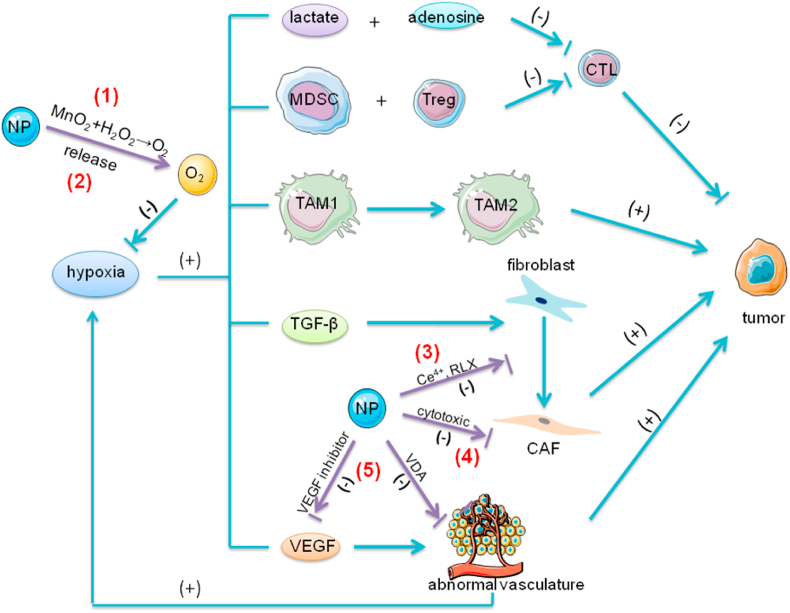
The tumor development and metastasis are closely related to the structure and function of the tumor microenvironment (TME). Recently, TME modulation strategies have attracted much attention in cancer immunotherapy. Despite the preliminary success of immunotherapeutic agents, their therapeutic effects have been restricted by the limited retention time of drugs in TME. Compared with traditional delivery systems, nanoparticles with unique physical properties and elaborate design can efficiently penetrate TME and specifically deliver to the major components in TME. In this review, we briefly introduce the substitutes of TME including dendritic cells, macrophages, fibroblasts, tumor vasculature, tumor-draining lymph nodes and hypoxic state, then review various nanoparticles targeting these components and their applications in tumor therapy. In addition, nanoparticles could be combined with other therapies, including chemotherapy, radiotherapy, and photodynamic therapy, however, the nanoplatform delivery system may not be effective in all types of tumors due to the heterogeneity of different tumors and individuals. The changes of TME at various stages during tumor development are required to be further elucidated so that more individualized nanoplatforms could be designed.

Keywords: AC-NPs, antigen-capturing nanoparticles; ANG2, angiopoietin-2; APCs, antigen-presenting cells; Ab, antibodies; Ag, antigen; AuNCs, gold nanocages; AuNPs, gold nanoparticles; BBB, blood-brain barrier; BTK, Bruton's tyrosine kinase; Bcl-2, B-cell lymphoma 2; CAFs, cancer associated fibroblasts; CAP, cleavable amphiphilic peptide; CAR-T, Chimeric antigen receptor-modified T-cell therapy; CCL, chemoattractant chemokines ligand; CTL, cytotoxic T lymphocytes; CTLA4, cytotoxic lymphocyte antigen 4; CaCO3, calcium carbonate; Cancer immunotherapy; DCs, dendritic cells; DMMA, 2,3-dimethylmaleic anhydrid; DMXAA, 5,6-dimethylxanthenone-4-acetic acid; DSF/Cu, disulfiram/copper; ECM, extracellular matrix; EGFR, epidermal growth factor receptor; EMT, epithelial-mesenchymal transition; EPG, egg phosphatidylglycerol; EPR, enhanced permeability and retention; FAP, fibroblast activation protein; FDA, the Food and Drug Administration; HA, hyaluronic acid; HB-GFs, heparin-binding growth factors; HIF, hypoxia-inducible factor; HPMA, N-(2-hydroxypropyl) methacrylamide; HSA, human serum albumin; Hypoxia; IBR, Ibrutinib; IFN-γ, interferon-γ; IFP, interstitial fluid pressure; IL, interleukin; LMWH, low molecular weight heparin; LPS, lipopolysaccharide; M2NP, M2-like TAM dual-targeting nanoparticle; MCMC, mannosylated carboxymethyl chitosan; MDSCs, myeloid-derived suppressor cells; MPs, microparticles; MnO2, manganese dioxide; NF-κB, nuclear factor κB; NK, nature killer; NO, nitric oxide; NPs, nanoparticles; Nanoparticles; ODN, oligodeoxynucleotides; PD-1, programmed cell death protein 1; PDT, photodynamic therapy; PFC, perfluorocarbon; PHDs, prolyl hydroxylases; PLGA, poly(lactic-co-glycolic acid); PS, photosensitizer; PSCs, pancreatic stellate cells; PTX, paclitaxel; RBC, red-blood-cell; RLX, relaxin-2; ROS, reactive oxygen species; SA, sialic acid; SPARC, secreted protein acidic and rich in cysteine; TAAs, tumor-associated antigens; TAMs, tumor-associated macrophages; TDPA, tumor-derived protein antigens; TGF-β, transforming growth factor β; TIE2, tyrosine kinase with immunoglobulin and epidermal growth factor homology domain 2; TIM-3, T cell immunoglobulin domain and mucin domain-3; TLR, Toll-like receptor; TME, tumor microenvironment; TNF-α, tumor necrosis factor alpha; TfR, transferrin receptor; Tregs, regulatory T cells; Tumor microenvironment; UPS-NP, ultra-pH-sensitive nanoparticle; VDA, vasculature disrupting agent; VEGF, vascular endothelial growth factor; cDCs, conventional dendritic cells; melittin-NP, melittin-lipid nanoparticle; nMOFs, nanoscale metal-organic frameworks; scFv, single-chain variable fragment; siRNA, small interfering RNA; tdLNs, tumor-draining lymph nodes; α-SMA, alpha-smooth muscle actin.


Conflict of interest statement

The authors declare that they have no known competing financial interests or personal relationships that could have appeared to influence the work reported in this paper.

Figures

Image 1
Graphical abstract
Fig. 1
Fig. 1
The role of DCs and the function of NPs in the tumor immunity. In response to endogenous and exogenous antigens DCs activate and maturate. They recognize these antigens and degrade them, then present them on the cell surface to naive T cells. The T cells become activated and transform to CTLs. The CTLs can attack tumor cells via direct killing or IFN-γ dependent pathways. (1) NPs modified with antigens and adjuvants specifically deliver antigens to DCs, (2) and promote DCs maturation and CTL activation by antigen presentation or the assistance of adjuvants. DCs present antigen fragments to naïve T cells. The CD4+ and CD8+ T cells become activated, undergo clonal expansion, and acquire cytotoxic abilities or helper functions (such as IFN-γ secretion). The addition of TLR ligands in NPs induces strong immune responses. Apart from extra addition of adjuvants in nanocomplex, Fe3O4 NPs as nano-immunopotentiators could promote the maturation of dendritic cells and immune responses; Abbreviations: Ag, antigen; DCs, dendritic cells; CTL, cytotoxic T lymphocyte; IFN-γ, interferon-γ; NPs, nanoparticles.
Fig. 2
Fig. 2
The role of TAMs and the function of NPs in the tumor immunity. In response to IFN-γ and LPS, TAMs transform to TAM1 phenotype and secrete high levels of IL-12, inhibiting tumor development. After exposure to IL-4 or IL-13 TAMs undergo the transition to TAM2 and produce IL-10, promoting tumor growth. In the hypoxic state in TME, TAM1 can repolarize to TAM2, which contributes to the immunosuppressive environment in TME. (1) NPs modified with HA, iron oxides or regorafenib can reprogram TAMs activities from TAM2 to TAM1 polarization. HA modulates the activation states of TAM by binding to CD44 on TAMs and activation of TLR4 pathways. Iron oxides attract macrophages and promote macrophage recruitment. After exposure to NPs, TAMs upregulate M1-related CD86 and TNF-α markers, and reduce the levels of M2-related IL-10 and CD206 markers. Iron oxides repolarize M2 to M1 and induce the Fenton reaction which can generate ROS and promote the apoptosis of tumor cells. The apoptotic tumor cells induce M1 polarization, which forms a feedback loop. The expression of TIE2, the receptor of ANG2, has been detected on TAM2. Regorafenib, an oral multi-kinase inhibitor, reduces TAM accumulation by ANG2/TIE2 blockade and modulates TAM polarization. (2) In addition, NPs can directly inhibit the survival and function of TAM2 by delivering siRNA or IBR. The anti-colony stimulating factor-1 receptor siRNA could specifically block M2 survival signals. IBR, an irreversible BTK inhibitor, can diminish the contribution of TAMs to tumorigenesis, thus reversing the immunosuppression established by TAMs.; Abbreviations: ANG2, angiopoietin-2; HA, hyaluronic acid; IFN-γ, interferon-γ; IBR, ibrutinib; IL, interleukin; LPS, lipopolysaccharide; NPs, nanoparticles; TAMs, tumor-associated macrophages; TIE2, tyrosine kinase with immunoglobulin and epidermal growth factor homology domain 2; TLR, toll-like receptor; TME, tumor microenvironment.
Fig. 3
Fig. 3
Graphic illustration of tdLNs and the function of NPs in the tumor immunity. TdLNs abound with both immunosupportive factors (e.g., DCs, T cells and B cells) and immunosuppressive factors (e.g., Tregs, MDSCs). TAAs are delivered to tdLNs via lymphatic drainage. DCs recognize these antigens and present them to T cells. The T cells produce IFN-γ and activate B cells to generate antibodies. These factors collectively contribute to anti-tumor immunity. MDSCs and Tregs inhibit the activation and function of T cells. (1) NPs encapsulating TAAs efficiently target tdLNs via lymphatic drainage. NPs with a medium size (10–100 nm) achieve maximum efficacy. (2) These TAAs then activate DCs and significantly promote the following T cell responses by antigen presentation or adjuvants. The addition of TLR ligands in NPs significantly promotes immune responses. (3) In addition, NPs with CpG, the TLR9 ligand, can reduce the number of MDSCs and Tregs. After exposure of CpG, DCs which express TLR9 produce proinflammatory cytokines and promote differentiation of MDSCs, blocking the inhibition of MDSCs on T cell proliferation.; Abbreviations: Ab, antibodies; IFN-γ, interferon-γ; tdLNs, tumor-draining lymph nodes; TAAs, tumor-associated antigens; DCs, dendritic cells; MDSCs, myeloid-derived suppressor cells; TLR, Toll-like receptor; Tregs, regulatory T cells; NPs, nanoparticles.
Fig. 4
Fig. 4
The effect of hypoxia and the role of NPs in tumor immunity. The hypoxic state in TME contributes to the immunosuppression in TME in multiple ways. First, the elevated levels of anaerobic metabolites, such as adenosine and lactate, impair the functions of CTL by affecting the production of IFN-γ. Second, hypoxia promotes the accumulation of immunosuppressive cells, such as MDSCs and Tregs, and also facilitates the conversion of macrophages from anti-tumorigenic phenotype TAM1 to pro-tumorigenic TAM2. Finally, hypoxia promotes the secretion of immunosuppressive factors, such as TGF-β and VEGF. TGF-β is a crucial factor in the transition of fibroblasts to CAF. VEGF promotes tumor angiogenesis. The excessive production of VEGF leads to the imbalance between the pro-angiogenic and anti-angiogenic factors, which results in rapid but aberrant tumor vessel formation, further exacerbating the hypoxic state in TME. (1) NPs modified with MnO2 react with H2O2 in TME and produce sufficient oxygen. (2) Another strategy is that NPs loaded with oxygen directly release oxygen in TME. PFC with extremely high oxygen solubility has the ability to load large amounts of oxygen. NPs encapsulating PFC exhibit high oxygen binding capacity and release oxygen in situ. (3) In addition, NPs with Ce4+ or RLX can inhibit the formation of CAF. Ce4+ prevents the TGF-β1-initiated and ROS-triggered formation of myofibroblasts which produce pro-invasive molecules promoting tumor invasion. RLX inhibits the differentiation of pancreatic stellate cells, the precursors of CAFs, by inhibiting pSmad2 signaling pathway. (4) NPs also can deliver cytotoxic drugs (such as chemotherapeutic drugs or navitoclax) to directly attack CAFs. Navitoclax, an inhibitor of Bcl-2, induces apoptosis of CAFs. (5) NPs regulate abnormal tumor vasculature by delivering VEGF inhibitor or VDA. The VEGF inhibitor can suppress the binding of VEGF to their receptors on endothelial cells and block the VEGF signaling pathway, inhibiting tumor angiogenesis. VDAs disrupt existing tumor blood vessels by the induction of endothelial cells apoptosis mediated by TNFα.; Abbreviations: Bcl-2, B-cell lymphoma 2; CAFs, cancer-associated fibroblasts; TAMs, tumor-associated macrophages; CTL, cytotoxic T lymphocytes; VEGF, vascular endothelial growth factor; MDSCs, myeloid-derived suppressor cells; TGF-β1, transforming growth factor β1; TME, tumor microenvironment; TNF-α, tumor necrosis factor alpha; Tregs, regulatory T cells; NPs, nanoparticles; VDA, vasculature disrupting agent; RLX, relaxin-2.

References

    1. Wu T., Dai Y. Tumor microenvironment and therapeutic response. Canc. Lett. 2017;387:61–68. - PubMed
    1. Denton A.E., Roberts E.W., Fearon D.T. Stromal cells in the tumor microenvironment. Adv. Exp. Med. Biol. 2018;1060:99–114. - PubMed
    1. Guo S., Deng C.X. Effect of stromal cells in tumor microenvironment on metastasis initiation. Int. J. Biol. Sci. 2018;14:2083–2093. - PMC - PubMed
    1. Hirata E., Sahai E. Tumor microenvironment and differential responses to therapy. Cold Spring Harb Perspect. Med. 2017;7:a026781. - PMC - PubMed
    1. Musetti S., Huang L. Nanoparticle-mediated remodeling of the tumor microenvironment to enhance immunotherapy. ACS Nano. 2018;12:11740–11755. - PubMed