. 2020 Dec 13;24(1):101938.
doi: 10.1016/j.isci.2020.101938. eCollection 2021 Jan 22.

Prune-1 drives polarization of tumor-associated macrophages (TAMs) within the lung metastatic niche in triple-negative breast cancer

Affiliations

Prune-1 drives polarization of tumor-associated macrophages (TAMs) within the lung metastatic niche in triple-negative breast cancer

Veronica Ferrucci et al. iScience. .

Abstract

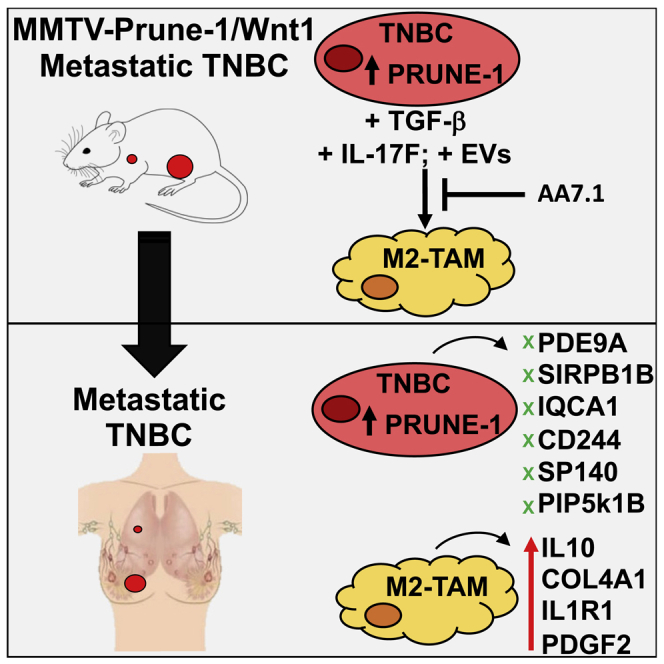
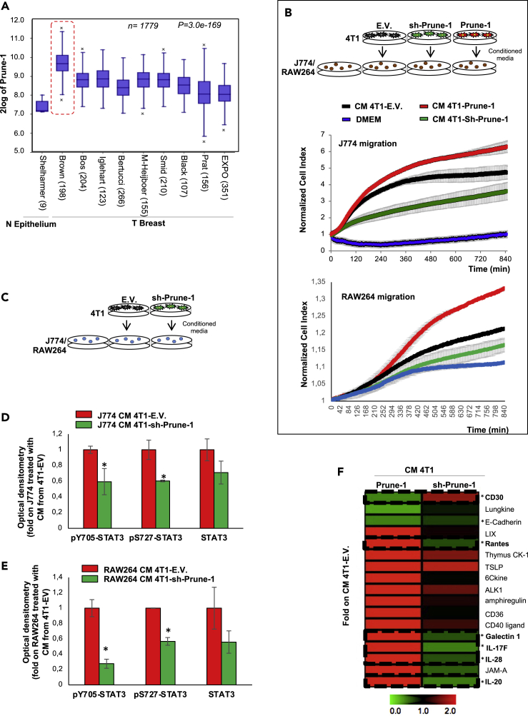
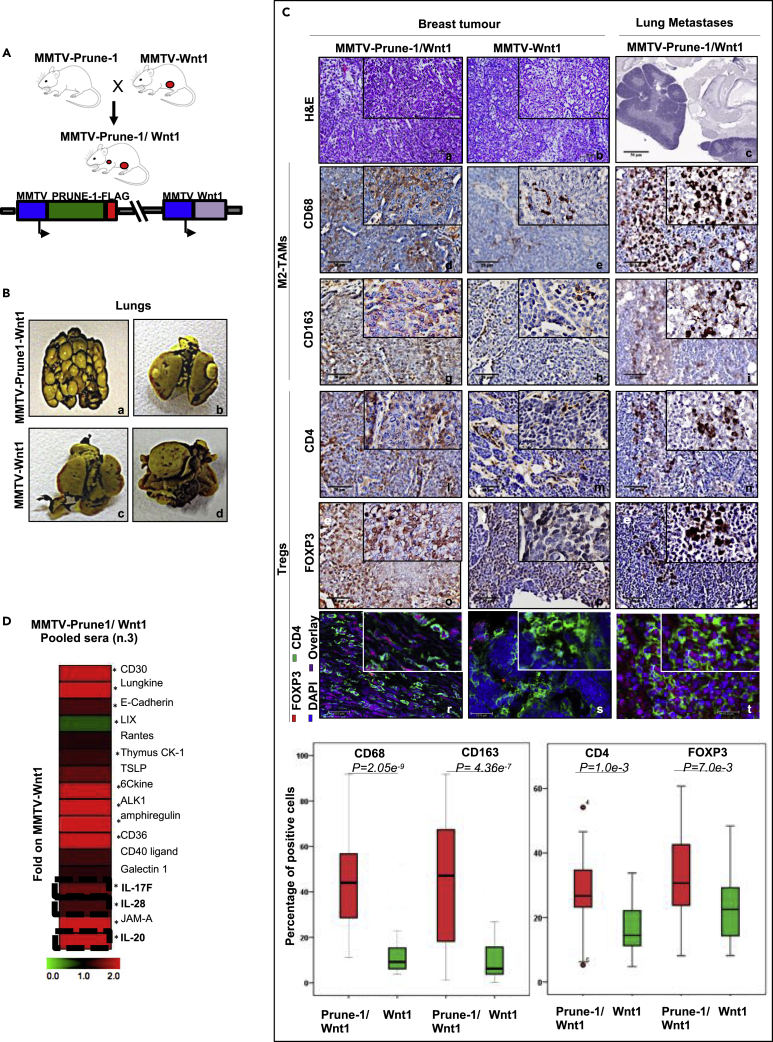
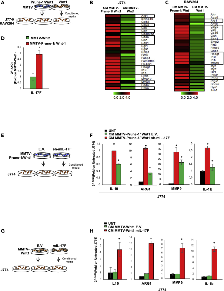
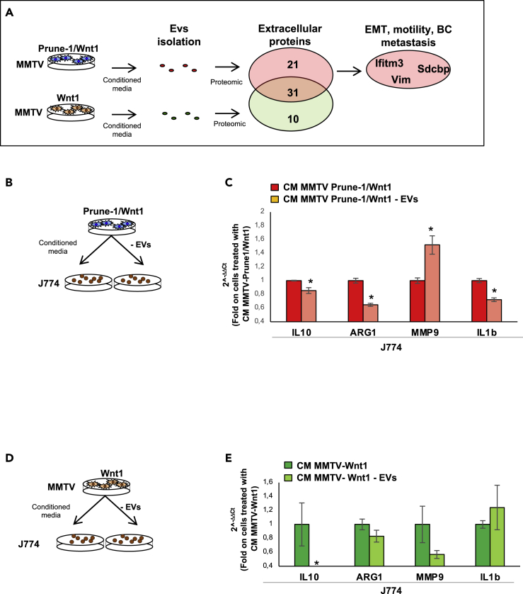
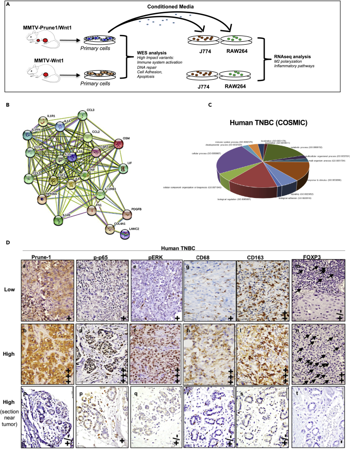
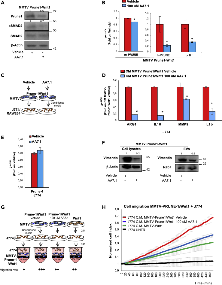
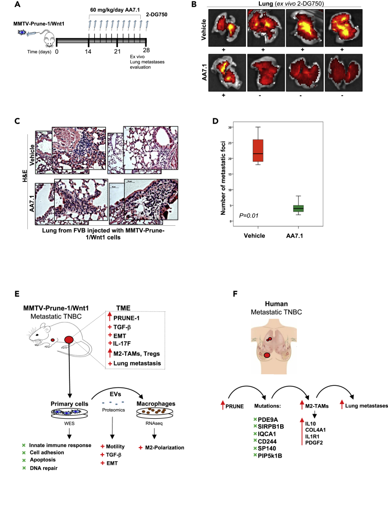
M2-tumor-associated macrophages (M2-TAMs) in the tumor microenvironment represent a prognostic indicator for poor outcome in triple-negative breast cancer (TNBC). Here we show that Prune-1 overexpression in human TNBC patients has positive correlation to lung metastasis and infiltrating M2-TAMs. Thus, we demonstrate that Prune-1 promotes lung metastasis in a genetically engineered mouse model of metastatic TNBC augmenting M2-polarization of TAMs within the tumor microenvironment. Thus, this occurs through TGF-β enhancement, IL-17F secretion, and extracellular vesicle protein content modulation. We also find murine inactivating gene variants in human TNBC patient cohorts that are involved in activation of the innate immune response, cell adhesion, apoptotic pathways, and DNA repair. Altogether, we indicate that the overexpression of Prune-1, IL-10, COL4A1, ILR1, and PDGFB, together with inactivating mutations of PDE9A, CD244, Sirpb1b, SV140, Iqca1, and PIP5K1B genes, might represent a route of metastatic lung dissemination that need future prognostic validations.

Keywords: Cancer; Immunology; Molecular Biology.


Conflict of interest statement

All the authors declare no competing financial interests.

Figures

None
Graphical abstract
Figure 1
Figure 1
Prune-1 protein is overexpressed in TNBC and promotes macrophage chemotaxis through STAT3 via soluble cytokines (A) Prune-1 overexpression in TNBC. RNA log2 expression analysis of Prune-1 levels of primary BC samples across different publicly available datasets, compared with normal epithelium (N Epithelium; Shelharmer dataset only). Data from 10 independent public-domain BC gene-expression datasets show the overexpression of Prune-1 in all of the BC samples compared with normal epithelium. Higher Prune-1 expression levels are seen for TNBC samples (i.e., Brown (Burstein et al., 2015); red dashed line) (n = 1779; p = 3.0 × 10−169). (B) Overexpression of Prune-1 in TNBC cells enhances macrophage chemotaxis in vitro. Normalized Cell Index as a measure of cell migration/chemotaxis of J774A.1 (i.e., J774; upper panel) and RAW264.7 (i.e., RAW264; lower panel) macrophages as generated by the xCELLigence RTCA software. Migration kinetics were monitored in response to conditioned media from Prune-1-overexpressing (4T1–Prune-1, red), Prune-1-silenced (4T1–Sh-Prune-1, green), and empty vector 4T1 cell clones (black), used as chemoattractants. Dulbecco's modified Eagle's medium was used as the negative control (blue). (C–E) Conditioned media (CM) from the 4T1 clones were collected after 24 h. J774 or Raw264 macrophages were starved for 6 h in serum-free medium and then grown in the conditioned media for 30 min (C). Densitometer analyses of immunoblotting for the indicated proteins in J774 (D) and Raw264 (E) macrophages grown for 30 min in conditioned media from Prune-1-silenced and control 4T1 clones are shown. Empty vector (EV) 4T1 clones and untreated (UNT) macrophages were used as the negative controls. β-Actin levels were used as the loading control. ∗p < 0.05 in Student's t test compared with J774 (D) or Raw264 (E) treated with conditioned media from 4T1 EV control clones. (F) Prune-1 induces the secretion of soluble proteins by TNBC cells. Densitometer analyses of the cytokines upregulated and downregulated in the conditioned media (CM) derived from Prune-1-overexpressing (4T1–Prune-1) and Prune-1-silenced (4T1–Sh-Prune-1) 4T1 clones (MultiExperiment Viewer, http://www.tm4.org/mev.html). Among the 17 cytokines modulated by Prune-1 in the conditioned media collected from Prune-1-overexpressing (4T1–Prune-1), one was upregulated (CD30) and five were downregulated (Rantes, Galectin-1, IL-17F, IL-28, IL-20) in the conditioned media from 4T1–Sh-Prune-1 cell clones, thus following an opposite trend. ∗p < 0.05 in Student's t test comparing cytokines levels in conditioned media of 4T1–Sh-Prune-1 cells with those in 4T1 Empty Vector control clones.
Figure 2
Figure 2
Prune-1 protein overexpressed in metastatic TNBC mouse model (MMTV–Prune-1/Wnt1) promotes M2-TAMs recruitment in both primary tumor and metastatic niche via soluble cytokines (A) Schematic diagram showing the cross between MMTV–Prune-1 and MMTV–Wnt1 mice to obtain the double transgenic MMTV–Prune-1/Wnt1 model. (B) Representative photographs of the lungs from MMTV–Prune-1/Wnt1 (a–b) and MMTV–Wnt1 (c–d) mice fixed in Bouin's solution. Metastatic foci (i.e., macrometastasis) are visible in the lungs from MMTV–Prune-1/Wnt1 (a–b). (C) Representative hematoxylin-eosin staining (a–c), immunohistochemistry (IHC; d–q), and immunofluorescence (IF; r–t) performed on sections of mammary tumors developed from MMTV–Prune-1/Wnt1 and MMTV–Wnt1 mice, and metastatic lungs from MMTV–Prune-1/Wnt1 mice, using antibodies against the following: CD68 (d–f) and CD163 (g–i), as markers for M2-TAMs; CD4 (l–n), as a marker for T cells; and FOXP3 (o–q), as a marker for Tregs. Double indirect IF was performed to detect Tregs (i.e., CD4+ FOXP3+, r–t). CD4: green; FOXP3: red; DAPI: blue. Quantification was performed using a quantitative pathology workstation (Mantra) with image analysis software (inform). Data were calculated from three independent tumors. Graphs were constructed using the IBM SPSS statistics software. Magnification, 5×, 20×, 40×. Scale bar: 50 μm, hematoxylin-eosin; 20 μm, IHC; 10 μm, IF. (D) Prune-1 induces secretion of soluble cytokines in vivo. Densitometer analyses of the cytokines upregulated and downregulated in the sera collected and pooled from three MMTV–Prune-1/Wnt1 mice and MMTV–Wnt1 mice. Among the cytokines modulated by Prune-1, significant upregulation of IL-17F, IL-28, and IL-20 was seen, with an opposite trend compared with the cytokines in the conditioned media from Prune-1-silenced 4T1 cells. Data are means ± standard deviation. Data were represented using MultiExperiment Viewer (http://www.tm4.org/mev.html). ∗, p < 0.05 in Student's t test comparing cytokines levels of pooled sera from n.3 MMTV-Prune-1/Wnt1 mice with those from n.3 MMTV-Wnt1 mice.
Figure 3
Figure 3
Prune-1 overexpressed in TNBC cells promotes M2-polarization of macrophages in vitro through IL-17F (A–C) J774A.1 and Raw264 macrophages were grown for 48 h in conditioned media collected (after 24 h) from MMTV–Prune-1/Wnt1 or MMTV–Wnt1 cells (A). Fold-change heatmap (http://www.tm4.org/mev.html) for upregulated M2-associated genes (fold change on untreated macrophages >2, p < 0.01) in J774A.1 (B) and Raw264 (C) macrophages grown in conditioned media from MMTV–Prune-1/Wnt1 versus MMTV–Wnt1 cells. The black boxes indicate the common genes upregulated in both J774A.1 and Raw264 macrophages. The color scale under the heatmap illustrates the fold-change values shown in the heatmap. (D) Real-time PCR analysis of IL-17F in MMTV–Prune-1/Wnt1 (red) and MMTV–Wnt1 (green) cells. ∗p < 0.05 in Student's t test comparing IL-17F levels of MMTV-Prune-1/Wnt1 with MMTV-Wnt1 cells. (E and F) J774A.1 macrophages were grown for 48 h in conditioned media collected (after 24 h) from MMTV–Prune-1/Wnt1 previously transfected with murine sh-IL17F plasmid or empty vector (EV) as negative control (E). Real-time PCR analysis of some M2-associated genes, including IL-10, Arg-1, MMP-9, and IL-1β, in J774A.1 macrophages grown for 48 h in conditioned media from MMTV–Wnt1 cells transfected with sh-IL-17F (red) or EV control (green) (F). Untreated J774A.1 macrophages were used as the negative controls (UNT, black). ∗p < 0.05 in Student's t test compared with untreated macrophages. (G and H) J774A.1 macrophages were grown for 48 h in conditioned media collected (after 24 h) from MMTV–Wnt1 previously transfected with murine IL-17F or EV as negative control (G). Real-time PCR analysis of some M2-associated genes, including Arg-1, MMP-9, and IL-1β, in J774A.1 macrophages grown for 48 h in conditioned media from MMTV–Wnt1 cells transfected with IL-17F (red) or EV control (green). Untreated J774A.1 macrophages were used as the negative controls (UNT, black) (H). ∗p < 0.05 in Student's t test compared with untreated macrophages.
Figure 4
Figure 4
Prune-1 overexpressed in TNBC cells promotes M2-polarization of macrophages in vitro through modulation of EV-protein content (A) Representative scheme for proteomic analyses performed on extracellular vesicles (EVs). EVs were isolated from media from murine primary MMTV–Prune-1/Wnt1 and MMTV–Wnt1 cells. Proteomic analyses were performed on isolated EVs using label-free quantitative mass spectrometry. Data show 31 extracellular proteins in common between EVs from media of MMTV–Prune-1/Wnt1 and MMTV–Wnt cells, and 21 and 10 mutually exclusive extracellular proteins from MMTV–Prune-1-Wnt cells (red) and MMTV–Wnt1 cells (green). Data are representative of two independent experiments. (B and C) J774 macrophages were grown for 48 h in conditioned media collected (after 24 h) from MMTV–Prune-1/Wnt1 cells depleted or not in EVs from the culture supernatant (B). Real-time PCR analysis of some M2-associated genes, including IL-10, Arg-1, MMP-9, and IL-1β, in J774 macrophages grown for 48 h in conditioned media from MMTV–Prune-1/Wnt1 depleted (red) or not (light red) in EVs (C). ∗p < 0.05 in Student's t test compared with macrophages treated with conditioned media from MMTV-Prune-1/Wnt1 cells not depleted in EVs. (D and E) J774 macrophages were grown for 48 h in conditioned media collected (after 24 h) from MMTV–Wnt1 cells depleted or not in EVs from the culture supernatant (D). Real-time PCR analysis of some M2-associated genes, including IL-10, Arg-1, MMP-9, and IL-1β, in J774 macrophages grown for 48 h in conditioned media from MMTV–Wnt1 depleted (light green) or not (dark green) in EVs (E). ∗p < 0.05 in Student's t test compared with macrophages treated with conditioned media from MMTV-Wnt1 cells not depleted in EVs.
Figure 5
Figure 5
Mutational spectrum in TNBC cells overexpressing Prune-1 regulating M2-macrophages polarization (A) Representative scheme for the experimental design. DNA from MMTV–Prune-1/Wnt1 and MMTV–Wnt1 cells was used for next-generation sequencing analyses through a whole-exome sequencing approach. J774A.1 and Raw264.7 macrophages were grown in conditioned media collected from MMTV–Prune-1/Wnt1 and MMTV–Wnt1 cells for 48 h. Total RNA was extracted from these macrophages, and RNAseq analyses were performed. (B) The inflammatory protein network generated via Search Tool for Retrieval of Interacting Genes/Proteins (STRING) database using the “core genes” defined as the common genes that are overexpressed in both the J774A.1 and RAW264.7 macrophages grown in media obtained from MMTV–Prune-1/Wnt1 cells (as compared with MMTV–Wnt1 cells) shared by at least four of five enriched gene sets from each canonical pathway sub-collection (i.e., Biocarta, Kegg, Pid, Reactome, Naba). The protein interaction network was generated using the STRING database (confidence: 0.4; https://string-db.org/cgi/network.pl?taskId=41jaTsLzWNqT). (C) Pie chart illustrating the Gene Ontology (GO) term analysis of deleterious variants of the 39 genes in MMTV–Prune-1/Wnt1 cells (compared with MMTV–Wnt1 cells) and in the public database of human basal TNBC (COSMIC, v91). (D) Representative immunostaining (IHC) from “tumor” (a–n) and “near tumor” (o–t) sections of our TNBC tissue cohort (n = 138) derived from patients who underwent mastectomy, quadrantectomy, or metastectomy at the “Giovanni Pascale” National Cancer Institute of Naples (Italy) from 2003 to 2010, with antibodies against the following: Prune-1 (a-b-o), phosphorylated-(Ser311)-p65 (c-d-p), phosphorylated-ERK1/2 (i.e., phospho-[Thr202/Tyr204]-ERK1 and phospho-[Thr185/Tyr187]-ERK2) (e-f-q), CD68 (g-h-r) and CD163 (i-l-s) (as markers for M2-TAMs), and FOXP3 (m-n-t) (as marker for Tregs). Magnification 40×. Scale bar: 50 μm; (−): negativity; (+): low immunopositivity; (++) medium immunopositivity; (+++): high immunopositivity.
Figure 6
Figure 6
The AA7.1 anti-Prune-1 agent affects macrophages polarization in vitro (A) Immunoblotting on total protein lysates of MMTV–Prune-1/Wnt1 cells treated with AA7.1 (100 μM, for 12 h) or PBS as the vehicle control, with the antibodies as indicated. (B) Real-time RT-PCR of total RNA extracted from MMTV–Prune-1/Wnt1 cells treated with AA7.1 (100 μM, for 12 h) or PBS as the vehicle control, to detect human and murine Prune-1 and murine IL-17F. ∗, p < 0.05 in Student's t test compared with vehicle-treated MMTV–Prune-1/Wnt1 cells. (C and D) Schematic representation of experimental design. J774A.1 macrophages were grown for 48 h in conditioned media collected from MMTV–Prune-1/Wnt1 cells treated with AA7.1 (100 μM, for 12 h) or PBS as the vehicle control (C). Real-time PCR analysis of some M2-associated genes, including IL-10, Arg-1, MMP-9, and IL-1β, in J774A.1 macrophages grown for 48 h in conditioned media collected from MMTV–Prune-1/Wnt1 cells treated with AA7.1 (blue) or PBS, as the vehicle control (red) (D). ∗p < 0.05 in Student's t test compared with macrophages grown in conditioned media collected from vehicle-treated MMTV–Prune-1/Wnt1 cells. (E) Real-time RT-PCR of total RNA extracted from J774A.1 macrophages treated with AA7.1 (100 μM, for 48 h). (F) Immunoblotting of total protein lysates or EVs of MMTV–Prune-1/Wnt1 cells treated with AA7.1 (100 μM) or PBS as the vehicle control, with the antibodies as indicated. (G and H) Co-culture experiments to measure migration rates of MMTV–Prune-1/Wnt1 cells (using 2% FBS as chemoattractant) in the presence of J774A.1 macrophages previously grown in conditioned media collected from MMTV–Prune-1/Wnt1 cells (untreated or treated with AA7.1) and MMTV–Wnt1 cells. Untreated macrophages were used as negative control (G). Normalized Cell Index as a measure of cell migration of MMTV–Prune-1/Wnt1 cells, as generated by the xCELLigence RTCA software. Migration kinetics were monitored (every 5 min) in response to the presence of untreated macrophages (black) or J774A.1 macrophages previously grown in conditioned media collected from vehicle-treated or AA7.1-treated MMTV–Prune-1/Wnt1 cells (red and blue, respectively) and MMTV–Wnt1 cells (green). The grading on migration rate is evaluated as the difference of Cell Index values observed at the end of the experiment. +++, 1.6; ++, 1.35; +/−, 1.04.
Figure 7
Figure 7
Hypothesized mechanisms of action of Prune-1 in the tumor microenvironment in the cross-talk between TNBC cells and macrophages (A–D) Schematic representation of the in-vivo trial. MMTV–Prune-1/Wnt1 cells (1 ×105) were injected via the tail vein in eight immunocompetent syngeneic mice (strain FVB). After 14 days from cell injection, the mice were grouped according to their weight and AA7.1 (60 mg/kg/day, IP) or PBS (as vehicle control) was administered daily. At 14 days from treatment start (i.e., 28 days from cell injection), the mice were injected with a fluorescent imaging probe (XenoLight RediJect 2-DG-750; Perkin Elmer) for ex-vivo targeting of the tumorigenic cells (A). Positive fluorescence signals were detected in the lungs derived from all of the control mice (treated with PBS as vehicle) and in one of four AA7.1-treated mice (i.e., 25%) (B). Hematoxylin/eosin staining performed on sections of lung tissue from AA7.1-treated mice compared with controls. Magnification: 4x, 20x, 40x; Scale bars: 200 μm, 50 μm, 20 μm (C). Box plot (generated with the SPSS software) showing differences in the number of metastatic foci detected in the lungs from AA7.1-treated mice (green), compared with the control group (red) (D). (E and F) The proposed mechanism of action of Prune-1 at the interplay between tumorigenic cells and TAMs in TME is shown for the murine model of metastatic TNBC (i.e., MMTV–Prune-1/Wnt1). Prune-1 acts in metastatic TNBC by promoting activation of intracellular pathways (i.e., TGF-β) and in a paracrine manner through the release of extracellular inflammatory molecules (i.e., IL-17F) and modulation of extracellular vesicle protein content (i.e., Sdcbp, Vim, Iftm3) involved in EMT and metastasis. Furthermore, mutational analyses in murine primary TNBC cells overexpressing Prune-1 (i.e., MMTV–Prune-1/Wnt1 cells) showed predicted deleterious variants in genes involved in activation of the innate immune response, apoptotic pathways, DNA repair, and cell adhesion. These gene variants were also found in patients with TNBC. In detail, in human, we identified deleterious variants for six genes that are mainly involved in the activation of the innate immune response. We also found upregulation of IL-10, COL4A1, ILR1, and PDGFB, the expression levels of which are negatively correlated with prognosis in patients with TNBC. Altogether, these actions induce recruitment of TAMs in the TNBC microenvironment and promote their polarization toward anti-inflammatory/pro-tumorigenic M2-status, thus preparing the system for lung metastasis within the premetastatic niche.

Similar articles

Cited by

References

    1. Adams T.A., Vail P.J., Ruiz A., Mollaee M., Mccue P.A., Knudsen E.S., Witkiewicz A.K. Composite analysis of immunological and metabolic markers defines novel subtypes of triple negative breast cancer. Mod. Pathol. 2017;31:288–298. - PMC - PubMed
    1. Bartholomeusz C., Gonzalez-Angulo A.M., Liu P., Hayashi N., Lluch A., Ferrer-Lozano J., Hortobagyi G.N. High ERK protein expression levels correlate with shorter survival in triple-negative breast cancer patients. Oncologist. 2012;17:766–774. - PMC - PubMed
    1. Bianchini G., Balko J.M., Mayer I.A., Sanders M.E., Gianni L. Triple-negative breast cancer: challenges and opportunities of a heterogeneous disease. Nat. Rev. Clin. Oncol. 2016;13:674–690. - PMC - PubMed
    1. Boye K., Grotterod I., Aasheim H.C., Hovig E., Maelandsmo G.M. Activation of NF-kappaB by extracellular S100A4: analysis of signal transduction mechanisms and identification of target genes. Int. J. Cancer. 2008;123:1301–1310. - PubMed
    1. Burstein M.D., Tsimelzon A., Poage G.M., Covington K.R., Contreras A., Fuqua S.A., Savage M.I., Osborne C.K., Hilsenbeck S.G., Chang J.C. Comprehensive genomic analysis identifies novel subtypes and targets of triple-negative breast cancer. Clin. Cancer Res. 2015;21:1688–1698. - PMC - PubMed